. 2022 Nov;192(11):1546-1558.
doi: 10.1016/j.ajpath.2022.07.006. Epub 2022 Aug 7.

Impaired Mitochondria Promote Aging-Associated Sebaceous Gland Dysfunction and Pathology

Affiliations

Impaired Mitochondria Promote Aging-Associated Sebaceous Gland Dysfunction and Pathology

Noha S Ahmed et al. Am J Pathol. 2022 Nov.

Abstract

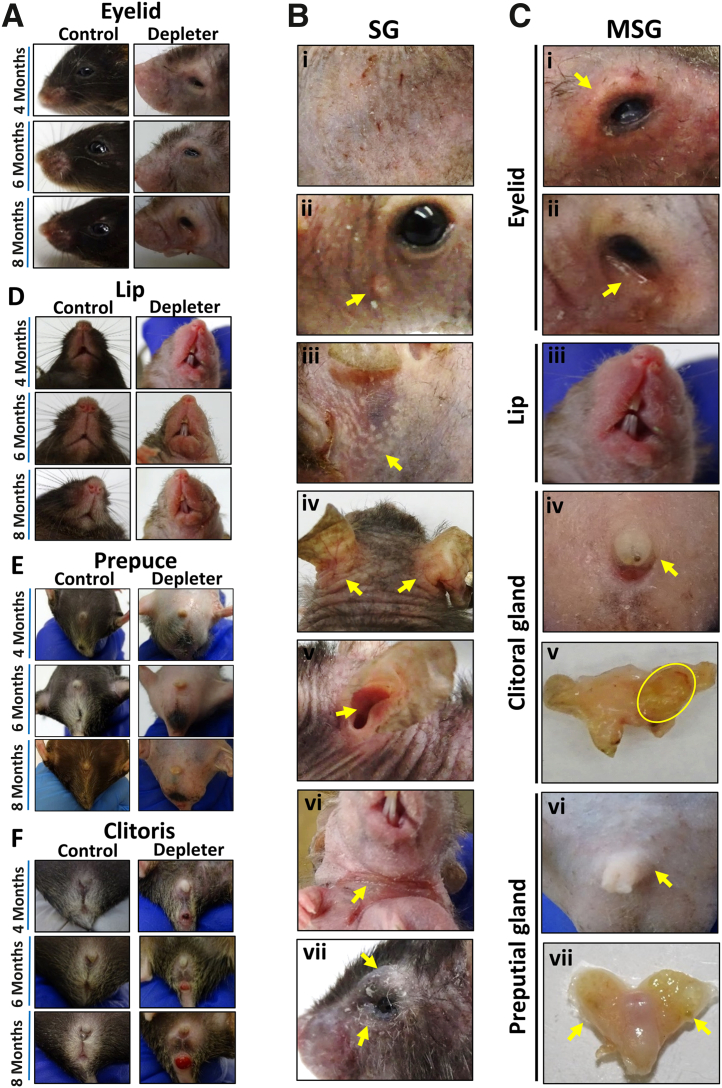
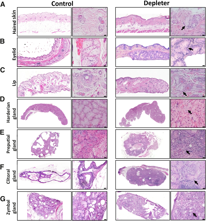
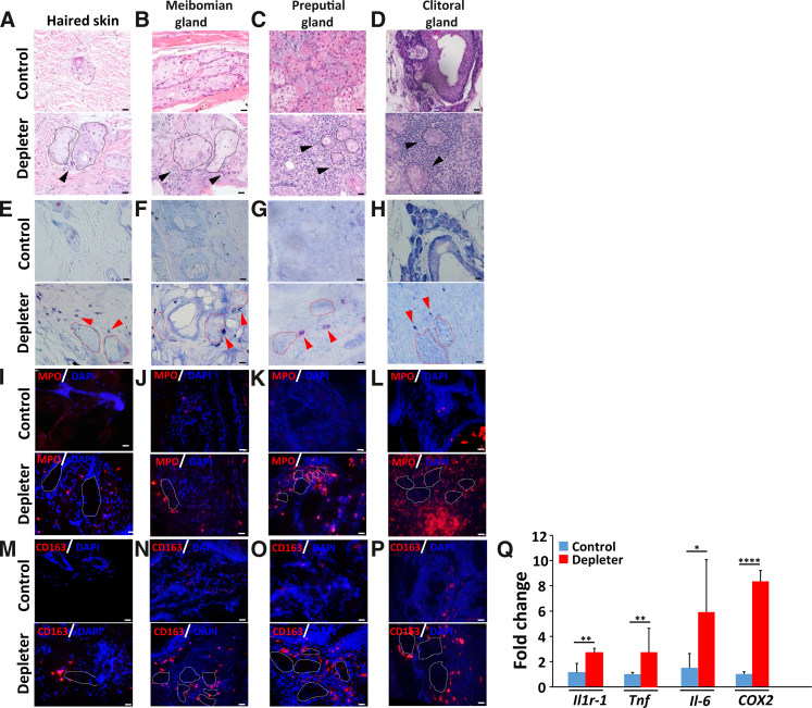
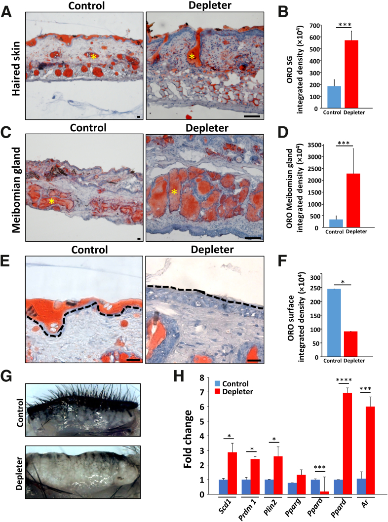
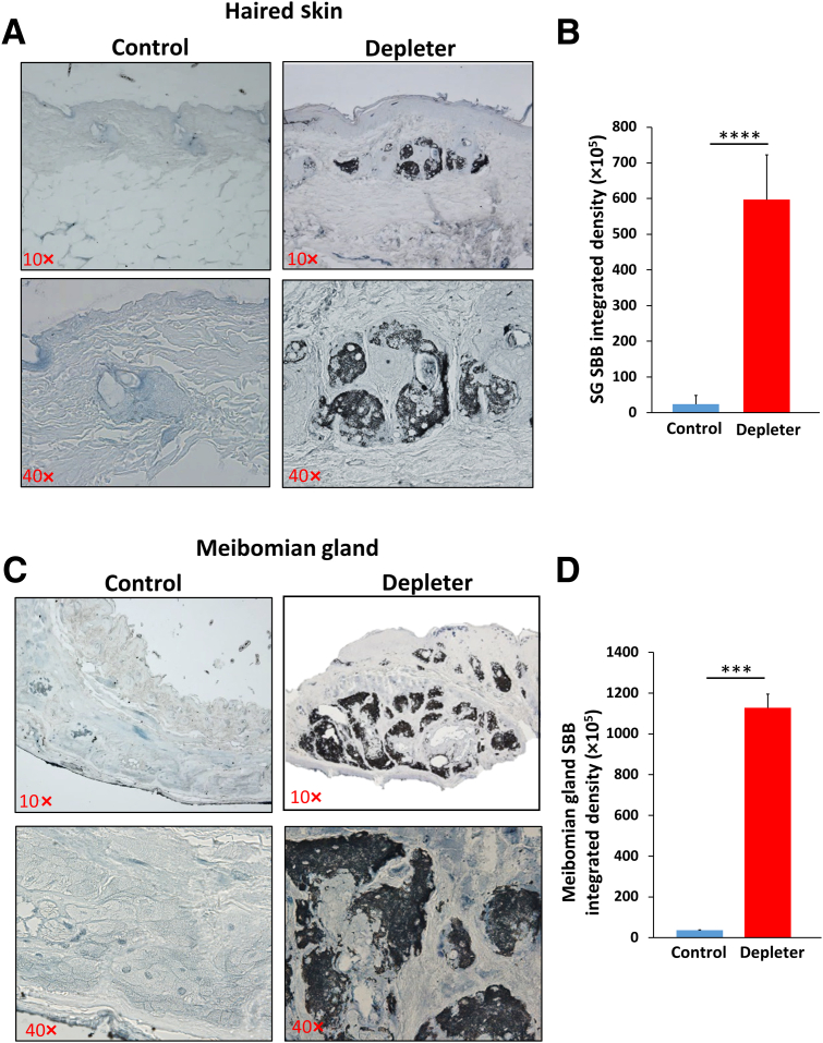
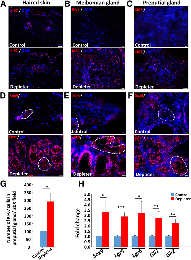
Mitochondrial dysfunction is one of the hallmarks of aging. Changes in sebaceous gland (SG) function and sebum production have been reported during aging. This study shows the direct effects of mitochondrial dysfunction on SG morphology and function. A mitochondrial DNA (mtDNA) depleter mouse was used as a model for introducing mitochondrial dysfunction in the whole animal. The effects on skin SGs and modified SGs of the eyelid, lip, clitoral, and preputial glands were characterized. The mtDNA depleter mice showed gross morphologic and histopathologic changes in SGs associated with increased infiltration by mast cells, neutrophils, and polarized macrophages. Consistently, there was increased expression of proinflammatory cytokines. The inflammatory changes were associated with abnormal sebocyte accumulation of lipid, defective sebum delivery at the skin surface, and the up-regulation of key lipogenesis-regulating genes and androgen receptor. The mtDNA depleter mice expressed aging-associated senescent marker. Increased sebocyte proliferation and aberrant expression of stem cell markers were observed. These studies provide, for the first time, a causal link between mitochondrial dysfunction and abnormal sebocyte function within sebaceous and modified SGs throughout the whole body of the animal. They suggest that mtDNA depleter mouse may serve as a novel tool to develop targeted therapeutics to address SG disorders in aging humans.


Figures

Figure 1
Figure 1
Phenotypic sebaceous gland (SG) and modified sebaceous gland (MSG) abnormalities due to mitochondrial dysfunction. Representative images were taken of mitochondrial DNA (mtDNA) depleter mice after doxycycline diet induction showing. Representative time-course images of mice taken over 4, 6, and 8 months of doxycycline induction for control and mtDNA depleter mice. A: Eyelid images showing no changes in control mice over time, and mtDNA depleter mice showing loss of eyelashes with erythematous scaly irregular lid margins. B: The spectrum of cutaneous phenotypes suggestive of SG dysfunction observed in the mtDNA depleter mouse model, including the following: i) scaly erythematous skin, ii) yellow umbilicated papules (yellow arrow) on the face, iii) deposition of yellow plaques in stretched skin (yellow arrow), iv) erythema and scaling behind ears (yellow arrows) and v) inside the ear (yellow arrow), vi) erythematous neck flexure accompanied with ulceration (yellow arrow), and vii) swelling, erythema, and scaling on eyelid margins (yellow arrows). C: The spectrum of phenotypes suggestive of MSG dysfunction observed in the mtDNA depleter mouse model, comprising the following: i) swollen, erythematous eyelids with telangiectasia (yellow arrow), ii) erythematous and irregular lower lid margin (yellow arrow), iii) enlarged and erythematous upper and lower lips, iv) enlarged external clitoris (yellow arrow) that shows v) enlargement of clitoral glands on dissection and abscess formation (yellow oval) in female mice, and vi) enlargement of prepuce externally (yellow arrow) with vii) enlarged preputial glands on dissection in male mice (yellow arrows). D: Lip images show no differences in control mice over time, whereas mtDNA depleter mice show lip erythema and swelling. E: Swelling of the prepuce in male mtDNA depleter mice. F: Swelling of the clitoris in female mtDNA depleter mice.
Figure 2
Figure 2
Mitochondrial dysfunction–induced histopathologic alterations in sebaceous and modified sebaceous glands. A: Representative hematoxylin and eosin–stained sections after 8 months of doxycycline induction of skin-associated sebaceous glands. BG: Modified sebaceous glands of eyelids (meibomian glands; B), lips (C), harderian gland (D), preputial gland (E), clitoral gland (F), and zymbal gland (G). Hyperplastic changes associated with sebocytes (black arrows) and variable amounts of mixed inflammatory infiltrate within the dermis/submucosa. Hyperplastic changes include increased cell and nuclear size, irregular cellular orientation within glands, and inflammation, most notably within clitoral and preputial glands. Scale bars = 50 μm (AG).
Figure 3
Figure 3
Impaired mitochondria induce inflammation. Representative images of skin sebaceous glands (dashed line) and modified sebaceous glands (dashed line), including meibomian, preputial, and clitoral glands. AD: Hematoxylin and eosin stain, showing inflammatory infiltrate (black arrowheads) in mitochondrial DNA (mtDNA) depleter mice centered around sebaceous glands (black dashed line). EH: Mast cells were evaluated using Giemsa stain (red arrowheads; Giemsa stain–positive cells) around sebaceous glands (red dashed line). IP: Granulocytes (myeloperoxidase-positive cells; IL) and M2-polarized macrophages (CD163-positive cells; MP) around sebaceous glands (white dashed line). Q: Quantitative RT-PCR analysis of inflammatory genes (Il1r1, Tnf, Il6, and COX2) in mRNA extracted from the preputial gland of male mice after 8 months of doxycycline induction showed higher expression in mtDNA depleter samples compared with control. Transcripts have been done in triplicates and were determined by normalizing to β-actin. Data are given as means ± SD (Q). ∗P ≤ 0.05, ∗∗P ≤ 0.01, and ∗∗∗∗P ≤ 0.0001 (t-test). Scale bars = 20 μm (AP).
Figure 4
Figure 4
Abnormal lipid accumulation in sebaceous glands (SGs) and modified sebaceous glands. Representative images of skin and eyelid stained with Oil Red O (ORO) for lipids, showing lipid accumulation in mitochondrial DNA (mtDNA) depleter mice after 8 months of doxycycline induction. A: Skin sebaceous glands (yellow asterisks). B: Quantification of ORO integrated density in SG compared with control littermate. C: Modified eyelid sebaceous glands (meibomian glands; yellow asterisks). D: Quantification of ORO integrated density in meibomian glands compared with control littermate. E: Oil Red O stain, showing no or minimal lipid layer on skin surface compared with continuous lipid layer in control skin (black dashed line). F: Quantification of ORO integrated density on the skin surface. G: Representative gross image, showing distension of meibomian glands in mtDNA depleter mice after 2 months of doxycycline induction compared with control. H: Quantitative RT-PCR analysis of lipogenesis-regulating genes (Scd1, Prdm1, Plin2, Ppara, Pparg, and Ppard) and androgen receptor gene (Ar) in mRNA extracted from the preputial gland of male mice after 8 months of doxycycline induction, showing higher expression in mtDNA depleter samples compared with control. Transcripts were done in triplicates and were determined by normalizing to β-actin. Data are given as means ± SD (B, D, F, and H). ∗P ≤ 0.05, ∗∗∗P ≤ 0.001, and ∗∗∗∗P ≤ 0.0001 (t-test). Scale bars = 20 μm (A, C, and E).
Figure 5
Figure 5
Mitochondrial dysfunction causes accumulation of senescent cells in sebaceous glands (SGs). Representative images stained with Sudan black B (SBB) for lipofuscin detection in skin sebaceous glands (A) and eyelid meibomian glands (C), showing characteristic deposition of blue-black pigments that represent positivity for lipofuscin in sebaceous glands of mitochondrial DNA depleter mice compared with control. Quantification of SBB integrated density in skin SG (B) and meibomian glands (D). Data are given as means ± SD (B and D). ∗∗∗P ≤ 0.001, ∗∗∗∗P ≤ 0.0001 (t-test). Original magnification, ×10 (A and C, top panels); ×40 (A and C, bottom panels).
Figure 6
Figure 6
Abnormal proliferation and changes in expression of stem cell markers. AC: Representative immunofluorescence staining of skin sebaceous glands and modified sebaceous glands (A), eyelid meibomian glands (B), and preputial gland (C) with proliferation marker Ki-67 (red) counterstained with nuclear stain DAPI (blue), showing higher expression in sebaceous glands of mitochondrial DNA (mtDNA) depleter mice compared with control. DF: Immunohistochemical staining of skin (D), eyelid meibomian glands (E), and preputial gland (F) sections with Sox9 (red) with nuclei counterstained with DAPI (blue) in mtDNA depleter mice compared with control, Sebaceous gland marked with the white dashed line. G: Quantitative analysis of Ki-67–stained cells in the preputial gland. H: Quantitative RT-PCR analysis of sebaceous gland stem cell expressed genes (Sox9, Lgr6, Lgr5, Gli1, and Gli2) in the preputial gland of mtDNA depleter mice compared with control. Transcripts were done in triplicates and were determined by normalizing to β-actin. Data are given as means ± SD (G and H). ∗P ≤ 0 .05, ∗∗P ≤ 0.01, and ∗∗∗P ≤ 0.001 (t-test). Scale bars: 50 μm (AC); 20 μm (DF).

References

    1. Schneider M.R., Paus R. Sebocytes, multifaceted epithelial cells: lipid production and holocrine secretion. Int J Biochem Cell Biol. 2010;42:181–185. - PubMed
    1. Thody A.J., Shuster S. Control and function of sebaceous glands. Physiol Rev. 1989;69:383–416. - PubMed
    1. Smith K.R., Thiboutot D.M. Thematic review series: skin lipids: sebaceous gland lipids: friend or foe? J Lipid Res. 2008;49:271–281. - PubMed
    1. Feingold K.R. The outer frontier: the importance of lipid metabolism in the skin. J Lipid Res. 2009;50:S417–S422. - PMC - PubMed
    1. Drake D.R., Brogden K.A., Dawson D.V., Wertz P.W. Thematic Review Series: skin lipids: antimicrobial lipids at the skin surface. J Lipid Res. 2008;49:4–11. - PubMed

Publication types

Substances