

Load-to-strength ratio at the radius is higher in adolescent and young adult females with obesity compared to normal-weight controls
- PMID: 35948256
- PMCID: PMC9826712
- DOI: 10.1016/j.bone.2022.116515
Load-to-strength ratio at the radius is higher in adolescent and young adult females with obesity compared to normal-weight controls
Abstract
Background: Among adolescents with extremity fractures, individuals with obesity have greater representation compared with individuals of normal-weight, despite having higher areal and volumetric bone mineral density (aBMD, vBMD) than their normal-weight counterparts. The relative increase in BMD in individuals with obesity may thus be insufficient to support the greater force generated upon falling. The load-to-strength ratio is a biomechanical approach for assessing the risk of fracture by comparing applied force to bone strength, with higher load-to-strength ratios indicating higher fracture risk.
Objective: To assess the load-to-strength ratio at the distal radius in adolescent and young adult females with severe obesity (OB) compared with normal-weight healthy controls (HC). We hypothesized that OB have a higher load-to-strength ratio compared to HC.
Methods: We examined bone parameters in 65 girls 14-21 years old: 33 OB and 32 HC. We used dual-energy X-ray absorptiometry (DXA) to assess body composition, high resolution peripheral quantitative CT (HR-pQCT) to estimate vBMD, and microfinite element analysis (μFEA) to assess bone strength at the distal radius. To quantify fracture risk, we computed the load-to-strength ratio, where the numerator is defined as the load applied to the outstretched hand during a forward fall and the denominator is the bone strength, as estimated by μFEA.
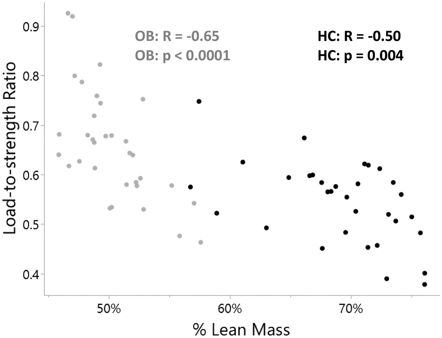
Results: Although OB had higher total vBMD than HC (368.3 vs. 319.9 mgHA/cm3, p = 0.002), load-to-strength ratio at the radius was greater in OB than HC after controlling for age and race (0.66 vs. 0.54, p < 0.0001). In OB, impact force and load-to-strength ratio were associated negatively with % lean mass (r = -0.49; p = 0.003 and r = -0.65; p < 0.0001 respectively) and positively with visceral fat (r = 0.65; p < 0.0001 and r = 0.36; p = 0.04 respectively).
Conclusions: Adolescent and young adult females with obesity have higher load-to-strength ratio at the distal radius due to higher forces applied to bone in a fall combined with incomplete adaptation of bone to increasing body weight. This is differentially affected by lean mass, fat mass, and visceral fat mass.
Keywords: Adolescents; Fracture; Load-to-strength ratio; Obesity; Radius.
Copyright © 2022 Elsevier Inc. All rights reserved.
Conflict of interest statement
Figures

References
-
- Hurt RT, et al., Obesity epidemic: overview, pathophysiology, and the intensive care unit conundrum. JPEN J Parenter Enteral Nutr, 2011. 35(5 Suppl): p. 4s–13s. - PubMed
-
- Marcus R, et al., Correlates of bone mineral density in the postmenopausal estrogen/progestin interventions trial. J Bone Miner Res, 1994. 9(9): p. 1467–76. - PubMed
-
- Ravn P, et al., Low body mass index is an important risk factor for low bone mass and increased bone loss in early postmenopausal women. Early Postmenopausal Intervention Cohort (EPIC) study group. J Bone Miner Res, 1999. 14(9): p. 1622–7. - PubMed
-
- Goulding A, Grant AM, and Williams SM, Bone and Body Composition of Children and Adolescents With Repeated Forearm Fractures. Journal of Bone and Mineral Research, 2005. 20(12): p. 2090–2096. - PubMed
-
- Macneil JA and Boyd SK, Bone strength at the distal radius can be estimated from high-resolution peripheral quantitative computed tomography and the finite element method. Bone, 2008. 42(6): p. 1203–13. - PubMed
Publication types
MeSH terms
Grants and funding
LinkOut - more resources
Full Text Sources
Medical
Miscellaneous
