

Neural and behavioral effects of modification of visual attention in body dysmorphic disorder
- PMID: 35948537
- PMCID: PMC9365821
- DOI: 10.1038/s41398-022-02099-2
Neural and behavioral effects of modification of visual attention in body dysmorphic disorder
Abstract
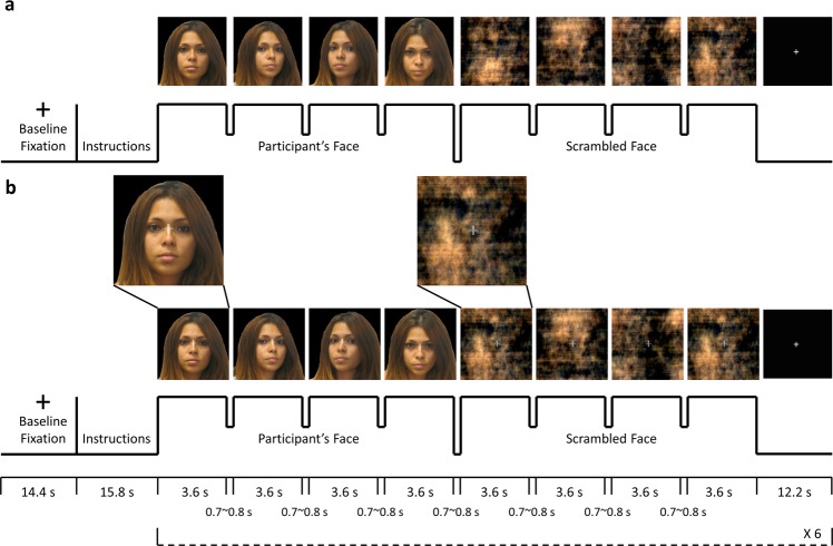
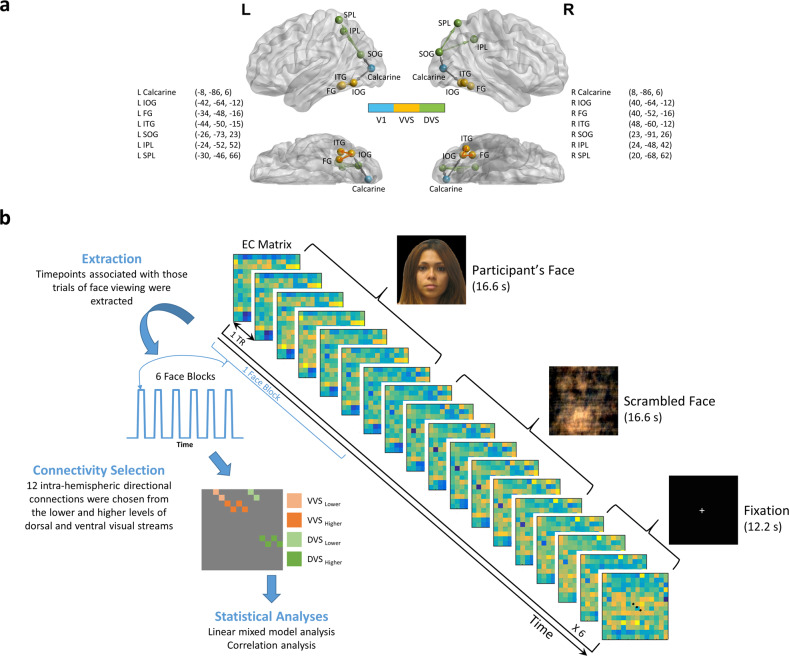
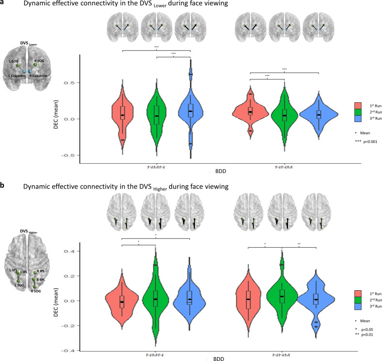
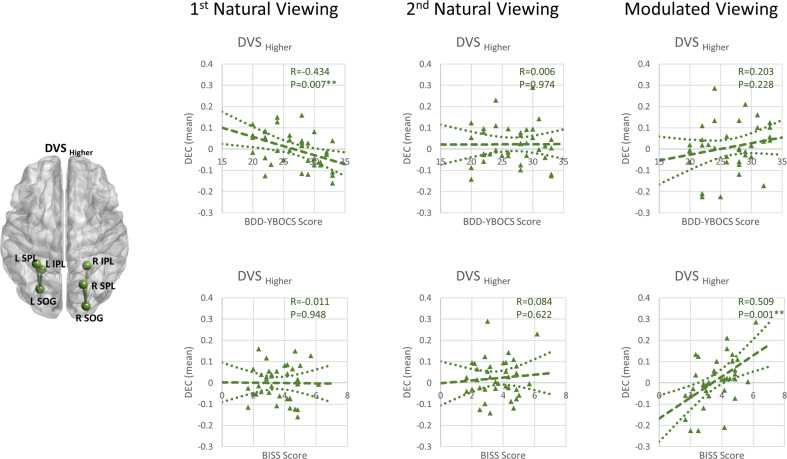
In individuals with body dysmorphic disorder (BDD), perceptual appearance distortions may be related to selective attention biases and aberrant visual scanning, contributing to imbalances in global vs. detailed visual processing. Treatments for the core symptom of perceptual distortions are underexplored in BDD; yet understanding their mechanistic effects on brain function is critical for rational treatment development. This study tested a behavioral strategy of visual-attention modification on visual system brain connectivity and eye behaviors. We acquired fMRI data in 37 unmedicated adults with BDD and 30 healthy controls. Participants viewed their faces naturalistically (naturalistic viewing), and holding their gaze on the image center (modulated viewing), monitored with an eye-tracking camera. We analyzed dynamic effective connectivity and visual fixation duration. Modulated viewing resulted in longer mean visual fixation duration compared to during naturalistic viewing, across groups. Further, modulated viewing resulted in stronger connectivity from occipital to parietal dorsal visual stream regions, also evident during the subsequent naturalistic viewing, compared with the initial naturalistic viewing, in BDD. Longer fixation duration was associated with a trend for stronger connectivity during modulated viewing. Those with more severe BDD symptoms had weaker dorsal visual stream connectivity during naturalistic viewing, and those with more negative appearance evaluations had weaker connectivity during modulated viewing. In sum, holding a constant gaze on a non-concerning area of one's face may confer increased communication in the occipital/parietal dorsal visual stream, facilitating global/holistic visual processing. This effect shows persistence during subsequent naturalistic viewing. Results have implications for perceptual retraining treatment designs.
© 2022. The Author(s).
Conflict of interest statement
The authors declare no competing interests.
Figures
Similar articles
-
Dynamic Effective Connectivity Patterns During Rapid Face Stimuli Presentation in Body Dysmorphic Disorder.Front Neurosci. 2022 May 24;16:890424. doi: 10.3389/fnins.2022.890424. eCollection 2022. Front Neurosci. 2022. PMID: 35685771 Free PMC article.
-
Effects of visual attention modulation on dynamic functional connectivity during own-face viewing in body dysmorphic disorder.Neuropsychopharmacology. 2021 Oct;46(11):2030-2038. doi: 10.1038/s41386-021-01039-w. Epub 2021 May 28. Neuropsychopharmacology. 2021. PMID: 34050267 Free PMC article.
-
Brain activation and connectivity in anorexia nervosa and body dysmorphic disorder when viewing bodies: relationships to clinical symptoms and perception of appearance.Brain Imaging Behav. 2021 Jun;15(3):1235-1252. doi: 10.1007/s11682-020-00323-5. Brain Imaging Behav. 2021. PMID: 32875486 Free PMC article.
-
Visual processing in anorexia nervosa and body dysmorphic disorder: similarities, differences, and future research directions.J Psychiatr Res. 2013 Oct;47(10):1483-91. doi: 10.1016/j.jpsychires.2013.06.003. Epub 2013 Jun 27. J Psychiatr Res. 2013. PMID: 23810196 Free PMC article. Review.
-
A hierarchy of visual processing deficits in body dysmorphic disorder: a conceptual review and empirical investigation.Cogn Neuropsychiatry. 2024 Mar;29(2):116-140. doi: 10.1080/13546805.2024.2326243. Epub 2024 Apr 2. Cogn Neuropsychiatry. 2024. PMID: 38563811 Review.
Cited by
-
Case Report: Physiological and psychological underpinnings of muscle dysmorphia using EEG, GSR, and eye-tracking.Front Psychol. 2025 Jul 21;16:1553997. doi: 10.3389/fpsyg.2025.1553997. eCollection 2025. Front Psychol. 2025. PMID: 40761458 Free PMC article.
-
A systematic review of neurocognition and social cognition in body dysmorphic disorder.Aust N Z J Psychiatry. 2025 Mar;59(3):224-247. doi: 10.1177/00048674241309747. Epub 2025 Jan 7. Aust N Z J Psychiatry. 2025. PMID: 39764591 Free PMC article.
-
Advancing Psychosocial Treatment for Body Dysmorphic Disorder: A State-of-the-Science Review.Behav Ther. 2024 Nov;55(6):1249-1288. doi: 10.1016/j.beth.2024.04.002. Epub 2024 Apr 10. Behav Ther. 2024. PMID: 39443065 Free PMC article. Review.
-
Body dysmorphic disorder.Nat Rev Dis Primers. 2024 Dec 5;10(1):92. doi: 10.1038/s41572-024-00577-z. Nat Rev Dis Primers. 2024. PMID: 39639018 Free PMC article. Review.
-
Learning of the same task subserved by substantially different mechanisms between patients with body dysmorphic disorder and healthy individuals.Cereb Cortex. 2024 May 2;34(5):bhae215. doi: 10.1093/cercor/bhae215. Cereb Cortex. 2024. PMID: 38798001 Free PMC article.
References
-
- Phillips KA. The broken mirror. NY: Oxford University Press; 2005.
-
- Phillips KA, McElroy SL, Keck PE, Jr, Hudson JI, Pope HG., Jr A comparison of delusional and nondelusional body dysmorphic disorder in 100 cases. Psychopharmacol Bull. 1994;30:179–86. - PubMed
Publication types
MeSH terms
Grants and funding
LinkOut - more resources
Full Text Sources
