. 2022 Aug 10;12(1):13610.
doi: 10.1038/s41598-022-17903-8.

Drinking hydrogen water improves photoreceptor structure and function in retinal degeneration 6 mice

Affiliations

Drinking hydrogen water improves photoreceptor structure and function in retinal degeneration 6 mice

Tsutomu Igarashi et al. Sci Rep. .

Abstract

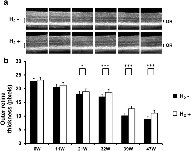
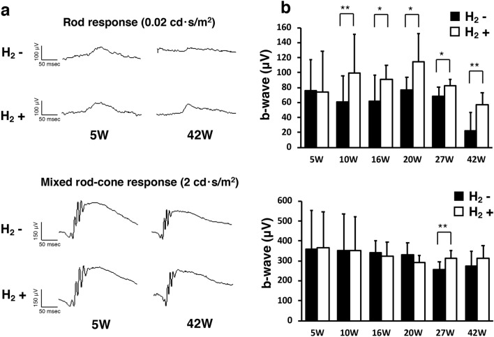
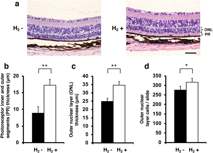
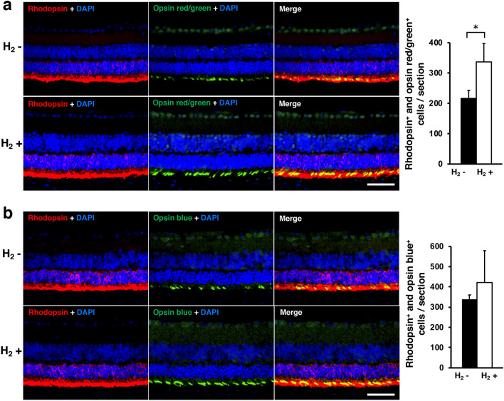
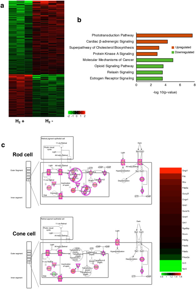
Retinitis pigmentosa (RP) is a genetically heterogeneous group of inherited retinal disorders involving the progressive dysfunction of photoreceptors and the retinal pigment epithelium, for which there is currently no treatment. The rd6 mouse is a natural model of autosomal recessive retinal degeneration. Given the known contributions of oxidative stress caused by reactive oxygen species (ROS) and selective inhibition of potent ROS peroxynitrite and OH·by H2 gas we have previously demonstrated, we hypothesized that ingestion of H2 water may delay the progression of photoreceptor death in rd6 mice. H2 mice showed significantly higher retinal thickness as compared to controls on optical coherence tomography. Histopathological and morphometric analyses revealed higher thickness of the outer nuclear layer for H2 mice than controls, as well as higher counts of opsin red/green-positive cells. RNA sequencing (RNA-seq) analysis of differentially expressed genes in the H2 group versus control group revealed 1996 genes with significantly different expressions. Gene and pathway ontology analysis showed substantial upregulation of genes responsible for phototransduction in H2 mice. Our results show that drinking water high in H2 (1.2-1.6 ppm) had neuroprotective effects and inhibited photoreceptor death in mice, and suggest the potential of H2 for the treatment of RP.


Conflict of interest statement

The authors declare no competing interests.

Figures

Figure 1
Figure 1
Preservation of H2 concentration in H2 water. (a) To maintain a high concentration of H2 in water, we developed water drinking valves. (b) The hydrogen concentration was 96.84 ± 1.03% before drinking and 70.68 ± 0.31% 1 week after drinking (C57BL/6J mice, n = 4). Bars depict mean ± standard deviation (SD).
Figure 2
Figure 2
Effect of H2 water on outer retinal thickness. (a) Representative retinal projections of OCT scans at 6–47 weeks of age. Double-headed arrows show the outer retina. OR outer retina. (b) Quantification of the outer retina thickness with/without H2 water. The outer retina thickness with H2 water (n = 10) was significantly greater than that without H2 water (n = 8) (p < 0.001 and 0.05). Bars depict mean ± standard deviation (SD). *p < 0.05, ***p < 0.001.
Figure 3
Figure 3
B-wave of ERGs in rd6 mice with/without H2 water. (a) Representative b-wave with/without H2 water at 0.02 cd·s/m2 and 2 cd·s/m2. (b) Quantification of the b-wave amplitude from 5 to 42 weeks of age. We found a significant difference between the control group (n = 8) and H2 group (n = 10). Bars depict means ± standard deviation (SD). *p < 0.05, **p < 0.01.
Figure 4
Figure 4
Thickness of photoreceptor inner and outer segments and outer nuclear layer and number of outer nuclear layer cells. (a) Images of representative slices from the control group (n = 4) and H2 group (n = 4). Photoreceptor inner and outer segments (PR) thickness and outer nuclear layer (ONL) are shown. (b) Photoreceptor inner and outer segments thickness with/without H2 water. Thickness was significantly greater with H2 water than without H2 water (p < 0.01). (c) Outer nuclear layer (ONL) thickness with/without H2 water. Thickness was significantly greater with H2 water than without H2 water (p < 0.01). (d) Outer nuclear layer cells/slide with/without H2 water. Thickness was significantly greater with H2 water than without H2 water (p < 0.05). Bars depict mean ± standard deviation (SD). Scale bar 50 μm. *p < 0.05, **p < 0.01.
Figure 5
Figure 5
Immunohistochemical analysis of rhodopsin and opsin. (a) Immunohistofluorescence co-staining for rhodopsin (red) and opsin red/green (green) is shown in representative retinal specimens with/without H2 water. Double-positive cells (yellow) were counted for eye cups in one slide. The number of double-positive cells was significantly greater in the control group (n = 4) than in the H2 group (n = 4; p < 0.05). (b) Immunohistofluorescence co-staining (yellow) for rhodopsin (red) and opsin blue (green) is shown in representative retinal specimens with/without H2 water. No significant difference was found (p = 0.35). Scale bar 50 μm. Bars depict mean ± standard deviation (SD).
Figure 6
Figure 6
RNA-seq analysis. (a) Heatmap based on differentially expressed genes. Each column represents a sample (H2 group: n = 3; control group: n = 4), and each row represents a gene. The expression level of each gene in a single sample is depicted according to the color scale. (b) Pathway analysis of the differentially expressed genes based on IPA. The top four most significant up- and downregulated pathways after drinking H2 water. (c) IPA pathway and heatmap of phototransduction. Red and green colors indicate that the genes are upregulated or downregulated, respectively.
Figure 7
Figure 7
Immunohistochemical analysis of GFAP, MOMA-2, and Iba-1. Immunohistofluorescence for GFAP (a), MOMA-2 (b), and Iba-1 (c) are shown in representative retinal specimens with/without H2 water (each group: n = 4). We found no significant difference between with/without H2 water (GFAP: p = 0.64, MOMA-2: p = 0.06). (c) Mean numbers of microglial cells counted in each retinal layer and total. No significant differences between with/without H2 water were evident. Layers were defined as: ganglion cell layer (GCL); inner plexiform layer (IPL); inner nuclear layer (INL); outer plexiform layer (OPL); outer nuclear layer (ONL); and layer of the photoreceptor outer segments (OS). Scale bar 50 μm. Bars depict means ± standard deviation (SD).

Similar articles

Cited by

References

    1. Hartong DT, Berson EL, Dryja TP. Retinitis pigmentosa. Lancet. 2006;368:1795–1809. doi: 10.1016/S0140-6736(06)69740-7. - DOI - PubMed
    1. Bunker CH, Berson EL, Bromley WC, Hayes RP, Roderick TH. Prevalence of retinitis pigmentosa in Maine. Am. J. Ophthalmol. 1984;97:357–365. doi: 10.1016/0002-9394(84)90636-6. - DOI - PubMed
    1. Hayakawa M, et al. Multicenter genetic study of retinitis pigmentosa in Japan: II Prevalence of autosomal recessive retinitis pigmentosa. Jpn. J. Ophthalmol. 1997;41:7–11. doi: 10.1016/S0021-5155(96)00019-6. - DOI - PubMed
    1. Nash BM, Wright DC, Grigg JR, Bennetts B, Jamieson RV. Retinal dystrophies, genomic applications in diagnosis and prospects for therapy. Transl. Pediatr. 2015;4:139–163. - PMC - PubMed
    1. Komeima K, Rogers BS, Lu L, Campochiaro PA. Antioxidants reduce cone cell death in a model of retinitis pigmentosa. Proc. Natl. Acad. Sci. USA. 2006;103:11300–11305. doi: 10.1073/pnas.0604056103. - DOI - PMC - PubMed

Publication types