

The a subunit isoforms of vacuolar-type proton ATPase exhibit differential distribution in mouse perigastrulation embryos
- PMID: 35948619
- PMCID: PMC9365772
- DOI: 10.1038/s41598-022-18002-4
The a subunit isoforms of vacuolar-type proton ATPase exhibit differential distribution in mouse perigastrulation embryos
Abstract
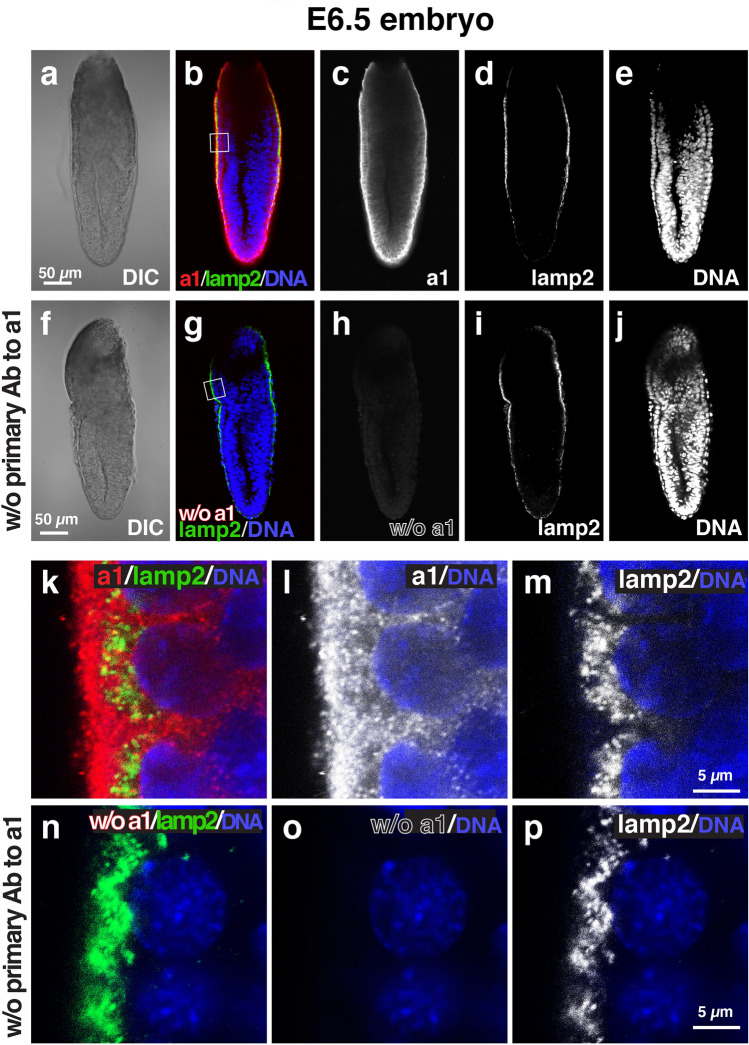
Vacuolar-type H+-ATPases (V-ATPases) are large multi-subunit complexes that play critical roles in the acidification of a variety of intracellular or extracellular compartments. Mammalian cells contain four isoforms of the membrane integral subunit a (a1-a4); these isoforms contain the information necessary to target the enzyme to different cellular destinations. They are also involved in regulating the efficiency of ATP hydrolysis and proton transport. Previously, we showed that early embryogenesis requires V-ATPase function, and the luminal acidic endocytic and lysosomal compartments in the visceral endoderm of mouse embryos at the pre-gastrulation stage (E6.5) are essential for both nutrition and signal transduction during early embryogenesis. In this study, we examined the expression and distribution of a subunit isoforms in mouse embryos at E6.5. We found that all four isoforms expressed and exhibited differential distribution in the E6.5 embryo. At this developmental stage, the embryos establish highly elaborate endocytic compartments called apical vacuoles, on which the a3 isoform specifically accumulated.
© 2022. The Author(s).
Conflict of interest statement
The authors declare no competing interests.
Figures


Similar articles
-
The function of vacuolar ATPase (V-ATPase) a subunit isoforms in invasiveness of MCF10a and MCF10CA1a human breast cancer cells.J Biol Chem. 2013 Nov 8;288(45):32731-32741. doi: 10.1074/jbc.M113.503771. Epub 2013 Sep 26. J Biol Chem. 2013. PMID: 24072707 Free PMC article.
-
The emerging roles of vacuolar-type ATPase-dependent Lysosomal acidification in neurodegenerative diseases.Transl Neurodegener. 2020 May 11;9(1):17. doi: 10.1186/s40035-020-00196-0. Transl Neurodegener. 2020. PMID: 32393395 Free PMC article. Review.
-
Vacuolar-type proton ATPase is required for maintenance of apicobasal polarity of embryonic visceral endoderm.Sci Rep. 2021 Sep 29;11(1):19355. doi: 10.1038/s41598-021-98952-3. Sci Rep. 2021. PMID: 34588579 Free PMC article.
-
Function of a subunit isoforms of the V-ATPase in pH homeostasis and in vitro invasion of MDA-MB231 human breast cancer cells.J Biol Chem. 2009 Jun 12;284(24):16400-16408. doi: 10.1074/jbc.M901201200. Epub 2009 Apr 14. J Biol Chem. 2009. PMID: 19366680 Free PMC article.
-
V-ATPase a3 Subunit in Secretory Lysosome Trafficking in Osteoclasts.Biol Pharm Bull. 2022;45(10):1426-1431. doi: 10.1248/bpb.b22-00371. Biol Pharm Bull. 2022. PMID: 36184499 Review.
Cited by
-
Transcriptional network governing extraembryonic endoderm cell fate choice.Dev Biol. 2023 Oct;502:20-37. doi: 10.1016/j.ydbio.2023.07.002. Epub 2023 Jul 7. Dev Biol. 2023. PMID: 37423592 Free PMC article.
-
Post-feeding transcriptomics reveals essential genes expressed in the midgut of the desert locust.Front Physiol. 2023 Aug 10;14:1232545. doi: 10.3389/fphys.2023.1232545. eCollection 2023. Front Physiol. 2023. PMID: 37692997 Free PMC article.
-
The cytosolic N-terminal domain of V-ATPase a-subunits is a regulatory hub targeted by multiple signals.Front Mol Biosci. 2023 Jun 16;10:1168680. doi: 10.3389/fmolb.2023.1168680. eCollection 2023. Front Mol Biosci. 2023. PMID: 37398550 Free PMC article. Review.
-
Human V-ATPase a-subunit isoforms bind specifically to distinct phosphoinositide phospholipids.J Biol Chem. 2023 Dec;299(12):105473. doi: 10.1016/j.jbc.2023.105473. Epub 2023 Nov 17. J Biol Chem. 2023. PMID: 37979916 Free PMC article.
References
-
- Sun-Wada GH, Wada Y. Vacuolar-type proton pump ATPases: Roles of subunit isoforms in physiology and pathology. Histol. Histopathol. 2010;25:1611–1620. - PubMed
Publication types
MeSH terms
Substances
LinkOut - more resources
Full Text Sources
Molecular Biology Databases
