. 2022 Aug;608(7924):750-756.
doi: 10.1038/s41586-022-05056-7. Epub 2022 Aug 10.

Pyramidal neuron subtype diversity governs microglia states in the neocortex

Affiliations

Pyramidal neuron subtype diversity governs microglia states in the neocortex

Jeffrey A Stogsdill et al. Nature. 2022 Aug.

Abstract

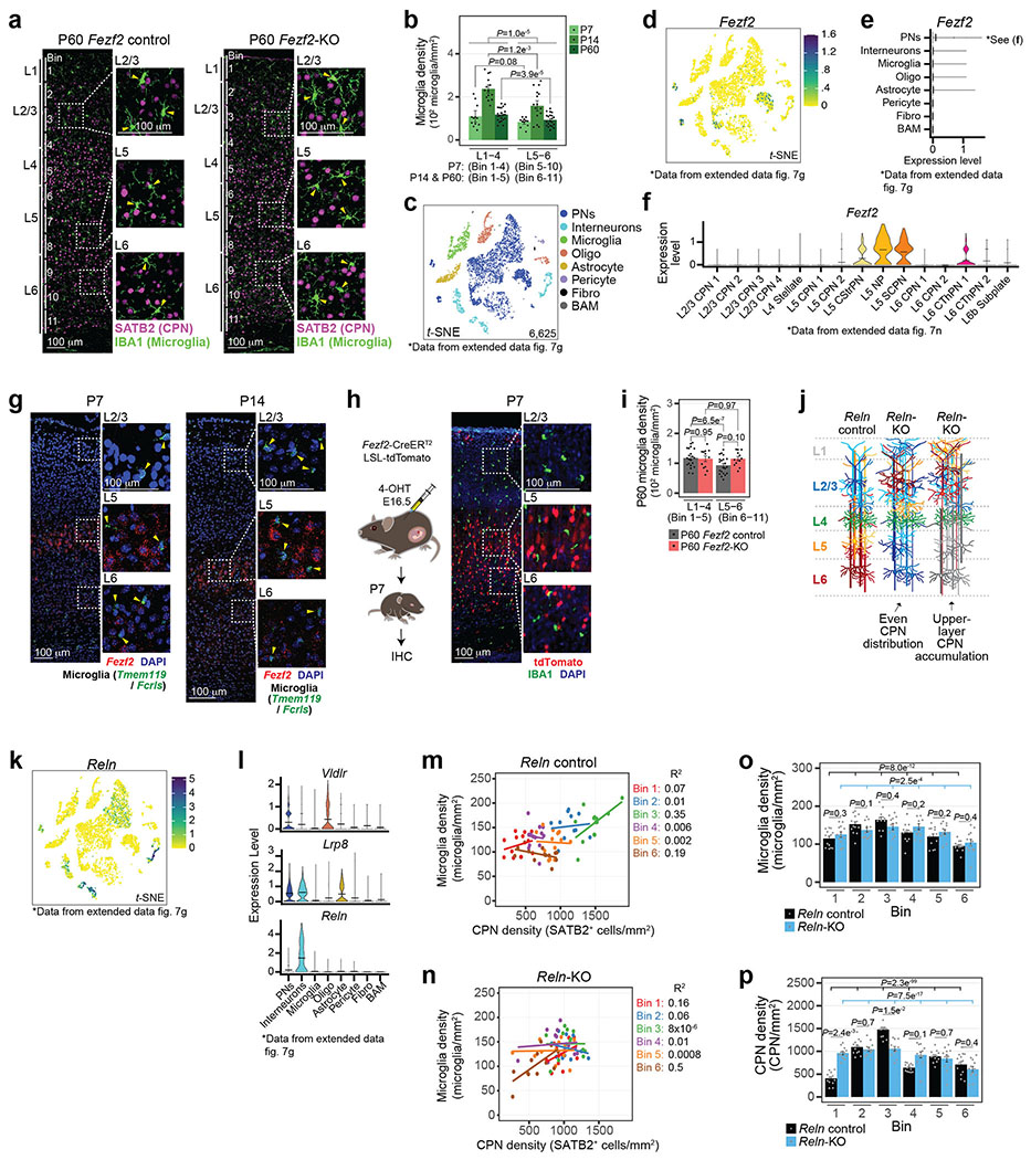
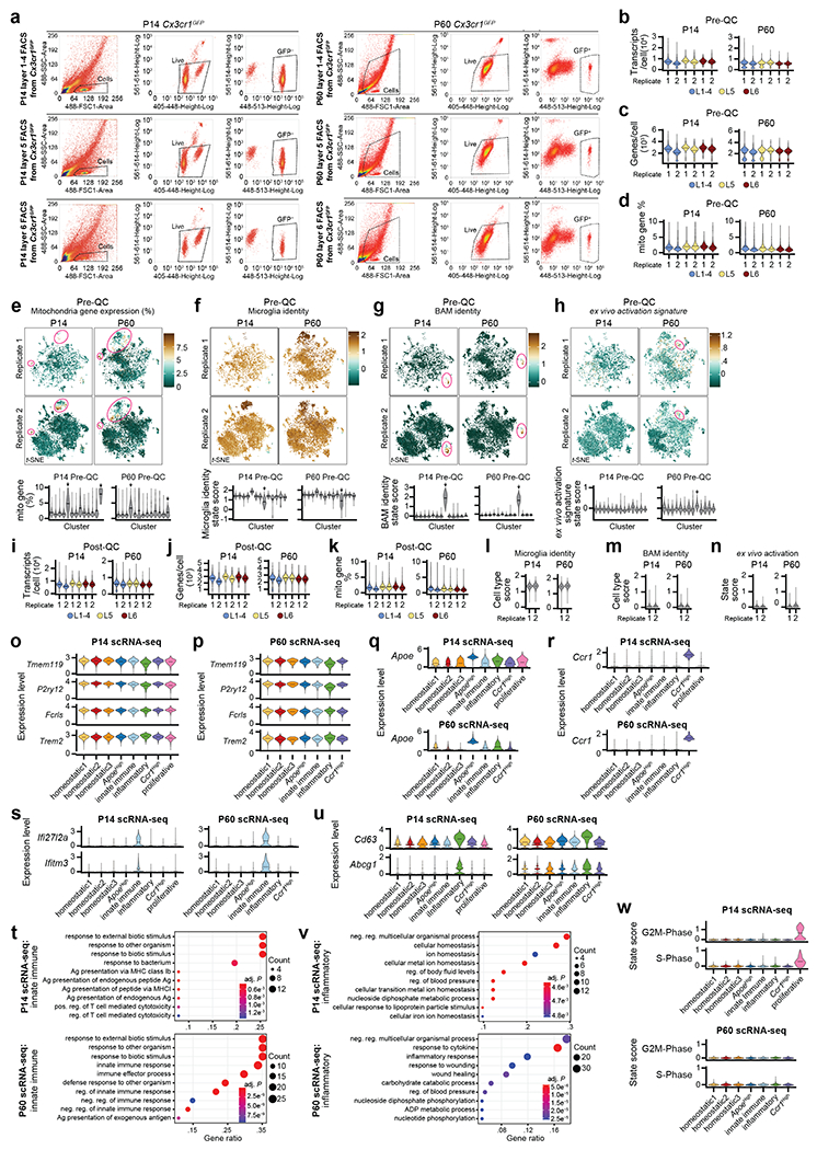
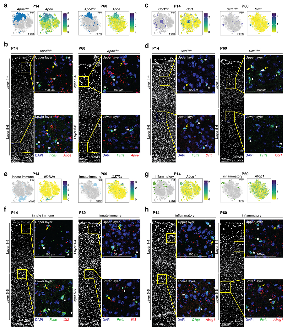
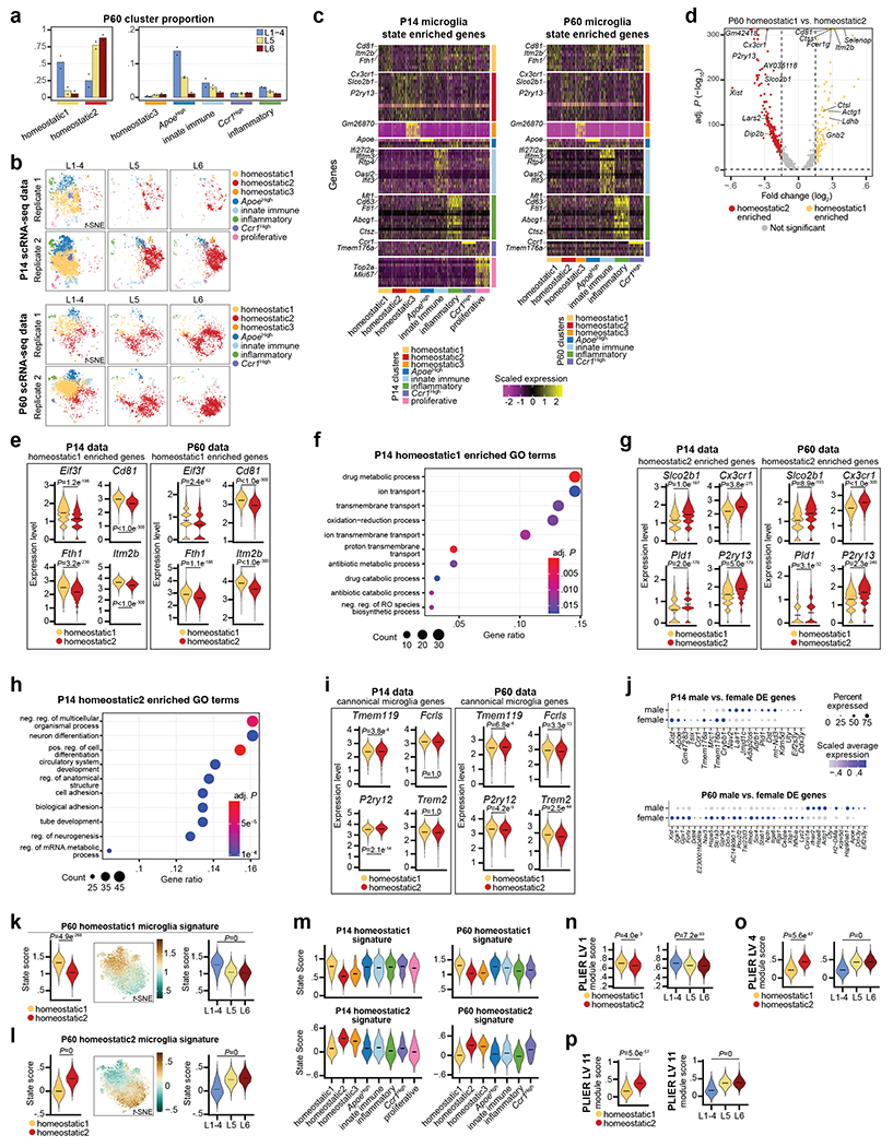
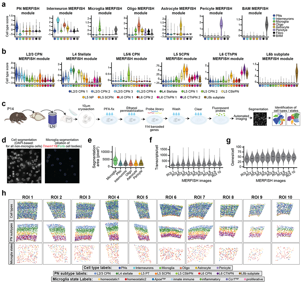
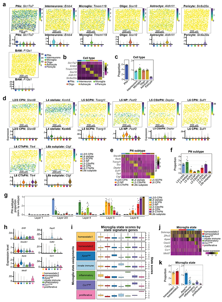
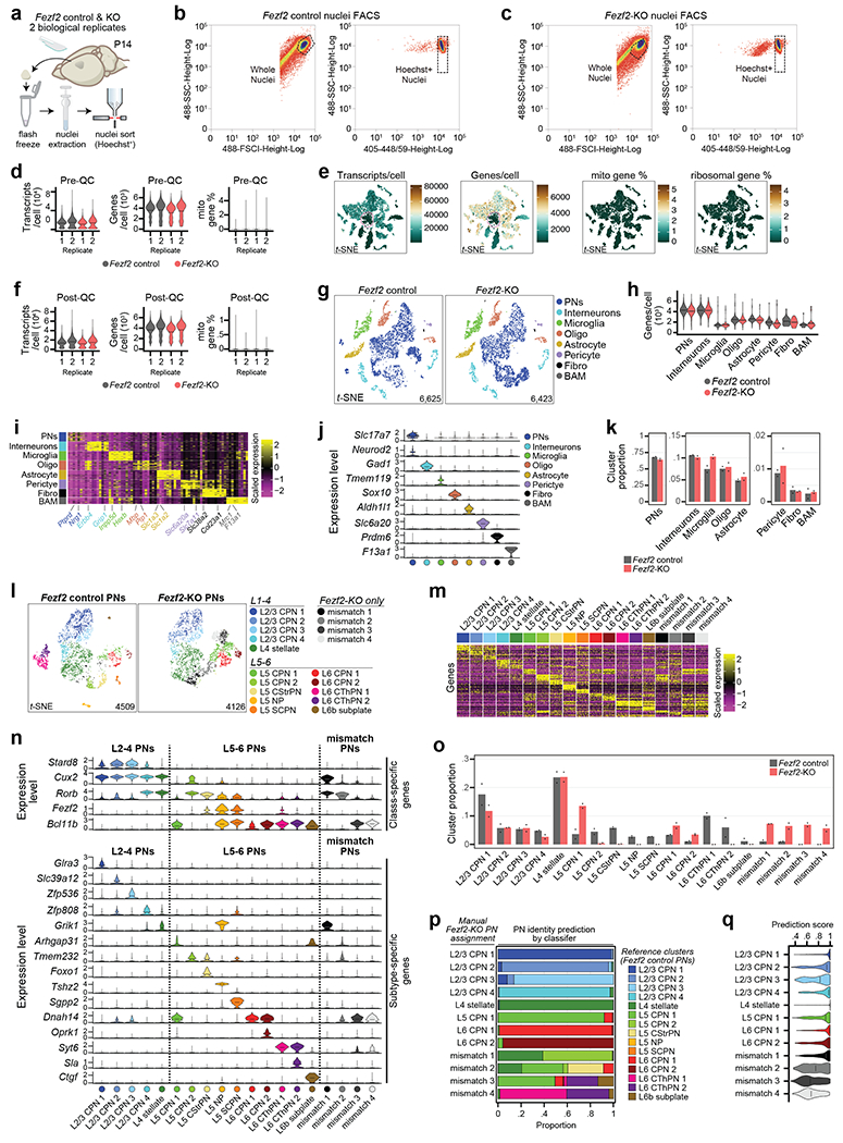
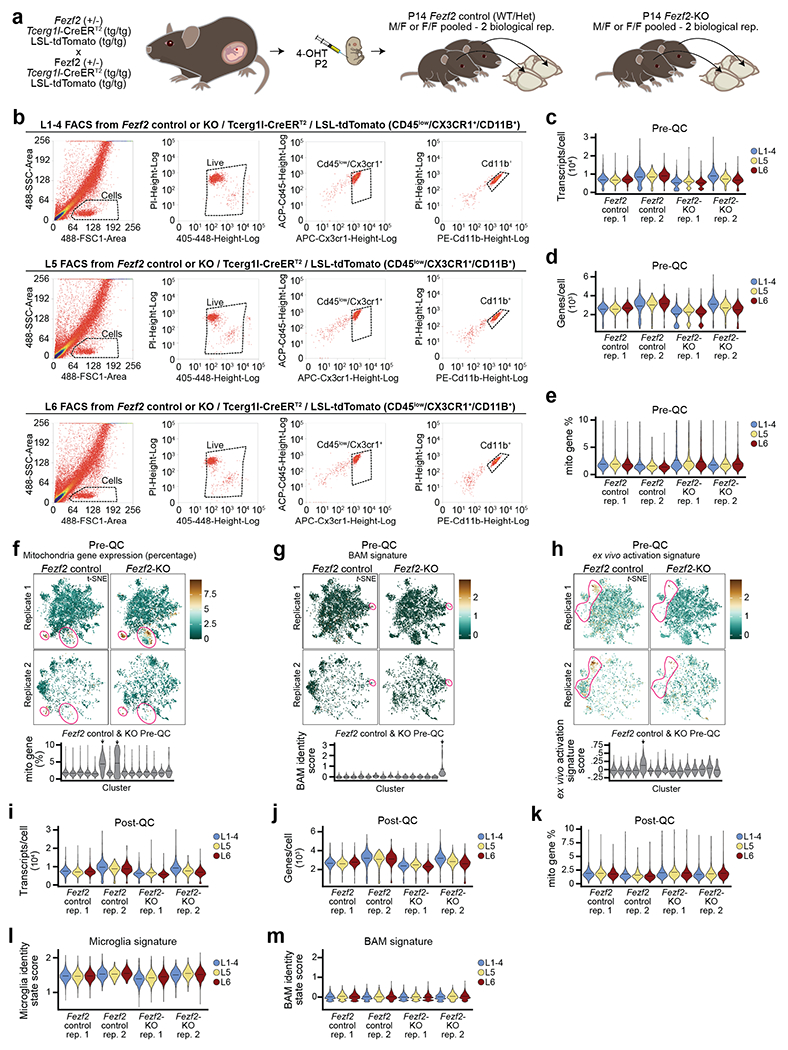
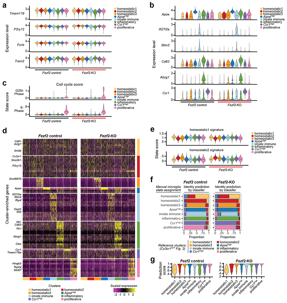
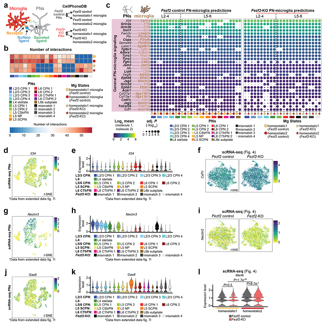
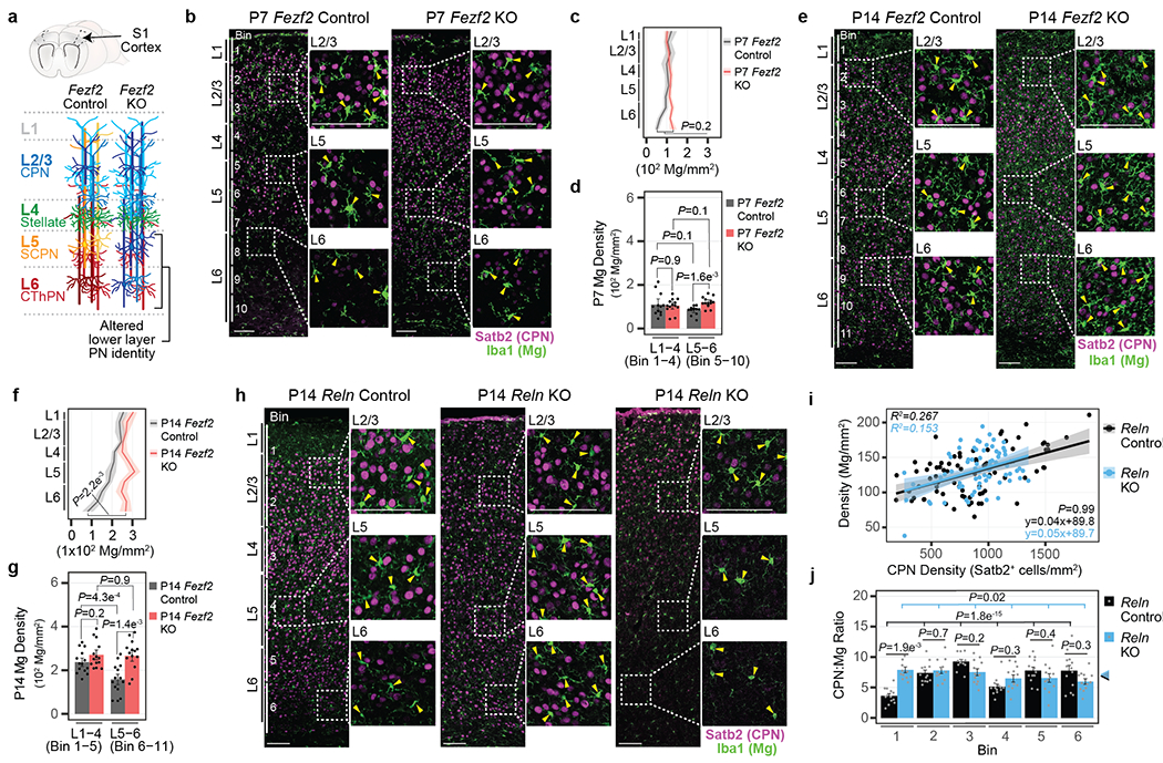
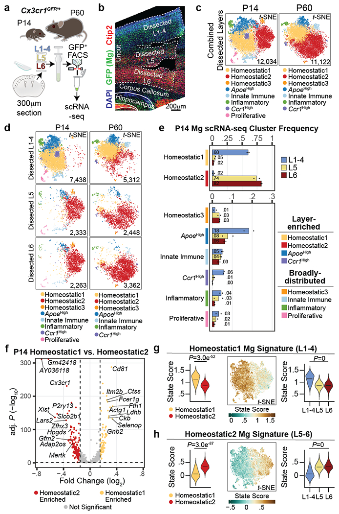
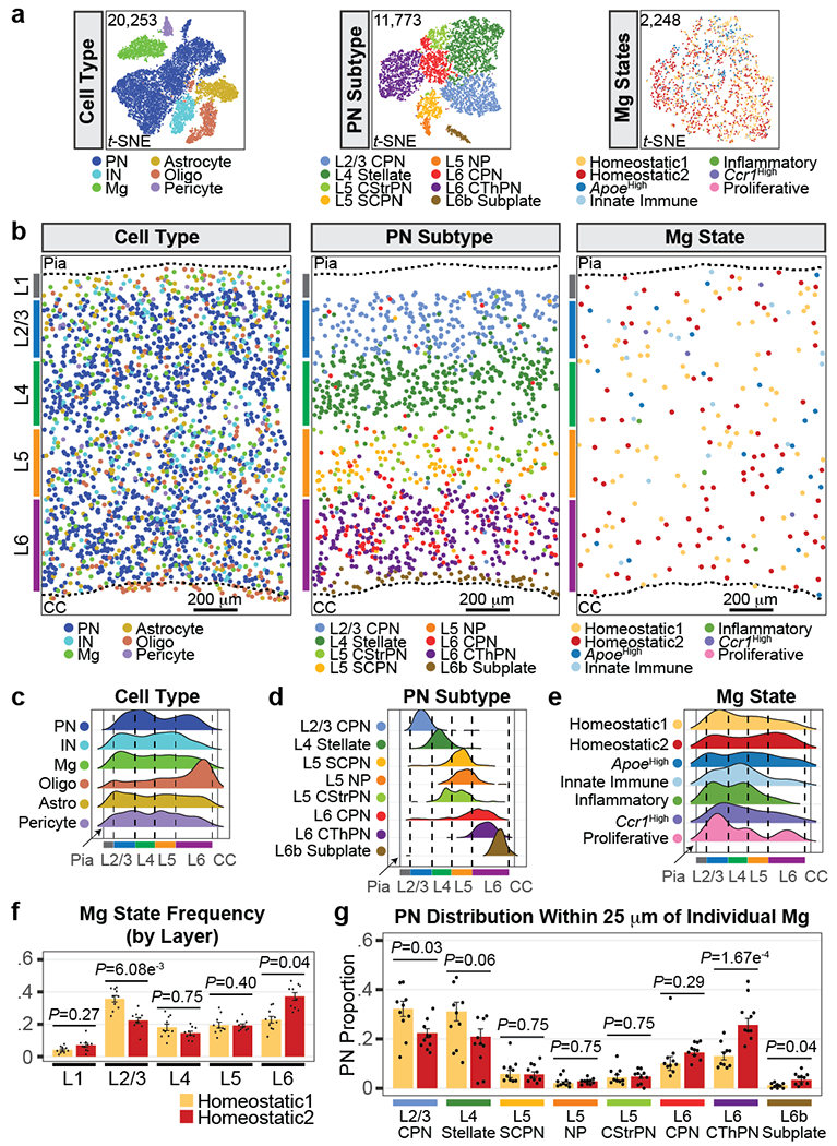
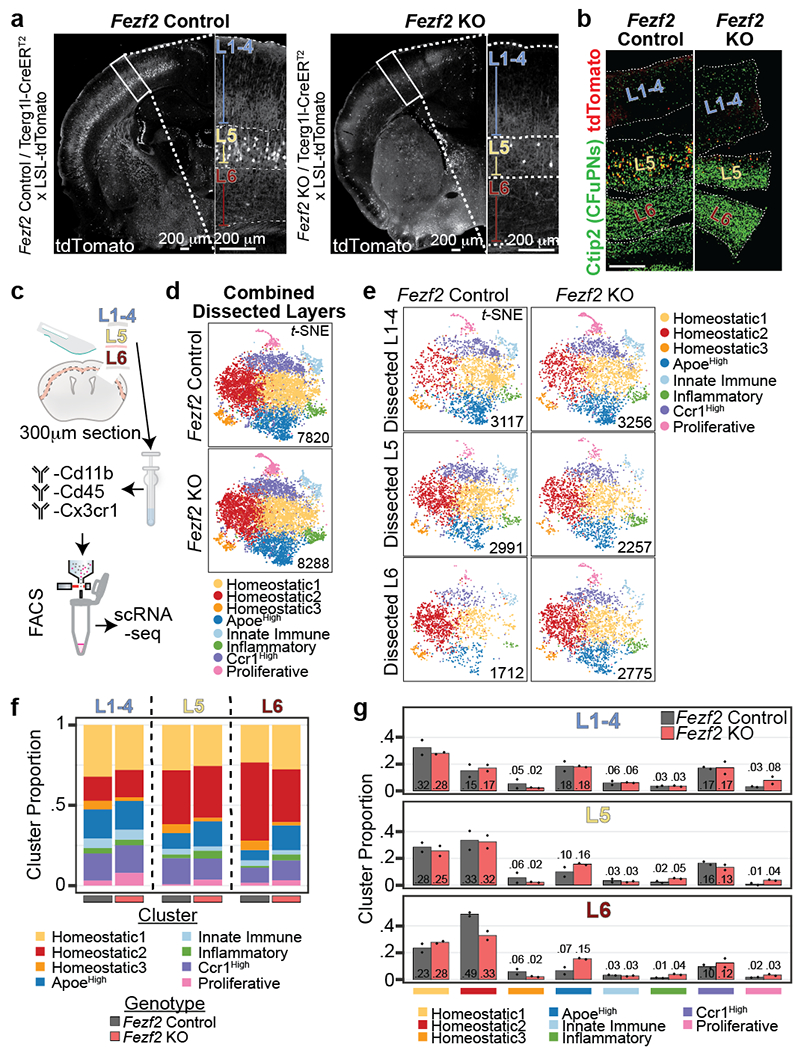
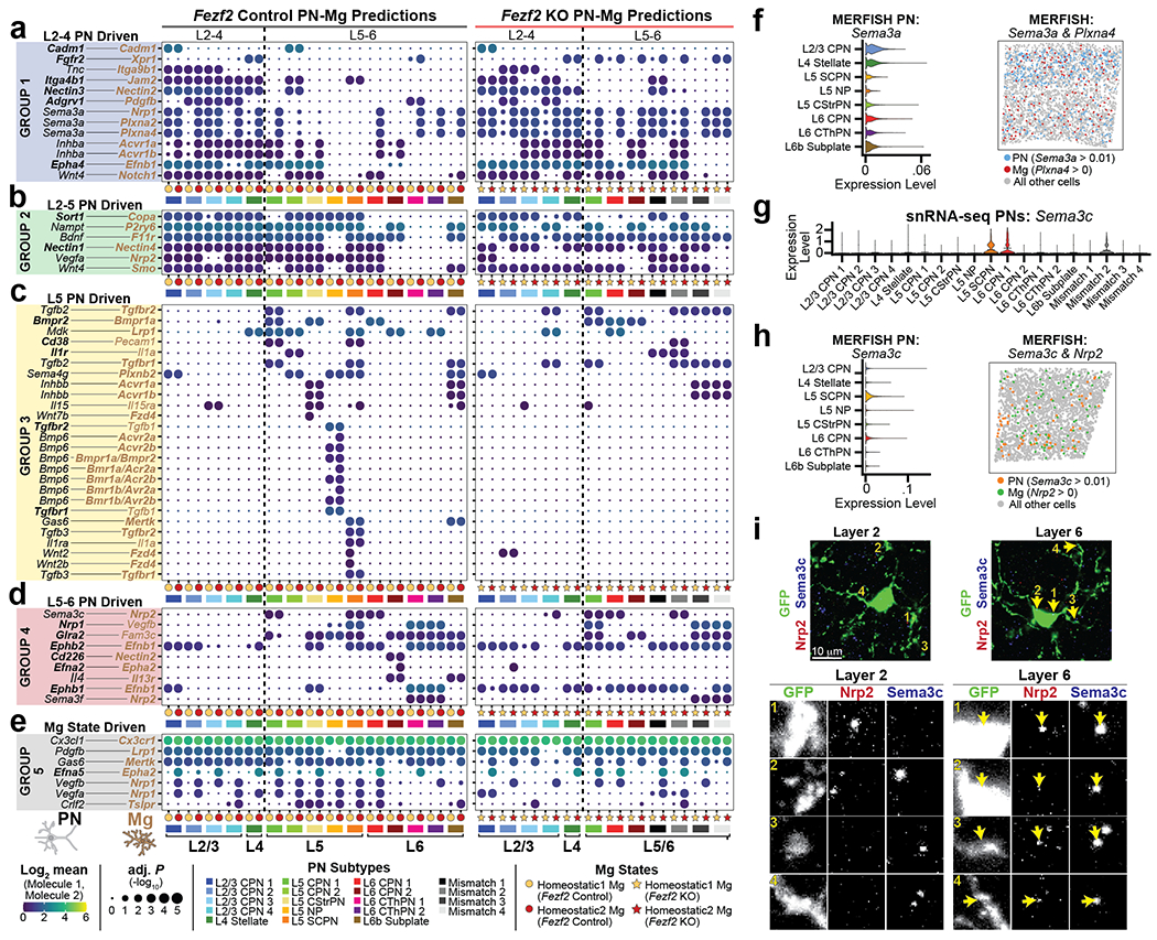
Microglia are specialized macrophages in the brain parenchyma that exist in multiple transcriptional states and reside within a wide range of neuronal environments1-4. However, how and where these states are generated remains poorly understood. Here, using the mouse somatosensory cortex, we demonstrate that microglia density and molecular state acquisition are determined by the local composition of pyramidal neuron classes. Using single-cell and spatial transcriptomic profiling, we unveil the molecular signatures and spatial distributions of diverse microglia populations and show that certain states are enriched in specific cortical layers, whereas others are broadly distributed throughout the cortex. Notably, conversion of deep-layer pyramidal neurons to an alternate class identity reconfigures the distribution of local, layer-enriched homeostatic microglia to match the new neuronal niche. Leveraging the transcriptional diversity of pyramidal neurons in the neocortex, we construct a ligand-receptor atlas describing interactions between individual pyramidal neuron subtypes and microglia states, revealing rules of neuron-microglia communication. Our findings uncover a fundamental role for neuronal diversity in instructing the acquisition of microglia states as a potential mechanism for fine-tuning neuroimmune interactions within the cortical local circuitry.


Conflict of interest statement

COMPETING INTEREST

P.A. is a SAB member in System 1 Biosciences and Foresite Labs and is a co-founder of FL60. J.A.S. is an employee of Sana Biotechnology, Inc. as of 09/2022.

Figures

Extended Data Figure 1:
Extended Data Figure 1:. Control experiments associated with Fezf2 and Reln KO and control cortices.
Related to Fig. 1 a, Representative micrograph of P60 Fezf2 Control and KO S1 cortex immunolabelled for Iba1 (green) and Satb2 (magenta). Enlarged representative layer images are boxed. Bin divisions for cell counts are indicated. Arrowhead = Mg cell body. Scale = 100 μm. b, Mg density grouped by bins corresponding to L1-4 and L5-6 for P7, P14 and P60 Control S1 cortices. Data are presented as mean values +/− SEM. n = 3 mice, 4 images/mouse (P7), 4 mice, 4 images/mouse (P14), and 4 mice, 6 images/mouse (P60). Statistics: Linear mixed models. c, t-SNE of snRNA-seq from P14 S1 cortex color-coded by cell type. PN = Pyramidal Neuron, IN = Inhibitory Neuron, Oligo = Oligodendrocyte-lineage cell, Fibro = Fibroblast, BAM = Border Associated Macrophage. Data is reproduced from Extended Data Fig. 7g (Fezf2 Control only). d-e, Expression feature plot (d) and violin plot (e) for Fezf2 transcript levels from snRNA-seq data. Fezf2 is excluded from Mg. Cell cluster labels are as in (c). Legend = expression value. Data is from Extended Data Fig. 7g. f, Violin plot of the expression level of Fezf2 among PN subtypes. Clusters are color-coded. Lines = mean. Data are presented as mean values with probability density. Data is from Extended Data Fig. 7n. g, Representative micrograph of RNA fluorescence in situ hybridization images from P7 (left) and P14 (right) S1 cortices marking Fezf2 (red), Fcrls/Tmem119 (Mg, green), and DAPI. Enlarged inserts show that Fezf2 is excluded from all Mg. Arrowheads = Mg. Scale = 100 μm. h, (Left) Schematic of Fezf2-CreERT2/LSL-tdTomato mice injected with 4-Hydroxytamoxifen at embryonic day 16.5 to mark and lineage-trace Fezf2-expressing cells. (Right) Representative widefield tile scan image of a P7 coronal brain section immunolabelled for Mg (Iba1, green), endogenous tdTomato (red) and DAPI. Enlarged inserts show that tdTomato signal is excluded from all Mg. Scale = 100 μm. i, Mg density grouped by bins corresponding to L1-4 and L5-6 of P60 Fezf2 Control and KO cortices. Data are presented as mean values +/− SEM. n = 4 mice, 6 images/mouse (P60). Statistics: linear mixed models. j, Cartoon schematic of S1 cortices of Reln Control and KO mice. Observed phenotypic variations among Reln KO cortices are schematized, with PN subytpes color-coded. k-l, Expression feature plot for Reln (k) and violin plots (l) for Reln and the genes that encode the cell surface receptors of Reln, Lrp8 and Vldlr. Reln, Lrp8 and Vldlr expression is excluded from nearly all Mg. Cell cluster labels are as in (c). Legend = expression value. Data are presented as mean values with probability density (l only). Data is from Extended Data Fig. 7g. m-n, Scatter plot of Mg density (y-axis) by CPN density (x-axis) per bin from Reln Control (m) and KO (n) S1 cortices. Dots = bin data points. Data are presented as a linear regression color-coded by bin. R2 is listed for each bin, n = 4 images/mouse, 3 = mice/genotype. o-p, Mg density (o) and CPN density (p) by cortical bin for Reln Control and KO S1 cortices. Data are presented as mean values +/− SEM. n = 4 images/mouse, 3 mice/genotype. Statistics: linear models.
Extended Data Figure 2:
Extended Data Figure 2:. Quality control and analysis of scRNA-seq of Mg from Cx3cr1GFP layer dissected cortices
Related to Fig. 2 a, FACS histogram plots of GFP+ cell sorting from P14 (left) and P60 (right) Cx3cr1GFP mice. S1 cortices were dissected and sorted separately for L1-4 (top), L5 (middle) and L6 (bottom). FACS gating for whole cells (left), live cells (Hoechst+, middle), and GFP+ cells (right) are plotted as dashed boxes. Only live GFP+ cells were used for scRNA-seq experiments. b-d, Violin plots of initial quality control (Pre-QC) metrics of the P14 (left) and P60 (right) Cx3cr1GFP scRNA-seq datasets. Transcripts/cell (b), genes/cell (c), and percent of mitochondrial gene expression/cell (d) are plotted. Data are split by replicate and color-coded by dissected layer. Data are presented as mean values with probability density. e, (Top) Expression feature plots for percent mitochondrial gene expression for the Pre-QC P14 (left) and P60 (right) Cx3cr1GFP scRNA-seq datasets. Feature plots are split by replicate for each age. Low-quality cells (mitochondrial gene > 10%) removed are circled. Legend = percentage. (Bottom) Violin plot of Pre-QC percent mitochondrial gene expression by initial clustering for the P14 (left) and P60 (right) Cx3cr1GFP scRNA-seq datasets. Low-quality clusters (mitochondrial gene > 10%) removed are denoted with arrows. Data are presented as mean values with probability density (bottom only). f-g, (Top) Expression feature plots for Mg (f) and BAM (g) cell type identity for the Pre-QC P14 (left) and P60 (right) Cx3cr1GFP scRNA-seq datasets. Feature plots are split by replicate for each genotype. BAMs removed are circled. (Bottom) Violin plot of Pre-QC Mg (f) and BAM (g) cell type identity by initial clustering for the P14 (left) and P60 (right) Cx3cr1GFP scRNA-seq datasets. BAM clusters removed are denoted with arrows. Data are presented as mean values with probability density (bottom only). Cell type scores (legend) are defined as a function of gene expression for core Mg and BAM genes identified in Marsh et al and Jordao et al (Supplementary Table 1). h, (Top) Expression feature plots for ex vivo activation signature for the Pre-QC P14 (left) and P60 (right) Cx3cr1GFP scRNA-seq datasets. Feature plots are split by replicate for each genotype. ex vivo-activated cells that were removed are circled. (Bottom) Violin plot of Pre-QC ex vivo activation signature by initial clustering for the P14 (left) and P60 (right) Cx3cr1GFP scRNA-seq datasets. ex vivo-activated clusters that were removed are denoted with arrows. Cell scores (legend) are defined as a function of gene expression for core ex vivo activation genes (Supplementary Table 1). i-k, Violin plots of final quality control (Post-QC) metrics of the P14 (left) and P60 (right) Cx3cr1GFP scRNA-seq datasets following the removal of BAMs, low-quality cells and ex vivo-activated cells. Transcripts/cell (i), genes/cell (j), and percent of mitochondrial gene expression/cell (k) are plotted. Data are split by replicate and color-coded by layer sample. Data are presented as mean values with probability density. l-m, Violin plots of the core Mg (l) and BAM (m) cell type scores split by replicate and age for the Post-QC P14 (left) and P60 (right) Cx3cr1GFP scRNA-seq datasets. Cell type scores are defined as a function of gene expression for core Mg and BAM genes (Supplementary Table 1). The final data strongly identify Mg. Data are presented as mean values with probability density. n, Violin plots of the ex vivo activation state score of all Post-QC cells from the P14 (left) and P60 (right) Cx3cr1GFP scRNA-seq datasets. Data are displayed as a state score, defined as a function of the core genes that are up-regulated in Mg through aberrant ex vivo activation (Supplementary Table 1). Data are split by replicate. Data are presented as mean values with probability density. o-p, Violin plots for canonical Mg genes plotted by color-coded cluster from the P14 (o) and P60 (p) Cx3cr1GFP scRNA-seq experiments. Data are presented as mean values with probability density. q-r, Violin plots for Apoe (q), and Ccr1 (r) expression in P14 (top) and P60 (bottom) Mg Cx3cr1GFP scRNA-seq clusters. Data are presented as mean values with probability density. s, Violin plots depicting expression of the interferon-induced genes Ifi27l2a (top) and Ifitm3 (bottom) in P14 (left) and P60 (right) Mg Cx3cr1GFP scRNA-seq clusters. Data are presented as mean values with probability density. t, Dot plot of the top 10 significant gene ontology terms enriched from the DE genes in the Innate Immune cluster compared to all other scRNA-seq clusters at P14 (top) and P60 (bottom). Legend: color = adj. P, dot size = number of genes. Statistics: hypergeometric test with BH procedure. u, Violin plots depicting expression of the inflammation-induced genes Cd63 (top) and Abcg1 (bottom) in P14 (left) and P60 (right) Mg Cx3cr1GFP scRNA-seq clusters. Data are presented as mean values with probability density. v, Dot plot of the top 10 significant gene ontology terms enriched from the DE genes in the Inflammatory cluster compared to all other scRNA-seq clusters at P14 (top) and P60 (bottom). Legend: color = adj. P, dot size = number of genes. Statistics: hypergeometric test with BH procedure. w, Violin plots for G2M (top) and S-phase (bottom) cell cycle phase scores for P14 (top) and P60 (bottom) Mg Cx3cr1GFP scRNA-seq clusters. Data are presented as mean values with probability density.
Extended Data Figure 3:
Extended Data Figure 3:. Identification of cortical non-homeostatic Mg in situ
Related to Fig. 2 a, t-SNE plot highlighting the ApoeHigh cluster (left) and feature plot of Apoe expression (right) in the P14 and P60 Cx3cr1GFP scRNA-seq datasets. Legend = expression value. b, Representative tile scan confocal RNA fluorescence in situ hybridization images from P14 (left) and P60 (right) S1 cortices marking Apoe (red), Fcrls (Mg, green) and DAPI. Enlarged inserts (yellow boxes) show Apoe-positive (arrow) and Apoe-negative (*) Mg at upper- and lower-layers of the cortex. Scale = 100 μm. c, t-SNE plot highlighting the Ccr1High cluster (left) and feature plot of Ccr1 expression (right) in the P14 and P60 Cx3cr1GFP scRNA-seq Mg datasets. Legend = expression value. d, Representative tile scan confocal RNA fluorescence in situ hybridization images from P14 (left) and P60 (right) S1 cortices marking Ccr1 (red), Fcrls (Mg, green) and DAPI. Enlarged inserts (yellow boxes) show Ccr1-positive (arrow) and Ccr1-negative (*) Mg at upper- and lower-layers of the cortex. Scale = 100 μm. e, t-SNE plot highlighting the Innate Immune cluster (left) and feature plot of Ifi27l2a expression (right) in the P14 and P60 Cx3cr1GFP scRNA-seq Mg datasets. Legend = expression value. f, Representative tile scan confocal RNA fluorescence in situ hybridization images from P14 (left) and P60 (right) S1 cortices marking Innate Immune Mg with Ifit3 (red), Fcrls (Mg, green) and DAPI. Enlarged inserts (yellow boxes) show Ifit3-positive (arrow) and Ifit3-negative (*) Mg at upper- and lower-layers of the cortex. Scale = 100 μm. g, t-SNE plot highlighting the Inflammatory cluster (left) and feature plot of Abcg1 expression (right) in the P14 and P60 Cx3cr1GFP scRNA-seq Mg datasets. Legend = expression value. h, Representative tile scan confocal RNA fluorescence in situ hybridization images from P14 (left) and P60 (right) S1 cortices marking Inflammatory Mg with Abcg1 (red), Fcrls or C1qa (Mg, green) and DAPI. Enlarged inserts (yellow boxes) show Abcg1-positive (arrow) and Abcg1-negative (*) Mg at upper- and lower-layers of the cortex. Scale = 100 μm.
Extended Data Figure 4:
Extended Data Figure 4:. Characterization of the transcriptional signature of Mg states
Related to Fig. 2 a, Bar plot of P60 Cx3cr1GFP scRNA-seq Mg cluster proportions normalized by dissected layer. Color coding indicates the layer from which the Mg were isolated. Data are presented as normalized mean values. n = 2 independent biological replicates. b, t-SNE of P14 (top) and P60 (bottom) Cx3cr1GFP scRNA-seq color-coded Mg state clusters split by dissected layer of origin and replicate. c, Heatmap of the top DE genes by Mg cluster in the P14 (left) and P60 (right) Cx3cr1GFP scRNA-seq datasets. Each row is a gene. Color-coded clusters are grouped. Legend = scaled expression value. d, Volcano plot of the DE genes between Homeostatic1 versus Homeostatic2 clusters in the P60 Cx3cr1GFP scRNA-seq dataset. Genes are color-coded by enrichment (log2 Fold Change > 0.15 and −log10 BH adj. P < 0.05). Homeostatic1-enriched (gold), Homeostatic2-enriched (red), not significant (grey). Several homeostatic state signature genes for each state are annotated. Vertical and horizontal dotted lines represent enrichment cut-offs. Statistics: generalized linear model with BH correction (MAST). e, Violin plots of Homeostatic1-enriched genes expressed in Homeostatic1 and Homeostatic2 clusters from P14 (left) and P60 (right) Cx3cr1GFP scRNA-seq datasets. Data are presented as mean values with probability density.; Statistics: generalized linear model with BH correction (MAST). f, Dot plot of the top 10 significant gene ontology terms enriched from the DE genes of the Homeostatic1 cluster compared to all other clusters in the P14 Cx3cr1GFP scRNA-seq dataset. Legend: color = BH adj. P, dot size = number of genes. Statistics: hypergeometric test with BH procedure. g, Violin plots of Homeostatic2-enriched Mg genes expressed in Homeostatic1 and Homeostatic2 clusters from P14 (left) and P60 (right) Cx3cr1GFP scRNA-seq dataset. Data are presented as mean values with probability density. Statistics: generalized linear model with BH correction (MAST). h, Dot plot of the top 10 significant gene ontology terms enriched from the DE genes of Homeostatic2 cluster compared to all other clusters in the P14 Cx3cr1GFP scRNA-seq dataset. Legend: color = BH adj. P, dot size = number of genes. Statistics: hypergeometric test with BH procedure. i, Violin plots of canonical Mg genes expressed in Homeostatic1 and Homeostatic2 clusters from the P14 (left) and P60 (right) Cx3cr1GFP scRNA-seq datasets. Data are presented as mean values with probability density. Statistics: generalized linear model with BH correction (MAST). j, Dot plot of the DE genes between male and female Mg from the P14 (top) and P60 (bottom) Cx3cr1GFP scRNA-seq datasets. The data represent analysis from the combined dissected layers. Legend: color = scaled average expression, dot size = percent of the cluster cells expressing the gene. Most sex-specific DE genes do not match the list of DE gene between Homeostatic state clusters. Statistics: generalized linear model with BH correction (MAST). k-l, Homeostatic1 (k) and Homeostatic2 (l) Mg signature state scoring of P60 Cx3cr1GFP scRNA-seq data: (Left) Violin plot of state scores for Mg in clusters Homeostatic1 and Homeostatic2. (Middle) Expression feature plot of state scores in t-SNE space. Legend: score value. (Right) Violin plot of state scores of homeostatic Mg split by dissected layer. Violin plot data are presented as mean values with probability density. Statistics: linear mixed models. Genes pertaining to the Homeostatic1 signature are listed in Supplemental Table 5. m, Violin plots of the Homeostatic1 (top row) and Homeostatic2 (bottom row) state scores split by all clusters from P14 (left) and P60 (bottom) Cx3cr1GFP scRNA-seq datasets. Data are presented as mean values with probability density. State scores are defined as a function of gene expression pertaining to the genes enriched in the Homeostatic signatures listed in Supplementary Table 5. n-p, Violin plots of gene module scores for PLIER latent variable (LV) 1 (n), LV 4 (o), and LV 11 (p) applied to Homeostatic1 and 2 Mg from the P14 scRNA-seq data. The data are split by state (left) and by dissected layer (right). Data are presented as mean values with probability density. Statistics: linear mixed models. Genes pertaining to the LV modules are listed in Supplementary Table 6.
Extended Data Figure 5:
Extended Data Figure 5:. Quality control metrics for the MERFISH datasets
Related to Fig. 3 a-b, Violin plots of cell-type scores for MERFISH gene modules targeting cell types (a) and PN subtypes (b). The data are from P14 Fezf2 Control snRNA-seq in Extended Data Fig. 7. Data are presented as mean values with probability density. State scores are defined as a function of gene expression for the genes grouped by cell type or PN subtype listed in Supplementary Table 7. c, Cartoon schematic of the MERFISH pipeline to map cell types and Mg states in the P14 S1 cortex. d, Representative micrographs depicting the automated segmentation of cells for MERFISH analysis. (Left) All nuclei were automatically segmented (white outlines) based on DAPI signal. (Right) Mg were separately segmented based on Tmem119 (red) and Fcrls (green) RNA puncta with a small dilation (blue outline) in order to maximize RNA detection and counts. See Methods for full cell segmentation details. e, Violin plots of the size of segmented cells split by the cell types identified in the MERFISH dataset. Data are presented as mean values with probability density. f-g, Violin plots representing the number of RNA transcripts/cell (f) and genes/cell (g) for the MERFISH dataset split by region of interest (ROI) samples. Data are presented as mean values with probability density. h, Spatial distribution MERFISH maps of cell types (top row), PN subtypes (middle row), and Mg states (bottom row) for each ROI from all imaged samples. Cropped images displayed are the analyzed regions of interest (S1 cortex) for each sample. Cell types and states are color-coded as indicated in the legend.
Extended Data Figure 6:
Extended Data Figure 6:. Identification of cell type and Mg state from MERFISH data
Related to Fig. 3 a, Spatial distribution feature (top row) and violin plots (bottom row) for the genes targeting cell types, via MERFISH. The spatial distribution feature plots are from a single representative image (ROI 1). Violin plots are split by cell type cluster. Spatial distribution feature plot legend = expression value. Violin plot legend = cell type. b, Heatmap of gene expression specific to cell types as captured by MERFISH. Cell type clusters are color coded (subset of 200 cells/cluster). Legend: scaled expression value. c, Mean cell type cluster proportion across MERFISH ROIs. Cell types are color coded. Data are presented as mean values +/− SEM. Dots = individual ROI frequencies. n = 10 ROI from 3 mice. d, Spatial distribution feature (top row) and violin plots (bottom row) for the genes targeting PN subtypes by MERFISH. Spatial distribution feature plots shown are a single representative image (ROI 1). Violin plots are split by PN subtype cluster. Spatial distribution feature plot legend = expression value. Violin plot legend = cell type. e, Heatmap of gene expression specific to PN subtypes as captured by MERFISH. PN subtype clusters are color coded (subset of 200 cells/cluster). Legend: scaled expression value. f, Mean PN subtype cluster proportion across MERFISH ROIs. PN subtypes are color-coded. Data are normalized to the total PN count. Dots = individual ROI proportions. Data are presented as mean values +/− SEM. n = 10 ROI from 3 mice. g, Mean layer proportion for each PN subtype identified by MERFISH. Data are normalized to the total count for each PN subtype. See methods for the calculation strategy for layer distinctions. PN subtypes are color-coded. Dots = individual sample proportions for each PN subtype by cortical layer. Data are presented as mean values +/− SEM. n = 10 ROI from 3 mice. h, Violin plots for representative genes targeting different Mg states by MERFISH. Mg states are color-coded and split by state. Data are presented as mean values with probability density. i, Box and whisker plots for MERFISH Mg state scoring. Mg state assignments are color coded and labelled at the left. See Methods for full details on MERFISH Mg state scoring. Mg state-enriched genes used for scoring are listed in Supplementary Table 7. Plots indicate median, 1st and 3rd quartiles, 1.5 inter-quartile range plus outlying points. n = all Tmem119+/Fcrls+ Mg from 10 ROI of 3 mice. j, Heatmap of gene expression enriched in Mg states captured by MERFISH. Mg state assignments are color coded (subset of 200 cells/cluster). Legend: scaled expression value. k, Mean Mg state assignment proportions across MERFISH ROIs. Mg state assignments are color coded. Dots = individual ROI proportions. Data are presented as mean values +/− SEM. n = 10 ROI from 3 mice.
Extended Data Figure 7:
Extended Data Figure 7:. Molecular profiles of cell types and PN subtypes in Fezf2 Control and KO cortices by snRNA-seq
Related to Fig. 4 a, Cartoon of S1 cortex dissection, nuclei extraction, and purification by FACS from P14 Fezf2 Control and KO mice. n = 2 biological replicates for each genotype (1 male and 1 female for each genotype). b-c, FACS scatter plots sorting nuclei from P14 Fezf2 Control (b) and KO (c) S1 cortices. FACS gating for whole nuclei (left) and Hoechst+ nuclei (right) are plotted as dashed boxes. Only Hoechst+ nuclei were used for snRNA-seq experiments. d, Violin plots of initial quality control (Pre-QC) metrics for the Fezf2 Control and KO snRNA-seq dataset. Data are split by replicate and color-coded by genotype. Transcripts/cell (left), genes/cell (middle), and percent of mitochondrial gene expression/cell (right) are plotted. Data are presented as mean values with probability density. e, Expression feature plots for Pre-QC metrics: transcripts/cell, genes/cell, percent mitochondrial gene expression/cell, and percent ribosomal gene expression/cell for the Pre-QC P14 Fezf2 Control and KO snRNA-seq data sets. Low-quality nuclei (circled) were those expressing very low genes/nucleus and did not return DE genes specific to known cell types. Legend: number (transcripts/nucleus and genes/nucleus), percent expression (mitochondrial and ribosomal gene %). f, Violin plots of quality control (Post-QC) metrics of the P14 Fezf2 Control and KO snRNA-seq datasets following the removal of low-quality nuclei. Transcripts/nucleus (left), genes/nucleus (middle), and percent of mitochondrial gene expression/nucleus (right) are plotted, split by replicate and genotype. Data are presented as mean values with probability density. g, t-SNE of Post-QC P14 Fezf2 Control (left) and KO (right) snRNA-seq datasets color-coded by cell type cluster. PN = Pyramidal Neurons, IN = Inhibitory Neuron, Oligo = Oligodendrocyte-lineage cells, Fibro = Fibroblast, BAM = Border Associated Macrophage. The number of sequenced nuclei is annotated for each genotype is noted. h, Violin plot of the number of genes/nuclei split by cell type cluster and genotype for the P14 Fezf2 Control and KO snRNA-seq dataset. Data are presented as mean values with probability density. i, Heatmap of gene expression for the top DE genes by cell type cluster from the P14 Fezf2 Control and KO snRNA-seq dataset. Each row is a gene and grouped by cell type cluster (color-coded). Legend = scaled expression value. j, Violin plots depicting the expression level of marker genes for each cell type cluster. Cell type clusters are color coded. Data are presented as mean values with probability density. k, Bar plot of mean cluster proportions by genotype for the P14 Fezf2 Control and KO snRNA-seq dataset. Data are normalized to the total cell count for each genotype. Color-coding indicates the genotype. n = 2 biological replicates for each genotype (1 male and 1 female for each genotype). l, t-SNE of PN subtype clusters from the P14 Fezf2 Control and KO snRNA-seq dataset split by genotype. PNs were subsetted Slc17a7+/Neurod2+ nuclei from (g). PN subtypes are color-coded and labelled by their cortical layer localization. The number of sequenced nuclei for each genotype is annotated. m, Heatmap of gene expression for the top 10 DE genes for each PN subtype cluster in the P14 Fezf2 Control and KO snRNA-seq datasets. Each row is a gene and grouped by PN subtype cluster (color-coded). Legend = scaled expression value. n, Violin plots depicting the expression of select markers genes (split by cluster): PN class-specific (top) and PN subtype-specific (bottom). Clusters are color-coded. Data are presented as mean values with probability density. Dashed line splits upper- and lower- cortical layer PN subtypes, as well as Fezf2 KO mismatch PNs. o, Bar plot of the mean PN subtype cluster proportion by genotype from P14 Fezf2 Control and KO snRNA-seq datasets. Data are normalized to the total PN count for each genotype. Color coding indicates the genotype. n = 2 biological replicates for each genotype (1 male and 1 female for each genotype). p, Stacked bar plots depicting the prediction results of the Fezf2 KO PN subtype clusters using Azimuth. PNs of the manually assigned subtype clusters (rows) from the P14 Fezf2 KO snRNA-seq data were re-classified based on the PN subtypes defined from the Control snRNA-seq data (color-coded). The re-classified data clusters are shown as proportions of the manual cluster size. q, Violin plots of the Azimuth prediction scores for the P14 Fezf2 KO re-classified clusters. Re-classified clusters are color-coded. Data are presented as mean values with probability density.
Extended Data Figure 8:
Extended Data Figure 8:. Cell sorting and quality control analysis for scRNA-seq of Mg from Fezf2 Control and KO layer dissected cortices
Related to Fig. 4 a, Cartoon schematic of the compound transgenic mouse line used to fluorescently label cortical L5b of Fezf2 Control and KO mice. 4-Hydroxytamoxifen (4-OHT, 50 mg/kg) was administered to pups on P2. Pups were sacrificed at P14 for dissection of the S1 cortex. n = 2 biological replicates (pooled 1 male and 1 female). b, Representative FACS scatter plots sorting Cd45low/Cx3cr1+/Cd11b+ Mg from P14 Fezf2 Control or KO/Tcerg1l-CreERT2/LSL-tdTomato mice. The S1 cortex was dissected and sorted separately for L1-4 (top), L5 (middle) and L6 (bottom). FACS gating for whole cell (far left), live cells (Hoechst+ middle left), Cd45low/Cx3cr1+ (middle right) and Cd11b+ (far right) is plotted as dashed boxes. Only Cd45low/Cx3cr1+/Cd11b+ Mg were used for scRNA-seq experiments. c-e, Violin plots of initial quality control (Pre-QC) metrics for the P14 Fezf2 Control (left) and KO (right) scRNA-seq datasets. Data are split by replicate and genotype, and color coded by dissected layer. Transcripts/cell (c), genes/cell (d), and percent of mitochondrial gene expression/cell (e) are plotted. Data are presented as mean values with probability density. f, (Top) Expression feature plots for percent mitochondrial gene expression for the Pre-QC P14 Fezf2 Control and KO scRNA-seq dataset. Feature plots are split by replicate for each genotype. Low-quality cells (mitochondrial gene > 10%) that were removed are circled. Legend = percentage. (Bottom) Violin plot of Pre-QC percent mitochondrial gene expression by initial clustering for the P14 Fezf2 Control and KO scRNA-seq dataset. Low-quality clusters (mitochondrial gene > 10%) removed are denoted with arrows. Violing plot data are presented as mean values with probability density. g-h, (Top) Expression feature plots for BAM cell type identity (g) and ex vivo activation signature (h) from the Pre-QC P14 Fezf2 Control and KO scRNA-seq dataset. Feature plots are split by replicate for each genotype. BAMs and ex vivo activated cells that were removed are circled. (Bottom) Violin plot of Pre-QC BAM (g) and ex vivo activated (h) signature scores by initial clustering for the P14 Fezf2 Control and KO scRNA-seq dataset. BAM and ex vivo activated clusters that were removed are denoted with arrows. Violin plot data are presented as mean values with probability density. Scores are calculated as a function of gene expression for core BAM and ex vivo activated genes, respectively (Supplementary Table 1). i-k, Violin plots of quality control (Post-QC) metrics of the P14 Fezf2 Control and KO scRNA-seq dataset following the removal of BAMs, low-quality, and ex vivo activated cells. Data are split by replicate, genotype, and dissected layer. Transcripts/cell (i), genes/cell (j), and percent of mitochondrial gene expression/cell (k) are plotted. Data are presented as mean values with probability density. l-m, Violin plots of the core Mg (l) and BAM (m) cell type scores from the post-QC Fezf2 Control and KO scRNA-seq dataset. Data are split by replicate and genotype, and color coded by dissected layer. The final data strongly identify Mg. Cell type scores are defined as a function of gene expression for core Mg and BAM genes (Supplementary Table 1). Data are presented as mean values with probability density.
Extended Data Figure 9:
Extended Data Figure 9:. Mg transcriptional signatures in the Fezf2 KO cortex
Related to Fig. 4 a-b, Violin plots for canonical Mg (a) and state-enriched (b) genes plotted by cluster and split by genotype from the P14 Fezf2 Control and KO layer dissected scRNA-seq dataset. Clusters are color coded. Data are presented as mean values with probability density. c, Violin plots for G2M (top) and S-phase (bottom) scores for the P14 Fezf2 Control and KO scRNA-seq data split by cluster. Cells are scored by cell cycle phase. Data are presented as mean values with probability density. d, Heatmap of gene expression for the top DE genes for each Mg state cluster in the P14 Fezf2 Control (left) and KO (right) scRNA-seq dataset. Each column is a gene and grouped by Mg state cluster (color-coded). Legend = scaled expression value. e, Violin plots of the Homeostatic1 (top row) and Homeostatic2 (bottom row) state scores split by all clusters from the P14 Fezf2 Control and KO scRNA-seq dataset. Clusters are color-coded. Data are presented as mean values with probability density. State scores are defined as a function of gene expression pertaining to the genes enriched in the Homeostatic signatures listed in Supplementary Table 5. f, Stacked bar plots depicting Mg state prediction results using Azimuth. Mg of the manually assigned clusters (rows) from the P14 Fezf2 Control (left) and KO (right) scRNA-seq dataset were re-classified based on the cell types defined in the P14 Cx3cr1GFP scRNA-seq dataset (color coded). The re-classified data are shown as proportions of the total cell count per manually assigned cluster. g, Violin plots of the Azimuth prediction scores for the P14 Fezf2 Control (top) and KO (bottom) re-classified clusters. Re-classified clusters are color coded. Data are presented as mean values with probability density.
Extended Data Figure 10:
Extended Data Figure 10:. Extended data for the ligand-receptor, PN-Mg interactome of the neocortex
Related to Fig. 5 a, Cartoon schematic of the ligand-receptor analysis to uncover novel PN-Mg subtype interactions using CellPhoneDB. The interactome predicts interactions between PN subtypes (snRNA-seq data, Extended Data Fig. 7) and homeostatic Mg (scRNA-seq data, Fig. 4) from P14 Fezf2 Control and KO mice. b, Heatmap of the number of significant ligand-receptor (L-R) interaction pairs between each PN subtype (column) and Mg state (row) cluster. Legend: PN subtype (colored boxes) and homeostatic Mg state (color-coded circles for the Fezf2 Control Mg, stars for the Fezf2 KO Mg). Scale = number of interactions. c, Dot plot for L-R expression between PN subtypes and homeostatic Mg states in Fezf2 Control (left) and KO (right) cortices. Legend: PN subtype (colored boxes) and layered homeostatic Mg (color-coded circles for the Fezf2 Control Mg, stars for the Fezf2 KO Mg) used in each pairwise comparison. −log10 adj. P values are indicated by circle size. Dot color indicates the average expression level of the interacting molecules from PN subtypes and Mg state. Receptor genes are bolded. Dotted vertical line divides upper- and lower-layer PN subtypes. Statistics: permutation-based BH adj. P. d-e, Expression feature (d) and violin plots (e) of Il34 levels in PN subtypes from the P14 Fezf2 Control and KO snRNA-seq dataset. Data originate from Extended Data Fig. 7l. Legend = expression (d) and color-coded PN-subtypes (e). Violin plot data are presented as mean values with probability density. f, Expression feature plot of Csf1r levels in Mg from the P14 Fezf2 Control (left) and KO (right) scRNA-seq datasets. Data originate from Fig. 4. Legend = Expression value. g-h, Expression feature (g) and violin plots (h) of Nectin3 levels in PN subtypes from the P14 Fezf2 Control and KO snRNA-seq dataset. Data originate from Extended Data Fig. 7l. Legend = expression (g) and color-coded PN subtypes (h). Violin plot data are presented as mean values with probability density. i, Expression feature plot of Nectin2 levels in Mg from the P14 Fezf2 Control (left) and KO (right) scRNA-seq dataset. Data originate from Fig. 4. Legend = Expression value. j-k, Expression feature (j) and violin plots (k) of Gas6 levels in PN subtypes from the P14 Fezf2 Control and KO snRNA-seq dataset. Data originate in Extended Data Fig. 7l. Legend = expression (j) and color-coded PN-subtypes (k). Violin plot data are presented as mean values with probability density. l, Violin plot of Mertk levels in Mg from the P14 Fezf2 Control and KO scRNA-seq dataset. Data denote the differential expression of Mertk between Homeostatic1 and Homeostatic2 and between genotypes. Data are presented as mean values with probability density. Statistics: generalized linear model with BH correction (MAST).
Figure 1:
Figure 1:. PN subtypes locally control Mg density in the cortex
a, Schematic of Fezf2 Control and KO S1 cortex. PN classes are color-coded. b, Representative micrograph of P7 Fezf2 Control and KO S1 cortex immunolabelled for Iba1 (green) and Satb2 (magenta). Enlarged images are boxed. Bin divisions are indicated. Arrowhead = Mg cell body. Scale = 100 μm. c-d, Mg density across cortical layers (c) or grouped by bins corresponding to L1-4 and L5-6 (d) from P7 Fezf2 Control and KO cortices. Data are presented as mean values +/− SEM. n = 3 mice/genotype, 4 images/mouse, Statistics: linear mixed models. e, Representative micrograph of P14 Fezf2 Control and KO S1 cortex immunolabelled for Iba1 (green) and Satb2 (magenta). Enlarged images are boxed. Bin divisions are indicated. Arrowhead = Mg cell body. Scale = 100 μm. f-g, Mg density across cortical layers (f) or grouped by bins (g) corresponding to L1-4 and L5-6 from P14 Fezf2 Control and KO cortices. Data are presented as mean values +/− SEM. n = 4 mice/genotype, 4 images/mouse, Statistics: linear mixed models. h, Representative micrograph of P14 Reln Control and KO S1 cortex immunolabelled for Iba1 (green) and Satb2 (magenta). Enlarged images are boxed. Bin divisions are indicated. Arrowhead = Mg cell body. Scale = 100 μm. i, Mg density by CPN density in Reln Control and KO cortices. Dots = bin data points. Data are presented as a linear regression +/− SEM. n = 3 mice/genotype, 4 images/mouse. Statistics: Linear regression. j, CPN:Mg ratio by bin for Reln Control and KO cortices. Colored arrowheads indicate the mean S1 cortex CPN:Mg ratio by genotype. Data are presented as mean values +/− SEM. Dots = bin ratio data points. n = 4 images/mouse, 3 mice/genotype. Statistics: linear mixed models.
Figure 2:
Figure 2:. Mg display cortical laminar heterogeneity
a, Schematic of S1 cortex layer dissection, GFP+ selection by FACS, and scRNA-seq from P14 and P60 Cx3cr1GFP mice. n = 2 biological replicates (pooled 1 male and 1 female) per age. b, Representative image of a dissected Cx3cr1GFP S1 cortex, immunostained for GFP (green), Ctip2 (CFuPNs, red) and DAPI (blue). Scale = 200 μm. c-d, t-SNE plots of combined dissected layers (c) or split by layer of origin (d) for P14 and P60 scRNA-seq datasets. Plots are color-coded by state cluster. Analyzed cell number is indicated. e, P14 Mg cluster proportions by dissected layer (color coded). n = 2 independent biological replicates. Data are presented as normalized mean values (displayed). f, Volcano plot of P14 Homeostatic1 versus Homeostatic2 DE genes. Genes are color-coded by enrichment (log2FC > 0.15 and −log10 BH adj. P < 0.05). Dotted lines represent enrichment cut-offs. Top signature genes are annotated. Statistics: generalized linear model with BH correction (MAST). g-h, Mg state signature scores for Homeostatic1 (g) and Homeostatic2 (h) from P14 scRNA-seq data: (Left) Violin plot of state score split by Homeostatic1 and Homeostatic2 cluster. Data are presented as mean values with probability density. (Middle) Expression feature plot of state scores in t-SNE space. Legend: score value. (Right) Violin plot of state scores of homeostatic Mg split by dissected layer. Data are presented as mean values with probability density. Statistics: FDR adjusted p-value from linear model (Propeller).
Figure 3:
Figure 3:. Cortical Mg display spatial heterogeneity
a, t-SNE plots of MERFISH data split by cell type (left), PN subtype (middle), and Mg state (right). Clusters are color-coded. Analyzed cell number is indicated. Mg states did not result in separate clusters likely because the MERFISH library only interrogates 32 genes, compared to the thousands of genes detected by scRNA-seq. b, Representative spatial MERFISH map of S1 cortex split by cell type (left), PN subtype (middle), and Mg state (right). Cells labels are color-coded as indicated. Cortical layers (colored bars), pia, and corpus callosum (CC) are annotated. Scale = 200 μm. c-e, Ridge plots of cell type (c), PN subtype (d), and Mg state (e) distributions along the dorsal-ventral axis of S1 cortex captured by MERFISH. Distributions are normalized as distance from the pia (left black solid line) to the CC (right black solid line). Cell types and states are color-coded. Cortical layer boundaries are indicated with dashed lines. n = 10 ROIs from 3 mice. f, Bar graph of homeostatic Mg state frequency by cortical layer. Mg states are color-coded. Data are normalized by the total Mg state count and presented as mean values +/− SEM. n = 10 ROIs from 3 mice. Statistics: linear models. g, PN subtype composition in the neighborhood of individual Mg (within 25 μm radius of the center of the cell body), by state (color-coded as in f). Mg for each state are pooled by sample (dots). Data are presented as mean values +/− SEM. n = 10 ROIs from 3 mice. Statistics: FDR-adj. Propeller.
Figure 4:
Figure 4:. Conversion of PN class identity alters the laminar proportion of homeostatic Mg
a, Representative micrographs of tdTomato signal in Fezf2 Control and KO P14 cortical hemisphere (left and right, respectively). Enlarged S1 cortex is boxed at right with layers indicated. L5b of Fezf2 KO contains few remaining tdTomato+ cells. Scale = 200 μm. b, Representative micrograph of immunohistochemistry for tdTomato (red) and Ctip2 (green) in Fezf2 Control (left) and KO (right) P14 dissected sections. Scale = 200 μm. c, Schematic of experimental design for layer-specific scRNA-seq from P14 Fezf2 Control and KO S1 cortex. n = 2 biological replicates (pooled 1 male and 1 female). d, t-SNE of Fezf2 Control (top) and KO (bottom) Mg scRNA-seq clusters color-coded by state. The number of analyzed Mg by genotype is indicated. e, t-SNE of Fezf2 Control (left) and KO (right) Mg scRNA-seq color-coded clusters split by dissected layer. The number of analyzed Mg from each layer by genotype is indicated. f-g, Stacked bar plot (f) and bar plot (g) of Mg state cluster proportions split by dissected layer and genotype from Fezf2 Control and KO mice. Data are normalized to the total cell count in each dissected layer by genotype. For (g) Data are presented as normalized mean values (displayed). n = 2 biological replicates (pooled 1 male and 1 female each) per genotype.
Figure 5:
Figure 5:. L-R interactome of PN-Mg communication in the neocortex
a-e, Dot plot of Ligand-Receptor (L-R) expression between PN subtypes and homeostatic Mg states in Fezf2 Control (left) and KO (right) cortices. PN data are from Extended Data Fig. 7. Mg data are from Fig. 4. L-R pairs are grouped by PN class interaction (a-e, colored boxes). Receptor genes are bolded. Legend: PN subtype = colored boxes, homeostatic Mg = colored circles (Fezf2 Control) and colored stars (Fezf2 KO). Circle size = −log10 BH adj. P values. Dot color = log2 mean expression level of the interacting L-R pairs. Dotted vertical line divides upper- and lower layers of neocortex. Statistics: permutation-based BH adj. P. f, (Left) Violin plot of Sema3a expression in PN subtypes. Data are presented as mean values with probability density. (Right) Thresholded spatial expression feature plot of PN Sema3a (blue) and Mg Plxna4 (red) from a representative MERFISH ROI. Gene expression thresholding is indicated. PNs and Mg not meeting threshold and all other cells are greyed out. Data originate from Fig. 3. g, Violin plot of Sema3c expression in PN subtypes (color-coded). Data originate from Extended Data Fig. 7l. Data are presented as mean values with probability density. h, (Left) Violin plot of Sema3c expression in PN subtypes. (Right) Spatial expression feature plot of thresholded PN Sema3c (orange) and thresholded Mg Nrp2 (green) from a representative MERFISH ROI. Gene expression thresholding is indicated. PNs and Mg not meeting threshold, and all other cell types, are greyed out. Data originate from Fig. 3. i, Top, representative micrographs of P14 Cx3cr1GFP L2 and L6 immunostained for Sema3c (blue), Nrp2 (red) and GFP (green). Bottom, enlarged numbered inserts split by channel, shown in grayscale. Arrows indicate co-localized Nrp2 and Sema3c puncta. Images are max-projection Z-stacks of 3 consecutive optical sections. Scale bar: 10 μm.

Comment in

References

    1. Bennett FC et al. A Combination of Ontogeny and CNS Environment Establishes Microglial Identity. Neuron 98, 1170–1183.e8 (2018). - PMC - PubMed
    1. Biase LMD et al. Local Cues Establish and Maintain Region-Specific Phenotypes of Basal Ganglia Microglia. Neuron 95, 341–356.e6 (2017). - PMC - PubMed
    1. Hammond TR et al. Single-Cell RNA Sequencing of Microglia throughout the Mouse Lifespan and in the Injured Brain Reveals Complex Cell-State Changes. Immunity 50, 253–271.e6 (2019). - PMC - PubMed
    1. Lawson LJ, Perry VH, Dri P & Gordon S Heterogeneity in the distribution and morphology of microglia in the normal adult mouse brain. Neuroscience 39, 151–170 (1990). - PubMed
    1. Colonna M & Butovsky O Microglia Function in the Central Nervous System During Health and Neurodegeneration. Annu Rev Immunol 35, 1–28 (2016). - PMC - PubMed