. 2022 Aug 10;12(1):13588.
doi: 10.1038/s41598-022-17918-1.

The prognosis of non-small cell lung cancer patients according to endobronchial metastatic lesion

Affiliations

The prognosis of non-small cell lung cancer patients according to endobronchial metastatic lesion

Yoonki Hong et al. Sci Rep. .

Abstract

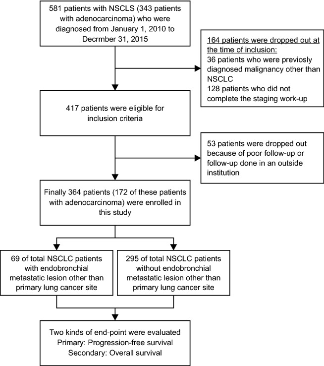
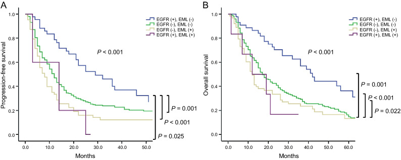
To evaluate the prognosis of non-small cell lung cancer (NSCLC) patients according to endobronchial metastatic lesion (EML), especially those not identified on positron emission tomography or computed tomography. We evaluated progression-free survival (PFS) and overall survival (OS) according to the presence of EML in patients with NSCLC who were diagnosed at a tertiary hospital between January 2010 and December 2019. A total of 364 patients were enrolled in this study. EML was found in 69 (19.0%) patients with NSCLC. In the patients with EML versus the patients without EML, median PFS was 7.0 (3.5-13.5) and 9.5 (5.5-17.5) months (P = 0.011), and median OS was 12.0 (6.0-30.0) versus 20.0 (10.0-39.0) months (P = 0.016), respectively. Median PFS and OS rates were highest in epidermal growth factor receptor (EGFR) (+) and EML (-) patients and lowest in EGFR (-) and EML (+) patients (P < 0.001). By multivariate cox regression analysis, PFS in overall patients with NSCLC was significantly associated with EML, EGFR mutation, performance status, and pleural effusion. NSCLC patients with EML had worse prognoses of PFS and OS than patients without EML.


Conflict of interest statement

The authors declare no competing interests.

Figures

Figure 1
Figure 1
Flowchart for enrolled patients Flowchart illustrates selection process for patients with NSCLC who were diagnosed during the study period. NSCLC non-small cell lung cancer, PET/CT positron emission tomography/computed tomography.
Figure 2
Figure 2
Median DFS, PFS and OS between NSCLC patients with and without EML other than primary cancer (N = 364) Median DFS was 47.0 (18.0–55.0) and 32.0 (19.0–49.0) months and in patients with EML compared to patients without EML (HR = 1.036, 95% CI 0.248–4.328, P = 0.912, (A); Median PFS (7.0 (3.5–13.5) vs. 9.5 (5.5–17.4) mo, HR = 0.681, 95% CI 0.411–0.833, P = 0.011, (B) and OS (12.0 (6.0–30.0) vs. 20.0 (10.0–39.0) mo, HR = 0.703, 95% CI 0.528–0.937, P = 0.016, (C) were significantly lower in patients with EML other than primary cancer. Median DFS (D), PFS (E) and OS (F) between NSCLC patients who have EML identified and not identified on PET/CT (N = 69). Median DFS 47.0 (43.0–55.0) mo vs. not checkable (HR = 0.224, 95% CI 0.014–3.590, P = 0.290, (D); Median PFS (5.5 (3.0–11.0) vs. 11.0 (7.0–27.0) mo, HR = 2.225, 95% CI 1.199–4.128, P = 0.011, (E); and OS 11.0 (5.5–26.0) vs. 18.0 (11.0–36.0) mo, HR = 1.644, 95% CI 0.885–3.054, P = 0.115, (F) between two groups. PFS was significantly higher in patients with EML not identified with PET/CT. DFS disease-free survival, EML endobronchial metastatic lesion, HR hazard ratio, mo months, NSCLC non-small cell lung cancer, OS overall survival rate, PFS progression-free survival rate, vs. versus.
Figure 3
Figure 3
Median PFS and OS according to EML when EGFR negative or positiveWhen compared PFS and OS according to EML in case of EGFR negative, PFS (A) and OS (B) were not significantly different as median PFS was 8.0 (4.0–16.0) versus 10.0 (5.0–22.0) months (HR = 0.705, 95% CI 0.523–1.013, P = 0.081, (A), and median OS was 12.0 (6.0–31.0) versus 17.0 (9.0–35.5) months (HR = 0.787, 95% CI 0.583–1.063, P = 0.118, (B). When EGFR was positive, PFS (C) and OS (D) were significantly lower in patients with EML compared to patients without EML as median PFS was 8.5 (3.0–14.0) versus 25.0 (12.5–39.5) months (HR = 0.277, 95% CI 0.103–0.747, P = 0.011, (C), and median OS was 15.5 (7.0–21.0) versus 34.0 (21.0–53.5) months (HR = 0.255, 95% CI 0.092–0.709, P = 0.009, (D).EGFR epidermal growth factor receptor, EML endobronchial metastatic lesion, HR hazard ratio, mo months, OS overall survival rate, PFS progression-free survival rate, vs. versus.
Figure 4
Figure 4
Median PFS according to EGFR and EML EGFR (−), EML (−) 11.0 (5.0–24.5); EGFR (+), EML (−) 25.0 (12.5–39.5); EGFR (−), EML (+) 8.0 (4.0–16.0); EGFR (+), EML (+) 8.5 (3.0–14.0) mo, respectively (P < 0.001). Median OS was also as follows; EGFR (−), EML (−) 17.0 (9.0–35.5); EGFR (+), EML (−) 34.0 (21.0–53.5); EGFR (−), EML (+) 12.0 (6.0–31.0); EGFR (+), EML (+) 15.5 (7.0–21.0) mo, respectively (P < 0.001). Both median PFS and OS were highest in EGFR (+) and EML (−). EGFR, epidermal growth factor receptor; EML endobronchial metastatic lesion, mo months, OS overall survival, PFS progression-free survival.

References

    1. Jemal A, et al. Global cancer statistics. CA Cancer J Clin. 2011;61(2):69–90. doi: 10.3322/caac.20107. - DOI - PubMed
    1. Silvestri GA, Hoffman B, Reed CE. One from column A: choosing between CT, positron emission tomography, endoscopic ultrasound with fine-needle aspiration, transbronchial needle aspiration, thoracoscopy, mediastinoscopy, and mediastinotomy for staging lung cancer. Chest. 2003;123(2):333–335. doi: 10.1378/chest.123.2.333. - DOI - PubMed
    1. Zaric B, et al. Advanced bronchoscopic techniques in diagnosis and staging of lung cancer. J Thorac Dis. 2013;5(suppl 4):S359–S370. - PMC - PubMed
    1. Lerner AD, Feller-Kopman D. Bronchoscopic techniques used in the diagnosis and staging of lung cancer. J Natl Compr Canc Netw. 2017;15(5):640–647. doi: 10.6004/jnccn.2017.0065. - DOI - PubMed
    1. Sharma P, Singh H, Basu S, Kumar R. Positron emission tomography-computed tomography in the management of lung cancer: an update. South Asian J Cancer. 2013;2(3):171–178. doi: 10.4103/2278-330X.114142. - DOI - PMC - PubMed

Publication types