

Astrocyte biomarker signatures of amyloid-β and tau pathologies in Alzheimer's disease
- PMID: 35948658
- PMCID: PMC9734046
- DOI: 10.1038/s41380-022-01716-2
Astrocyte biomarker signatures of amyloid-β and tau pathologies in Alzheimer's disease
Abstract
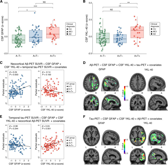
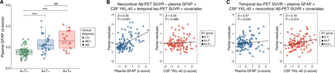
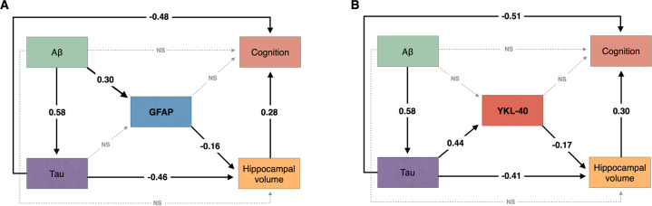
Astrocytes can adopt multiple molecular phenotypes in the brain of Alzheimer's disease (AD) patients. Here, we studied the associations of cerebrospinal fluid (CSF) glial fibrillary acidic protein (GFAP) and chitinase-3-like protein 1 (YKL-40) levels with brain amyloid-β (Aβ) and tau pathologies. We assessed 121 individuals across the aging and AD clinical spectrum with positron emission tomography (PET) brain imaging for Aβ ([18F]AZD4694) and tau ([18F]MK-6240), as well as CSF GFAP and YKL-40 measures. We observed that higher CSF GFAP levels were associated with elevated Aβ-PET but not tau-PET load. By contrast, higher CSF YKL-40 levels were associated with elevated tau-PET but not Aβ-PET burden. Structural equation modeling revealed that CSF GFAP and YKL-40 mediate the effects of Aβ and tau, respectively, on hippocampal atrophy, which was further associated with cognitive impairment. Our results suggest the existence of distinct astrocyte biomarker signatures in response to brain Aβ and tau accumulation, which may contribute to our understanding of the complex link between reactive astrogliosis heterogeneity and AD progression.
© 2022. The Author(s).
Conflict of interest statement
SG has served as a scientific advisor to Cerveau Therapeutics. KB has served as a consultant, at advisory boards, or at data monitoring committees for Abcam, Axon, Biogen, JOMDD/Shimadzu. Julius Clinical, Lilly, MagQu, Novartis, Prothena, Roche Diagnostics, and Siemens Healthineers, and is a co-founder of Brain Biomarker Solutions in Gothenburg AB (BBS), which is a part of the GU Ventures Incubator Program. HZ has served at scientific advisory boards and/or as a consultant for Abbvie, Alector, Annexon, AZTherapies, CogRx, Denali, Eisai, Nervgen, Pinteon Therapeutics, Red Abbey Labs, Passage Bio, Roche, Samumed, Siemens Healthineers, Triplet Therapeutics, and Wave, has given lectures in symposia sponsored by Cellectricon, Fujirebio, Alzecure and Biogen, and is a co-founder of Brain Biomarker Solutions in Gothenburg AB (BBS), which is a part of the GU Ventures Incubator Program. All other authors declare no competing interests.
Figures
Comment in
-
Reactive astrocyte biomarkers mirror Alzheimer disease pathology.Nat Rev Neurol. 2022 Oct;18(10):575. doi: 10.1038/s41582-022-00713-x. Nat Rev Neurol. 2022. PMID: 35999471 No abstract available.
References
MeSH terms
Substances
Grants and funding
- AACSF-20-648075/ALZ/Alzheimer's Association/United States
- NIRP-12-259245/ALZ/Alzheimer's Association/United States
- MOP-11-51-31/Gouvernement du Canada | Canadian Institutes of Health Research (Instituts de Recherche en Santé du Canada)
- 33397/Fondation Brain Canada (Fondation Neuro Canada)
- 312410/2018- 2/Ministry of Science, Technology and Innovation | Conselho Nacional de Desenvolvimento Científico e Tecnológico (National Council for Scientific and Technological Development)
- 435642/2018-9/Ministry of Science, Technology and Innovation | Conselho Nacional de Desenvolvimento Científico e Tecnológico (National Council for Scientific and Technological Development)
- R01 AG073267/AG/NIA NIH HHS/United States
- R01AG073267/U.S. Department of Health & Human Services | National Institutes of Health (NIH)
- R01AG075336/U.S. Department of Health & Human Services | National Institutes of Health (NIH)
- 2020-VICO-279314/Fonds de Recherche du Québec - Santé (Fonds de la recherche en sante du Quebec)
- R01 AG075336/AG/NIA NIH HHS/United States
- 88887.627297/2021-00/Coordenação de Aperfeiçoamento de Pessoal de Nível Superior (Brazilian Federal Agency for the Support and Evaluation of Graduate Education)
- NIRG-12-92090/ALZ/Alzheimer's Association/United States
LinkOut - more resources
Full Text Sources
Medical
Research Materials
Miscellaneous
