. 2022 Nov;27(11):4754-4769.
doi: 10.1038/s41380-022-01701-9. Epub 2022 Aug 10.

White matter damage as a consequence of vascular dysfunction in a spontaneous mouse model of chronic mild chronic hypoperfusion with eNOS deficiency

Affiliations

White matter damage as a consequence of vascular dysfunction in a spontaneous mouse model of chronic mild chronic hypoperfusion with eNOS deficiency

Xingyong Chen et al. Mol Psychiatry. 2022 Nov.

Abstract

Vascular cognitive impairment and dementia (VCID) is the second most common form of dementia after Alzheimer's disease (AD). Currently, the mechanistic insights into the evolution and progression of VCID remain elusive. White matter change represents an invariant feature. Compelling clinical neuroimaging and pathological evidence suggest a link between white matter changes and neurodegeneration. Our prior study detected hypoperfused lesions in mice with partial deficiency of endothelial nitric oxide (eNOS) at very young age, precisely matching to those hypoperfused areas identified in preclinical AD patients. White matter tracts are particularly susceptible to the vascular damage induced by chronic hypoperfusion. Using immunohistochemistry, we detected severe demyelination in the middle-aged eNOS-deficient mice. The demyelinated areas were confined to cortical and subcortical areas including the corpus callosum and hippocampus. The intensity of demyelination correlated with behavioral deficits of gait and associative recognition memory performances. By Evans blue angiography, we detected blood-brain barrier (BBB) leakage as another early pathological change affecting frontal and parietal cortex in eNOS-deficient mice. Sodium nitrate fortified drinking water provided to young and middle-aged eNOS-deficient mice completely prevented non-perfusion, BBB leakage, and white matter pathology, indicating that impaired endothelium-derived NO signaling may have caused these pathological events. Furthermore, genome-wide transcriptomic analysis revealed altered gene clusters most related to mitochondrial respiratory pathways selectively in the white matter of young eNOS-deficient mice. Using eNOS-deficient mice, we identified BBB breakdown and hypoperfusion as the two earliest pathological events, resulting from insufficient vascular NO signaling. We speculate that the compromised BBB and mild chronic hypoperfusion trigger vascular damage, along with oxidative stress and astrogliosis, accounting for the white matter pathological changes in the eNOS-deficient mouse model. We conclude that eNOS-deficient mice represent an ideal spontaneous evolving model for studying the earliest events leading to white matter changes, which will be instrumental to future therapeutic testing of drug candidates and for targeting novel/specific vascular mechanisms contributing to VCID and AD.


Conflict of interest statement

The authors declare no competing interests.

Figures
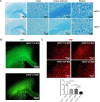
Fig. 1
Fig. 1. Severe myelin loss in middle-aged eNOS-deficient mice.
A Representative images of LFB (coronal, 16 μm) staining taken from mice at 12 months of age. Scaled bars are indicated in each picture. B Representative images of MBP immunohistochemistry at 12 months of age (12 MO). Scaled bars: 500 μm. C Representative images of MBP immunohistochemistry from the frontal cortical regions at 12 months of age. Scaled bars: 100 μm. Quantification was performed based on the mean fluorescent intensity from the frontal cortical regions and expressed as mean ± s.e.m; N = 5–6 mice/group.
Fig. 2
Fig. 2. Gait disbalance, impaired associated recognition memory and cortical pyramidal neurodegeneration at early mid-age.
A Gait performance by CatWalk testing. The test started once the mouse entered the visual field of the camera, and stopped once the mouse disappeared. The averaged running speed and additional parameters (e.g., altered sequence patterns and interlimb coordination) were recorded, analyzed, and expressed as mean ± s.e.m; N = 5–7 mice/group per genotype (11–12 MO). B The novel objective recognition (NOR) test was used to assess associated recognition memory in eNOS+/+, eNOS+/−, and eNOS−/− mice (9 MO). The eNOS−/− mice showed a significant reduction in discrimination index (DI) and recognition index (RI), evident by their inability to discriminate and recognize between the novel and familiar objects, compared to littermate eNOS+/+ mice (mean ± s.e.m; N = 6–9). C Representative images of LFB and Nissl double-stained sections of brains of eNOS−/− and control littermates showing frontal cortical regions at 12 months of age. Red double arrows and the corresponding Roman numerals indicate cortical layers I through V. Red arrowheads indicate degenerating neuron with swollen soma. Scaled bars are indicated in each picture.
Fig. 3
Fig. 3. Elevated reactive oxygen species (ROS) and astrogliosis.
A Representative images of dihydroethidium (DHE)-stained frontal forebrain cortical regions from mice at 12 months of age. Roman numerals in yellow indicate cortical layers II/III and V where we detected the most elevated ROS signals. Lower right panel images with zoned the area showing intracellular DHE signals presumably deriving from the clustered mitochondria in the cytoplasm. Quantification was performed based on the mean fluorescent intensity from the frontal cortical regions and expressed as mean ± s.e.m; N = 5-6 mice/group. B Representative images of GFAP immunohistochemistry on coronal brain sections of mice at 12 and 24 months of age. C Quantification of GFAP and Iba-1 immunofluorescence intensity based on 5–6 mice/group and expressed as mean ± s.e.m.
Fig. 4
Fig. 4. Functional recovery after sodium nitrate feeding.
A Schematic diagram illustrating sodium nitrate (SN) feeding schedules in three cohorts of mice. B Representative FITC-dextran angiograms of whole brain (left two panels) from dorsal view from eNOS mice at 3 months of age. Red boxed indicates the parietal cortical zone from the dorsal view. The right two panels show the representative FITC angiograms taken from 100 mm coronal sections of frontal brains from the same mice as in the left two panels. C Representative images of hydroxyprobe images. D Representative images of Evans blue angiograms of young eNOS+/+ and eNOS−/− mice (3 months of age) taken at 2.0 to −1.82 mm Bregman, showing BBB leakage detected at a young age which can be prevented by sodium nitrate (SN) feeding in drinking water for 6 weeks starting from 8 weeks old (SN eNOS−/−, N = 3 mice/groups). Evans Blue (EB, 2 % in sterile water) was injected in 150 μL volume through mouse tail vein. Mice were euthanized 5 min later without perfusion and whole brains removed and vibratome-processed to 100 μm serial sections for fluorescent imaging using an rhodamine filter. E Quantification of Evans blue fluorescent signals based on the angiograms presented in panel. Data are presented as mean ± s.e.m based on N = 3 mice each group. FH Examples of compound axon action potentials (CAPs) recorded in the corpus callosum in eNOS+/+ (WT) mice, eNOS+/− mice, eNOS−/− mice and sodium nitrite-treated (SN) eNOS−/− mice. G Summary of the amplitudes of the myelinated axon-generated fast N1 component in the four groups of mice. H Summary of the amplitudes of the unmyelinated axon-generated slow N2 component in the four groups of mice.
Fig. 5
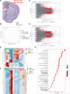
Fig. 5. Transcriptional profiling identifies white matter changes in heterozygous eNOS+/− mice.
A Diagram of the specific brain region tissue for micro punch. B PCA plot of the RNA-seq data from white matter and gray matter tissues isolated from the brains of wild-type eNOS+/+ and heterozygous eNOS+/− mice. MA plots of the pairwise comparisons between eNOS+/+ or eNOS+/−, C in gray matter and D in white matter tissues; log fold changes (LFCs) are plotted against the mean of normalized counts to determine the variance between two treatments in terms of gene expression. Red nodes on the graph represent statistically significant data points i.e., p.adj < 0.05 and LFC > 1.5. Gray nodes are data points that are not statistically significant. Numerical values in parentheses for the significant legend indicate the number of genes that meet the prior condition. Dashed lines indicate the cutoff LFC values. E Heatmap showing the alteration of significant genes belonging to gene ontology (GO) term regulation of long-term synaptic potentiation (GO:1900271), in white matter and gray matter tissues isolated from the brains of eNOS+/+ and eNOS+/− mice (significant genes p.adj < 0.05 and LFC > 1.5) in the white matter of eNOS+/− compared to eNOS+/+. F Heatmap showing the progression of significant genes belonging to gene ontology (GO) term cellular respiration (GO:0045333), in white matter and gray matter tissues isolated from the brains of eNOS+/+ and eNOS+/− mice (significant genes, p.adj < 0.05 and LFC > 1.5). G GO enrichment dot plot showing the number of genes affected in the GO terms related to synaptic plasticity and cognition in white matter tissue isolated from the brains of eNOS+/+ and eNOS+/− mice (significant genes, p.adj < 0.05 and LFC > 1.5).
Fig. 6
Fig. 6. Potential negative impact of the aberrantly upregulated BMP4 signaling upon eNOS deficiency on BBB.
A qPCR graphs showing increased expression of the two genes of the large transforming growth factor TGFβ family: Tgf-β1: transforming growth factor-β1; Bmp4: bone morphogenetic protein 4. Data are presented as mean ± s.d.m., normalized by Gapdh gene expression. N = 3–4 mice/group. B Representative western blots and graph of semi-quantitative analysis based on densitometry. C Microscopic image of BMP4 immunosignals taken from eNOS−/− brain (5 MO). D Representative fluorescent images taken from the forebrain of BMP4-CFP reporter mouse on an eNOS+/− background (5 MO) using wide DAPI filter. Quantification graphs were based on the BMP4-CFPepositive cells and on the fluorescent intensity of the positive cells, respectively (N = 3 mice/genotype, comparison between eNOS+/+ and eNOS+/−). E Microscopic image of BMP4 immunosignals on the same brain section taken from eNOS−/− brain for Evans blue angiography. F Transendothelial electrical resistance measurement (TEER values) of the bEnd.3 cells (monolayer BBB model) treated with recombinant rBMP4 (25 ng/mL; mean ± s.e.m; N = 3 independent experiments). G Representative western blots of tight junction marker proteins of rBMP4-treated bEnd.3 cells (N = 3 independent experiments).

Similar articles

Cited by

References

    1. Snyder HM, Corriveau RA, Craft S, Faber JE, Greenberg S, Knopman D, et al. Vascular contributions to cognitive impairment and dementia including Alzheimer’s disease. Alzheimers Dement. 2015;11:710–7. - PMC - PubMed
    1. Schneider JA, Arvanitakis Z, Bang W, Bennett DA. Mixed brain pathologies account for most dementia cases in community-dwelling older persons. Neurology. 2007;69:2197–204. - PubMed
    1. Iadecola C. The pathobiology of vascular dementia. Neuron. 2013;80:844–66. - PMC - PubMed
    1. Attems J, Jellinger K. The overlap between vascular disease and Alzheimer’s disease lessons from pathology. BMC Med. 2014;12:206. - PMC - PubMed
    1. Hofman A, Ott A, Breteler MM, Bots ML, Slooter AJ, van Harskamp F, et al. Atherosclerosis, apolipoprotein E, and prevalence of dementia and Alzheimer’s disease in the Rotterdam study. Lancet. 1997;349:151–4. - PubMed