. 2022 Aug 10;22(1):214.
doi: 10.1186/s12906-022-03694-y.

Harpagoside attenuates local bone Erosion and systemic osteoporosis in collagen-induced arthritis in mice

Affiliations

Harpagoside attenuates local bone Erosion and systemic osteoporosis in collagen-induced arthritis in mice

Ju-Young Kim et al. BMC Complement Med Ther. .

Abstract

Background: Rheumatoid arthritis (RA) is a chronic inflammatory autoimmune disease that causes local bone erosion and systemic osteoporosis. Harpagoside (HAR), an iridoid glycoside, has various pharmacological effects on pain, arthritis, and inflammation. Our previous study suggests that HAR is more deeply involved in the mechanism of bone loss caused by inflammatory stimuli than hormonal changes. Here, we identified the local and systemic bone loss inhibitory effects of HAR on RA and its intracellular mechanisms using a type 2 collagen-induced arthritis (CIA) mouse model.

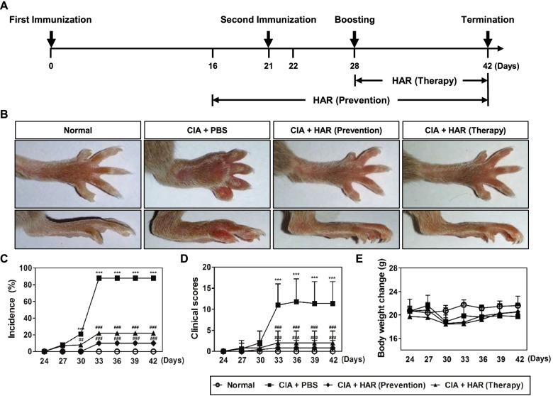
Methods: The anti-osteoporosis and anti-arthritic effects of HAR were evaluated on bone marrow macrophage in vitro and CIA in mice in vivo by obtaining clinical scores, measuring hind paw thickness and inflammatory cytokine levels, micro-CT and histopathological assessments, and cell-based assay.

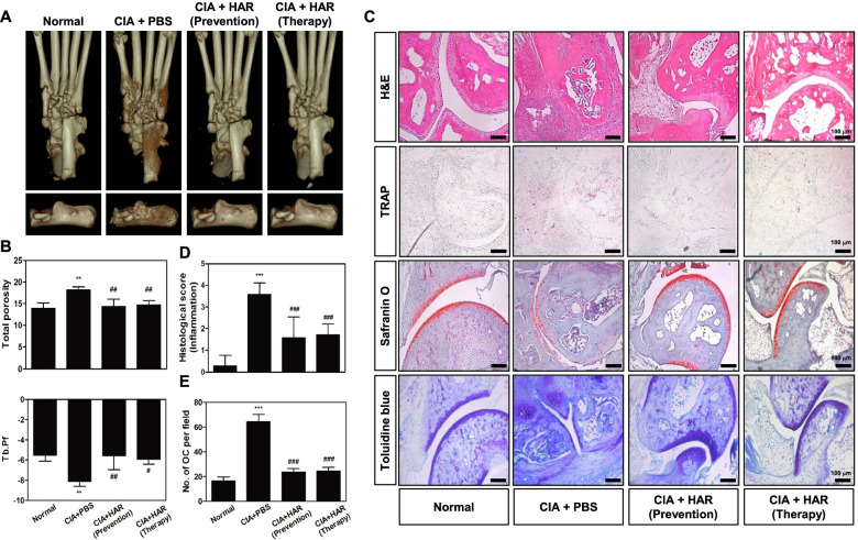
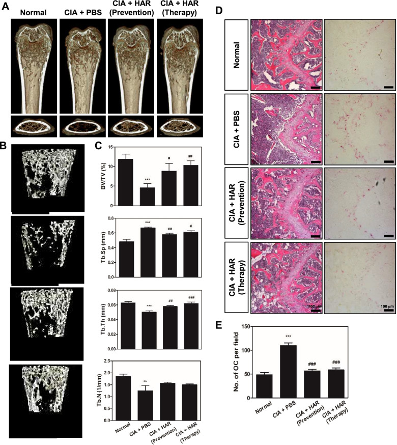
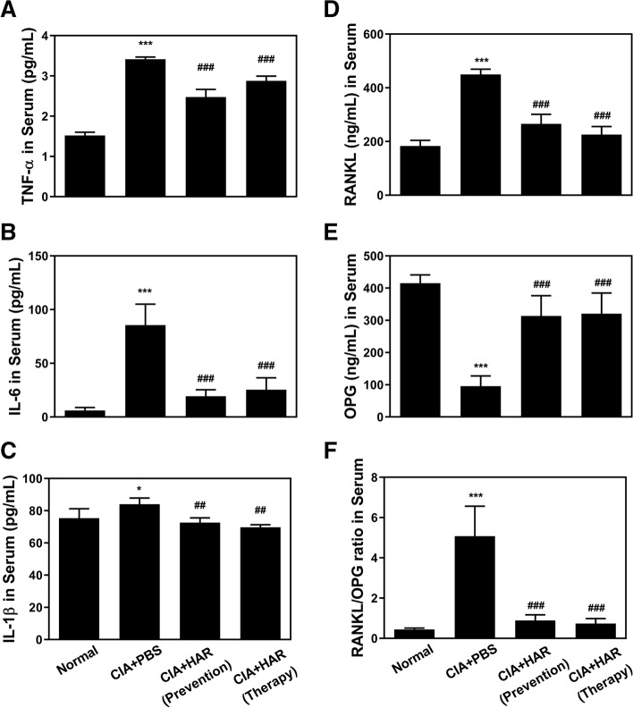
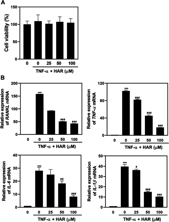
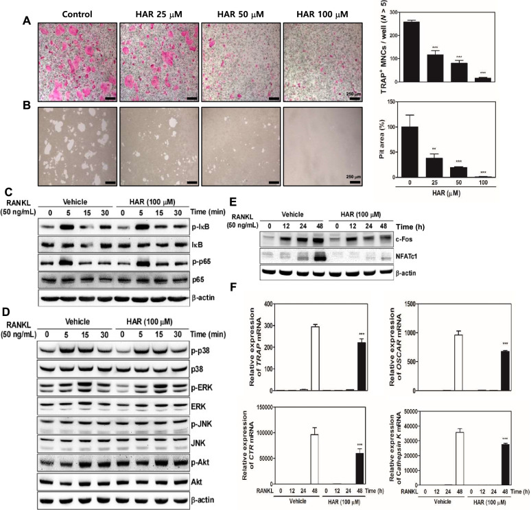
Results: HAR markedly reduced the clinical score and incidence rate of CIA in both the prevention and therapy groups. Histological analysis demonstrated that HAR locally ameliorated the destruction of bone and cartilage and the formation of pannus. In this process, HAR decreased the expression of inflammatory cytokines, such as tumor necrosis factor-α, interleukin (IL)-6, and IL-1β in the serum of CIA mice. Additionally, HAR downregulated the expression of receptor activator of nuclear factor-κB ligand and upregulated that of osteoprotegerin. HAR suppressed systemic bone loss by inhibiting osteoclast differentiation and osteoclast marker gene expression in a CIA mouse model.

Conclusions: Taken together, these findings show the beneficial effect of HAR on local symptoms and systemic bone erosion triggered by inflammatory arthritis.

Keywords: Collagen-induced arthritis model; Harpagoside; Inflammation; Osteoclasts; Rheumatoid arthritis.


Conflict of interest statement

The authors declare that this article involves no conflict of interest.

Figures

Fig. 1
Fig. 1
Effect of harpagoside (HAR) on the progression of collagen-induced arthritis (CIA). A Study design to induce CIA in mice. Eight-week-old male DBA/1 mice were first immunized with Col 2 and CFA on day 0 and then immunized intraperitoneally with Col 2 on day 21. Next, booster injection with LPS was given on day 28. CIA mice were orally administrated with HAR (10 mg/kg) every day from day 16 to 42 in the prevention group and from day 28 to 42 in the therapy group. B Representative photographs of normal, PBS-, and HAR-treated CIA mice. C The incidence of arthritis, D clinical scores, and E body weight change were determined on the indicated days. ***p < 0.001 versus the normal group; ##p < 0.01 and ###p < 0.001 versus the CIA + PBS group
Fig. 2
Fig. 2
Effect of harpagoside (HAR) on the severity of arthritis and histopathological changes in collagen-induced arthritis (CIA) mice. On day 42 after the initial immunization with Col 2, systemic bone loss was analyzed by micro-CT, and bone destruction and cartilage damage of CIA mice were analyzed by histological staining of the ankle joint. A Representative 3D images of the hind paw (up) and calcaneus (down). B The total porosity, trabecular thickness (Tb.Th), and trabecular pattern factor (Tb.Pf) of the calcaneus. C The ankle joint was sectioned and stained with H&E, TRAP, Safranin O, and toluidine blue. Scale bar, 100 μm. D The pathological severity scores of inflammation, pannus formation, cartilage damage, and bone damage in the ankle joint of CIA mice. E The number of osteoclasts per field was counted using the histomorphometric methods. **p < 0.01 and ***p < 0.001 versus the normal group; # p < 0.05, ##p < 0.01, and ###p < 0.001 versus the CIA + PBS group
Fig. 3
Fig. 3
Effect of harpagoside (HAR) on bone erosion, bone destruction, and TRAP formation in collagen-induced arthritis (CIA) mice. A Mice were sacrificed on day 42 after the first immunization, and radiographs of the confocal and transverse planes of proximal femur were obtained from micro-CT apparatus. B Micro-CT images of specific regions of the trabecular bone for the assessment of various bone parameters. C The bone volume per tissue volume (BV/TV), trabecular separation (Tb.Sp), trabecular thickness (Tb,Th), and trabecular number (Tb.N) of femur were determined using the micro-CT data and analyzed by INFINITT-Xelis Software. After micro-CT scans, dissected femurs were fixed, decalcified, embedded, and sectioned. Sections were stained with (D) hematoxylin and eosin (H&E) (left) and TRAP (right). Scale bar, 100 μm. E The number of osteoclasts per field was counted using the histomorphometric methods. **p < 0.01 and ***p < 0.001 versus the normal group; #p < 0.05, ##p < 0.01, and ###p < 0.001 versus the CIA + PBS group
Fig. 4
Fig. 4
Effect of harpagoside (HAR) on pro-inflammatory mediators and bone markers in serum of collagen-induced arthritis (CIA) mice. Serum was prepared from CIA mice on day 42. The expression levels of (A) TNF-α, (B) IL-6, (C) IL-1β, (D) RANKL, and (E) OPG were determined using murine ELISA assay, and the ratio of (F) RANKL/OPG was calculated. *p < 0.05 and ***p < 0.001 versus the normal group; ##p < 0.01 and ###p < 0.001 versus the CIA + PBS group
Fig. 5
Fig. 5
Effect of harpagoside (HAR) on the production of proinflammatory cytokines, RANKL, TNFα, IL-6, and IL-1β in TNFα-stimulated SW982 synovial cells. SW982 synovial cells were treated various doses of HAR (0, 25, 50 and 100 μM) for 1 h and then incubated with TNFα (50 ng/mL) for 24 h. A Cell viability was determined using the XTT assay. B The total RNA was extracted and real-time RT-PCR was performed to measure the transcripts of RANKL, TNFα, IL-6, and IL-1β. The mRNA levels of these genes were normalized to GAPDH and represented as fold change over the TNFα-untreated, HAR-untreated cells. ***p < 0.001 versus TNFα-untreated, HAR-untreated cells; #p < 0.05, ##p < 0.01 and ###p < 0.001 versus TNFα-treated, HAR-untreated cells
Fig. 6
Fig. 6
Effect of harpagoside (HAR) on RANKL-induced osteoclast differentiation, bone resorption, and signal pathway in bone marrow macrophages (BMMs). A BMMs were cultured for 4 days in the presence of M-CSF (30 ng/mL) and RANKL (50 ng/mL) with HAR (0–100 μM). Then, the cells were fixed with 3.7% formalin, permeabilized with 0.1% Triton X-100, and stained with TRAP. Scale bar, 250 μm. TRAP-positive multi-nucleated osteoclasts (TRAP+ MNCs) with more than five nuclei were counted. B Mature osteoclasts were seeded on hydroxyapatite-coated plates and treated with the indicated concentrations of HAR for 24 h. The attached cells on the plates were removed and photographed under a light microscope. Pit areas were quantified using Image J. C-E BMMs were pretreated with HAR (100 μM) or vehicle (DMSO) for 1 h before RANKL (50 ng/mL) stimulation at the indicated times. The cell lysates were analyzed by western blotting with the indicated antibodies. F The mRNA expressions of TRAP, OSCAR, CTR, and Cathepsin K were analyzed by real-time RT-PCR. **p < 0.01 and ***p < 0.001 versus the vehicle

Similar articles

Cited by

References

    1. Schett G. Autoimmunity as a trigger for structural bone damage in rheumatoid arthritis. Mod Rheumatol. 2017;27:193–197. doi: 10.1080/14397595.2016.1265907. - DOI - PubMed
    1. Tanaka S. Emerging anti-osteoclast therapy for rheumatoid arthritis. J Orthop Sci. 2018;23:717–721. doi: 10.1016/j.jos.2018.06.001. - DOI - PubMed
    1. Kumar G, Roger PM. From crosstalk between immune and bone cells to bone erosion in infection. Int J Mol Sci. 2019;20:5154. doi: 10.3390/ijms20205154. - DOI - PMC - PubMed
    1. Jung SM, Kim KW, Yang CW, Park SH, Ju JH. Cytokine-mediated bone destruction in rheumatoid arthritis. J Immunol Res. 2014;2014:263625. doi: 10.1155/2014/263625. - DOI - PMC - PubMed
    1. Schett G, Gravallese E. Bone erosion in rheumatoid arthritis: mechanisms, diagnosis and treatment. Nat Rev Rheumatol. 2012;8:656–664. doi: 10.1038/nrrheum.2012.153. - DOI - PMC - PubMed