. 2022 Aug 11;24(1):47.
doi: 10.1186/s12968-022-00879-9.

An inline deep learning based free-breathing ECG-free cine for exercise cardiovascular magnetic resonance

Affiliations

An inline deep learning based free-breathing ECG-free cine for exercise cardiovascular magnetic resonance

Manuel A Morales et al. J Cardiovasc Magn Reson. .

Abstract

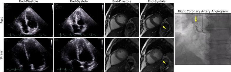
Background: Exercise cardiovascular magnetic resonance (Ex-CMR) is a promising stress imaging test for coronary artery disease (CAD). However, Ex-CMR requires accelerated imaging techniques that result in significant aliasing artifacts. Our goal was to develop and evaluate a free-breathing and electrocardiogram (ECG)-free real-time cine with deep learning (DL)-based radial acceleration for Ex-CMR.

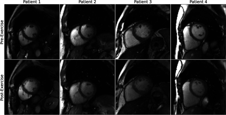
Methods: A 3D (2D + time) convolutional neural network was implemented to suppress artifacts from aliased radial cine images. The network was trained using synthetic real-time radial cine images simulated using breath-hold, ECG-gated segmented Cartesian k-space data acquired at 3 T from 503 patients at rest. A prototype real-time radial sequence with acceleration rate = 12 was used to collect images with inline DL reconstruction. Performance was evaluated in 8 healthy subjects in whom only rest images were collected. Subsequently, 14 subjects (6 healthy and 8 patients with suspected CAD) were prospectively recruited for an Ex-CMR to evaluate image quality. At rest (n = 22), standard breath-hold ECG-gated Cartesian segmented cine and free-breathing ECG-free real-time radial cine images were acquired. During post-exercise stress (n = 14), only real-time radial cine images were acquired. Three readers evaluated residual artifact level in all collected images on a 4-point Likert scale (1-non-diagnostic, 2-severe, 3-moderate, 4-minimal).

Results: The DL model substantially suppressed artifacts in real-time radial cine images acquired at rest and during post-exercise stress. In real-time images at rest, 89.4% of scores were moderate to minimal. The mean score was 3.3 ± 0.7, representing increased (P < 0.001) artifacts compared to standard cine (3.9 ± 0.3). In real-time images during post-exercise stress, 84.6% of scores were moderate to minimal, and the mean artifact level score was 3.1 ± 0.6. Comparison of left-ventricular (LV) measures derived from standard and real-time cine at rest showed differences in LV end-diastolic volume (3.0 mL [- 11.7, 17.8], P = 0.320) that were not significantly different from zero. Differences in measures of LV end-systolic volume (7.0 mL [- 1.3, 15.3], P < 0.001) and LV ejection fraction (- 5.0% [- 11.1, 1.0], P < 0.001) were significant. Total inline reconstruction time of real-time radial images was 16.6 ms per frame.

Conclusions: Our proof-of-concept study demonstrated the feasibility of inline real-time cine with DL-based radial acceleration for Ex-CMR.

Keywords: Coronary artery disease; Deep learning; Exercise MRI; Inline; Radial golden angle.


Conflict of interest statement

Xiaoying Cai and Kelvin Chow are employees of Siemens Medical Solutions USA, Inc. Reza Nezafat has a research agreement with Siemens. Hassan Haji-valizadeh is an employee of Canon Medical Research USA, Inc.

Figures

Fig. 1
Fig. 1
Deep learning model and generation of training data. A The deep learning model consists of a three-dimensional U-Net trained to filter out streaking artifacts from complex-valued real-time radial cine n×n images with nt time frames. The inputs and outputs to the U-Net are the real and imaginary components concatenated along the spatial dimension, and the loss during training was the mean square error (MSE) between inputs and outputs. During inference, the generated real and imaginary parts with suppressed artifacts are re-combined. B Synthetic real-time image training pairs were generated from electrocardiogram (ECG)-gated segmented Cartesian k-space data acquired at rest in 503 patients. First, the k-space data were reconstructed using the generalized autocalibrating partial parallel acquisition (GRAPPA) technique. Spatiotemporal interpolation followed by coil combination was then applied to the reconstructed images to simulate reference (i.e., ground-truth) real-time images. In addition, prior to coil combination, the interpolated images were also used to simulate undersampled real-time radial cine images by applying an inverse and forward non-uniform fast Fourier transform (NUFFT) with 12 radial lines. The coil-combined images with streaking artifacts were used as inputs during training
Fig. 2
Fig. 2
Inline implementation of real-time cine with deep learning-based radial acceleration. Multi-coil raw radial k-space data acquired from the scanner is processed in the Image Reconstruction Environment (ICE) on the vendor reconstruction computer. Using the International Society for Magnetic Resonance in Medicine Raw Data (ISMRMRD) format, the collected data is transferred to the Framework for Image Reconstruction (FIRE) server using a FireEmitter functor. The FIRE server is located in the vendor reconstruction computer. The data is then transferred from the FIRE server to an external server via a connecting Secure Shell Protocol (SSH) tunnel. In the external sever, a Docker containing all Python dependencies such as PyTorch is used to process the raw k-space data in a single 32 GB Graphics Processing Unit (GPU). The deep-learning radial acceleration with parallel reconstruction (DRAPR) technique was implemented in the Docker. First, a non-uniform fast Fourier transform (NUFFT) is used to grid and reconstruct undersampled multi-coil radial k-space data. GPU parallelization is done in PyTorch by treating frames and coils as batch and channel dimensions. This approach enables application of the NUFFT at 10 ms per frame. Coil sensitivity and combination is subsequently performed in PyTorch at negligible computational cost. These coil-combined images are send to the U-Net for de-aliasing, which requires 6.6 ms per frame. The total processing time of 16.6 ms is about half the 37.7 ms temporal resolution of collected frames. Images are then returned to the FIRE server via the same SSH tunnel, and to ICE using a FireInjector functor. Finally, the reconstructed de-aliased images are finalized into DICOM format and returned to the scanner computer console for immediate display
Fig. 3
Fig. 3
Exercise imaging protocol. Rest scans consisted of breath-hold, ECG-gated Cartesian segmented cine followed by free-breathing ECG-free real-time radial cine. After rest imaging, subjects were removed from the scanner bore and were exercised in the supine position using a cycle ergometer. Work rate was started at 25 W and was increased by + 25 W every 2 min while subjects maintained a constant pedaling speed of 75 rpm. After reaching target heart rate or exhaustion, subjects were immediately placed back inside the scanner bore for post-exercise stress imaging. This consisted of a repetition of the real-time cine sequence
Fig. 4
Fig. 4
Bland–Altman comparisons of left-ventricular (LV) parameters. Measures derived from breath-hold ECG-gated segmented cine are compared to those derived from the free-breathing ECG-free real-time cine reconstructed with the GRASP and deep-learning radial acceleration with parallel reconstruction (DRAPR) techniques. Solid and dotted lines represent the mean difference and mean difference ± 1.96 × standard deviation of the difference. A The mean differences in LV end-diastolic and end-systolic volumes with golden angle radial sparse parallel (GRASP) were 4.4 mL and 6.4 mL, while the mean difference in LV ejection fraction was − 4.3%. B The mean differences in LV end-diastolic and LV end-systolic volumes with DRAPR were 3 mL and 7 mL, while the mean difference in ejection fraction was − 5%
Fig. 5
Fig. 5
Subjective evaluation of image quality. Three readers evaluated artifacts across all cine scans. Images were classified as having a 1-non-diagnostic, 2-severe, 3-moderate, and 4-minimal artifact level. The mean score of breath-hold ECG-gated segmented cine images was 3.0. The mean score of free-breathing ECG-free real-time cine images reconstructed with GRASP was 3.7 at rest, with 9.1% of images graded as severe. The mean score during post-exercise stress was 3.5, and the percentage of images graded as severe increased to 17.9%. Real-time cine images at rest reconstructed with DRAPR had a mean score of 3.3, with 10.6% of images graded as severe. During post-exercise stress, images had a mean score of 3.1. The percentage of images graded as severe increased to 15.4%. None of the real-time radial cine images reconstructed with GRASP or DRAPR were labeled as non-diagnostic. *p < 0.01. GRASP: golden-angle radial sparse parallel; DRAPR: deep-learning radial acceleration with parallel reconstruction
Fig. 6
Fig. 6
Representative real-time cine images of subjects at rest. The NUFFT was used to grid and reconstruct radial k-space data. Artifacts were subsequently suppressed using the GRASP and DRAPR techniques. Images were classified as having a 1-non-diagnostic, 2-severe, 3-moderate, and 4-minimal artifact level. All NUFFT images were 1-non-diagnostic. The five subjects shown at end-diastole represent the range of mean scores at rest. Subjects 1–4 are patients. The mean artifact levels with DRAPR for subjects 1–5 were 3.0, 3.3, 3.3, 4.0 and 4.0, accordingly. The body mass index (BMI) for subjects 1–5 was 38, 33, 26, 23 and 21 lbs/in2, accordingly
Fig. 7
Fig. 7
Representative real-time cine images of subjects at post-exercise stress. The NUFFT was used to grid and reconstruct radial k-space data. Artifacts were subsequently suppressed using the GRASP and DRAPR techniques. Images were classified as having a 1-non-diagnostic, 2-severe, 3-moderate, and 4-minimal artifact level. All NUFFT images were 1-non-diagnostic. The five subjects shown at end-diastole represent the range of mean scores at post-exercise stress. Subjects 3–4 are healthy. Subjects 2 and 5 correspond to Subjects 1 and 4 in Fig. 6, accordingly. The mean artifact levels with DRAPR for subjects 1–5 were 2.3, 3.0, 3.0, 3.3 and 3.7, accordingly. The body mass index (BMI) for subjects 1–5 was 32, 38, 23, 29 and 24 lbs/in2, accordingly. The peak heart rate post-exercise for subjects 1–5 was 110, 108, 97, 132, and 135 bpm, accordingly
Fig. 8
Fig. 8
Inline real-time cine with deep learning-based radial acceleration in coronary artery disease (CAD). Four patients are shown whose recruitment criteria included being symptomatic and having a stress test for suspicion of CAD. Patients underwent an exercise CMR protocol. Top images were collected at rest, and bottom images were collected after supine exercise using a CMR compatible cycle ergometer. Images are shown at end-diastole. Patient 4 at pre-exercise corresponds to subject 4 in Fig. 6, and at post-exercise corresponds to subject 5 in Fig. 7, accordingly. The BMI for patients 1–4 was 22, 27, 26, and 24 lbs/in2, accordingly. During rest, the mean artifact levels were 3.0, 3.7, 4.0 and 4.0. The resting heart rates were 83, 62, 70 and 49 bpm. During post-exercise stress, the mean artifact levels were 3.3, 3.3, 3.3 and 3.7. The peak heart rates post-exercise were 115, 132, 107 and 134 bpm, accordingly
Fig. 9
Fig. 9
Illustrative clinical case. A 60-year-old male with worsening upper chest heaviness and shortness of breath with exertion. Stress echo showed focal systolic dysfunction with hypokinesis of the left-ventricular basal inferoseptum and inferior walls at both rest and stress. Similarly, during an exercise CMR protocol, real-time cine with deep learning-based radial acceleration showed hypokinesis of the left-ventricular basal inferoseptal wall during rest and at post-exercise stress (arrows). Subsequent coronary angiography revealed a heavily calcified right coronary artery (arrow) with serial subtotal occlusions

Similar articles

Cited by

References

    1. Arai AE, Schulz-Menger J, Berman D, Mahrholdt H, Han Y, Bandettini WP, et al. Gadobutrol-enhanced cardiac magnetic resonance imaging for detection of coronary artery disease. J Am Coll Cardiol. 2020;76(13):1536–1547. doi: 10.1016/j.jacc.2020.07.060. - DOI - PubMed
    1. Greenwood JP, Herzog BA, Brown JM, Everett CC, Plein S. Cardiovascular magnetic resonance and single-photon emission computed tomography in suspected coronary heart disease. Ann Intern Med. 2016;165(11):830–831. doi: 10.7326/L16-0480. - DOI - PubMed
    1. Members WC, Lawton JS, Tamis-Holland JE, Bangalore S, Bates ER, Beckie TM, et al. 2021 ACC/AHA/SCAI guideline for coronary artery revascularization: a report of the American College of Cardiology/American Heart Association Joint Committee on Clinical Practice Guidelines. J Am Coll Cardiol. 2022;79(2):e21–e129. doi: 10.1016/j.jacc.2021.09.006. - DOI - PubMed
    1. Nagel E, Greenwood JP, McCann GP, Bettencourt N, Shah AM, Hussain ST, et al. Magnetic resonance perfusion or fractional flow reserve in coronary disease. N Engl J Med. 2019;380(25):2418–2428. doi: 10.1056/NEJMoa1716734. - DOI - PubMed
    1. Schwitter J, Wacker CM, Wilke N, Al-Saadi N, Sauer E, Huettle K, et al. MR-IMPACT II: Magnetic Resonance Imaging for Myocardial Perfusion Assessment in Coronary artery disease Trial: perfusion-cardiac magnetic resonance vs. single-photon emission computed tomography for the detection of coronary artery disease: a comparative multicentre, multivendor trial. Eur Heart J. 2013;34(10):775–781. doi: 10.1093/eurheartj/ehs022. - DOI - PubMed

Publication types

MeSH terms