. 2022 Sep;36(9):e24564.
doi: 10.1002/jcla.24564. Epub 2022 Aug 10.

Effect of artificial skin membrane on the expression of miR-155 and miR-506-3p in patients with second-degree burns

Affiliations

Effect of artificial skin membrane on the expression of miR-155 and miR-506-3p in patients with second-degree burns

Fei Li et al. J Clin Lab Anal. 2022 Sep.

Abstract

Objective: To investigate the effect of artificial skin on the expression of miR-155 and miR-506-3p in patients with second-degree burns.

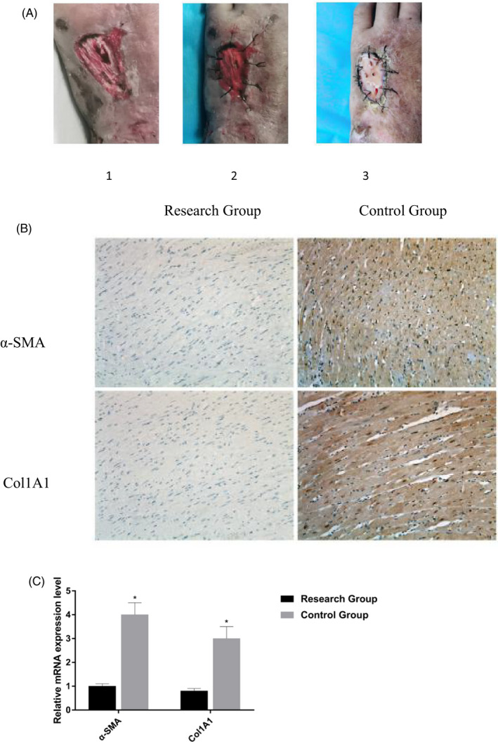
Methods: The study subjects included 50 patients with second-degree burns treated from July 2019 to July 2021. The control group received routine nursing, while the research group received both routine and artificial skin intervention simultaneously. The changes in wound tissue fibrosis and prognosis were observed. The expression levels of miR-155 and miR-506-3p and their downstream regulatory factors were detected and correlated with the rehabilitation of patients after artificial skin treatment.

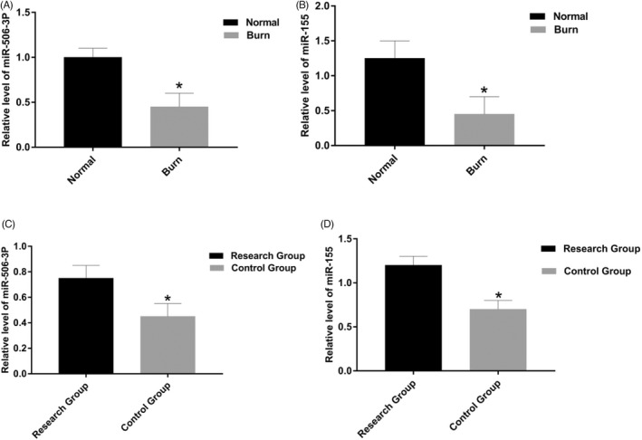
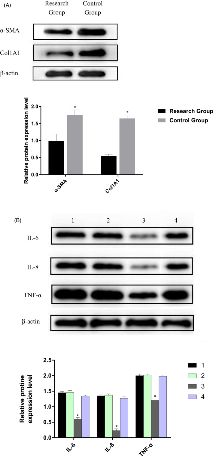
Results: After treating second-degree burns with artificial skin membranes, the patient's wound tissue fibrosis and inflammation level improved. At the same time, the expression levels of miR-155 and miR-506-3p in related tests were higher than those in patients with available treatment.

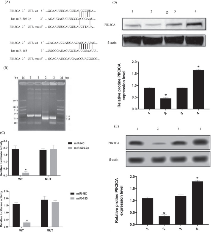
Conclusion: The effect of artificial skin membrane on the wound healing of second-degree burn patients may be realized by influencing the expression levels of miR-155 and miR-506-3p and their related signaling pathways.

Keywords: artificial skin; burn; microRNA-506-3p; microRNA155.


Conflict of interest statement

The authors declare that they have no conflicts of interest.

Figures

FIGURE 1
FIGURE 1
Comparison of wound and the changes of Immunohistochemistry. (A) After the same debridement application, the study group was given haifukang artificial skin film combined. 1. The wound surface of the patient's preoperative second‐degree burn. 2. Haifukang artificial skin film was used to directly cover the wound. 3. The second‐degree burn wound was well vascularized 4 weeks after artificial skin membrane transplantation. (B) Immunohistochemistry results showed that the protein expression levels of Col1A1 and α‐SMA in the control group were higher than those in the research group. (C) The mRNA expression levels of Col1A1 and α‐SMA decrease in the research group.
FIGURE 2
FIGURE 2
Expression of miR‐506‐3p and miR‐155 by qRT‐PCR. (A) The expression of miR‐506‐3p in burn tissues decreased (p < 0.05). (B) The expression of miR‐155 in burn tissues decreased (p < 0.05). (C) In cured tissues, the expression of miR‐506‐3p in control group is lower than research group (p < 0.05). (D) In cured tissues, the expression of miR‐155 in control group is lower than research group (p < 0.05).
FIGURE 3
FIGURE 3
Western blot analysis detected the expression of fibrosis marker proteins and inflammatory factors in each group. (A) The expression of Col1A1 and α‐SMA in patient second‐degree burns tissues after artificial skin membrane treatment was significantly lower than that before treatment (p < 0.05). (B) The levels of TNF‐α, IL‐6 and IL‐8 decreased significantly in research group, and the difference was statistically significant (p < 0.05). 1. The pre‐treatment research group. 2. The pre‐treatment control group. 3. The research group after treatment. 4. The control group after treatment.
FIGURE 4
FIGURE 4
miR‐506‐3p and miR‐155 can target regulation of the PI3K‐Akt pathway. (A) PIK3CA, was identified as one of the potential targets of miR‐506‐3p and miR‐155. (B) Recombinant plasmids P miR‐Report‐PIK3CA‐WT and P miR‐Report‐PIK3CA‐MUt. The length of p miR‐Report was 6470 bp, and PCR showed two fragments of the recombinant plasmid: PIK3CA‐WT 330 bp/PIK3CA‐MUT 235 bp. Agarose gel electrophoresis showed that the exogenous gene was successfully inserted. (C) miR‐506‐3p mimic and miR‐155 mimic reduced PIK3CA‐WT's luciferase activity, yet it had very little influence on PIK3CA‐MUT's luciferase activity. (D) 1. mimic‐N.C; 2. miR‐506‐3p mimic; 3. inhibitors‐N.C; 4. miR‐506‐3p inhibitor. After transfection of miR‐506‐3P mimic into Hsas4 cells, the expression of PIK3CA protein was lower than that of mimic‐N.C (p < 0.05), PIK3CA protein expression in miR‐506‐3p inhibitor group was higher than that in inhibitors‐NC (p < 0.05). (E) 1. mimic‐N.C; 2. miR‐155 mimic; 3. inhibitors‐N.C; 4. miR‐155 inhibitor. After transfection of miR‐155 mimic into Hsas4 cells, the expression of PIK3CA protein was lower than that of mimic‐N.C (p < 0.05), PIK3CA protein expression in miR‐155 inhibitor group was higher than that in inhibitors‐NC (p < 0.05).
FIGURE 5
FIGURE 5
(A) After overexpression of miR‐155, the proliferation level of Hsas4 cells in the research group was lower than that in the control group at the same time point. However, after the addition of IGF‐1 (An Activator of PI3K/AKT Signaling pathway), compared with the research group, the proliferation level of Hsas4 cells in the IGF‐1 group increased. (B) After overexpression of miR‐506‐3P, the proliferation level of Hsas4 cells in the research group was lower than that in the control group at the same time point. However, after the addition of IGF‐1 (An Activator of PI3K/AKT Signaling pathway), compared with the research group, the proliferation level of Hsas4 cells in the IGF‐1 group increased.

Similar articles

Cited by

References

    1. Song KX, Liu S, Zhang MZ, et al. Hyperbaric oxygen therapy improves the effect of keloid surgery and radiotherapy by reducing the recurrence rate. J Zhejiang Univ Sci B. 2018;19(11):853‐862. - PMC - PubMed
    1. Tredget EE, Shupp JW, Schneider JC. Scar management following burn injury. J Burn Care Res. 2017;38(3):146‐147. doi:10.1097/BCR.0000000000000548 - DOI - PMC - PubMed
    1. Wang X, Wang R, Li F, Wu Y, Liu Y, Zhang W. Relationship between miR‐21 and miR‐182 levels in peripheral blood and gastric cancer tissue. Oncol Lett. 2017;14:1427‐1432. - PMC - PubMed
    1. Zhang J, Li Y, Bai X, Li Y, Shi J, Hu D. Recent advances in hypertrophic scar. Histol Histopathol. 2018;33(1):27‐39. doi:10.14670/HH-11-908 - DOI - PubMed
    1. Liang PF, Zhang PH, Zhang MH, et al. Repair methods and clinical effects of full‐thickness burn wounds deep to tendon or even bone in fingers. Zhonghua shao shang za zhi = Zhonghua shaoshang zazhi = Chinese Journal of Burns. 2021;37:1‐8. - PMC - PubMed