. 2022 Dec 7;30(12):3639-3657.
doi: 10.1016/j.ymthe.2022.08.002. Epub 2022 Aug 10.

Modular capsid decoration boosts adenovirus vaccine-induced humoral immunity against SARS-CoV-2

Affiliations

Modular capsid decoration boosts adenovirus vaccine-induced humoral immunity against SARS-CoV-2

Matthew D J Dicks et al. Mol Ther. .

Abstract

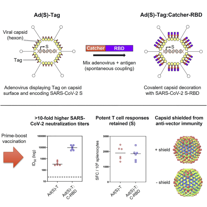
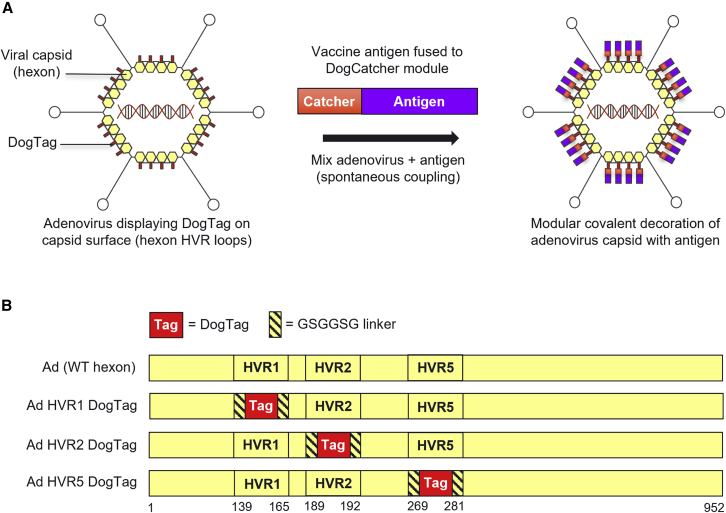
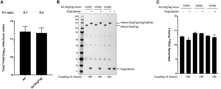
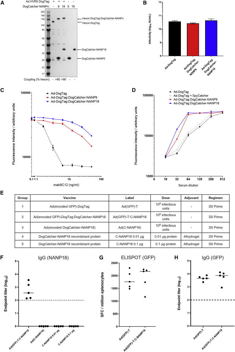
Adenovirus vector vaccines have been widely and successfully deployed in response to coronavirus disease 2019 (COVID-19). However, despite inducing potent T cell immunity, improvement of vaccine-specific antibody responses upon homologous boosting is modest compared with other technologies. Here, we describe a system enabling modular decoration of adenovirus capsid surfaces with antigens and demonstrate potent induction of humoral immunity against these displayed antigens. Ligand attachment via a covalent bond was achieved using a protein superglue, DogTag/DogCatcher (similar to SpyTag/SpyCatcher), in a rapid and spontaneous reaction requiring only co-incubation of ligand and vector components. DogTag was inserted into surface-exposed loops in the adenovirus hexon protein to allow attachment of DogCatcher-fused ligands on virus particles. Efficient coverage of the capsid surface was achieved using various ligands, with vector infectivity retained in each case. Capsid decoration shielded particles from vector neutralizing antibodies. In prime-boost regimens, adenovirus vectors decorated with the receptor-binding domain of severe acute respiratory syndrome coronavirus-2 (SARS-CoV-2) spike induced >10-fold higher SARS-CoV-2 neutralization titers compared with an undecorated vector encoding spike. Importantly, decorated vectors achieved equivalent or superior T cell immunogenicity against encoded antigens compared with undecorated vectors. We propose capsid decoration using protein superglues as a novel strategy to improve efficacy and boostability of adenovirus-based vaccines and therapeutics.

Keywords: SARS-CoV-2; adenovirus; protein superglue; vaccine vector; vector engineering.


Conflict of interest statement

Declaration of interests M.D.J.D., L.M.R., R.A.R., and L.A.H.B. are employees of SpyBiotech Ltd. S.B. is CSO and co-founder of SpyBiotech Ltd. S.J.D. and M.H. are co-founders of SpyBiotech Ltd, and M.H. is also an author on a number of patents relating to protein superglues, including the DogTag/DogCatcher technology. C.G., J.M.J.-G., K.J.D., and M.H.M. declare no competing interests.

Figures

None
Graphical abstract
Figure 1
Figure 1
Modular covalent decoration of the adenovirus capsid via insertion of DogTag into hexon HVR loops (A) Modular display of DogCatcher-fused antigenic ligands on the surface of the adenovirus capsid via covalent coupling with DogTag inserted into hexon HVR surface loops. Attachment of antigens to the capsid achieved by simple co-incubation of adenovirus and antigen components in a rapid and spontaneous reaction. (B) Design of modified adenovirus hexon sequences with DogTag inserted into either HVR1, HVR2, or HVR5 flanked by flexible linkers. Amino acid residue numbers corresponding to deletion/insertion sites at HVR1, HVR2, or HVR5 in Ad5 hexon are indicated.
Figure 2
Figure 2
DogTag is highly reactive with DogCatcher following insertion into adenovirus hexon HVR loops, with decorated virions retaining infectivity (A) Yield comparison of GFP-expressing Ad vector preparations displaying DogTag on hexon HVR5 (Ad-DogTag) versus Ad vectors with an unmodified hexon (Ad). Data show mean +SD, n = 3, infectious yield from 1,500 cm2 adherent 293A cells. Mean P:I ratios (ratio of total viral particles calculated by UV spectrophotometry to infectious units calculated by GFP focus assay) for vector batches are indicated above each bar. (B) SDS-PAGE and Coomassie staining analysis of Ad virions displaying DogTag at HVR1, HVR2, or HVR5 (1E+10 viral particles) incubated with DogCatcher (5 μM) at 4°C for 16 h. Gel shift observed upon covalent coupling of DogCatcher to virion associated hexon-DogTag. (C) Vector infectivity (GFP focus assay) performed in 293A cells on the samples from (B). Data show mean +SD of triplicate wells.
Figure 3
Figure 3
Capsid display of DogCatcher-NANP18 ligand shields the particles from anti-vector neutralizing antibodies and elicits potent ligand-specific humoral immunity (A) SDS-PAGE and Coomassie staining analysis of Ad virions displaying DogTag at HVR5, (1E+10 viral particles) incubated with DogCatcher-NANP9 (5 μM) or DogCatcher-NANP18 (5 μM) at 4°C for 16 h. (B) Vector infectivity (GFP focus assay) performed in 293A cells on the samples from (A). Data show mean +SD of triplicate wells. (C) Vector neutralization assay using anti-hexon mAb 9C12. Ad particles encoding a GFP transgene with or without a capsid ligand were added to 293A cells in the presence of a varying concentration of neutralizing mAb. Vector transduction was measured via fluorescence of encoded GFP expressed in the cells. (D) Vector neutralization assay using anti-Ad5 serum. Ad particles encoding a GFP transgene with or without a capsid ligand were added to 293A cells in the presence of a varying concentration of serum from mice immunized with Ad5. (C) and (D) show mean + range of duplicate wells. (E) BALB/c mice (five per group) were immunized intramuscularly as described (vector-encoded antigens in brackets). Note that Ad(C-NANP18) has an unmodified hexon (no DogTag). The DogCatcher-NANP18 protein dose in group 2 was calculated to be < 0.05 μg per mouse. (F) Serum IgG antibody responses to NANP18 in groups 2–5 measured by endpoint ELISA 14 days post immunization. (G) CD8+ T cell responses in the spleen to encoded epitope EGFP200-208 were measured in groups 1 and 2 by overnight ex vivo IFNγ-ELISpot 14 days post immunization. SFC, spot forming cells. (H) Serum IgG antibody responses to encoded GFP in groups 1 and 2 were measured by endpoint ELISA 14 days post immunization. In (F)–(H), bars show median responses. Dashed line represents limit of detection.
Figure 4
Figure 4
RBD can be fused to DogCatcher, displayed on adenovirus particles, and is an effective capsid shield (A) Design of DogCatcher-RBD protein (B) and SDS-PAGE and Coomassie staining analysis of Ad virions displaying DogTag at HVR5 (1E+10 viral particles) incubated with DogCatcher-RBD (3.5 μM) at 4°C for 16 h. (C) Vector infectivity (GFP focus assay) performed in 293A cells on the samples from (B). Data show mean +SD of triplicate wells. (D) Vector neutralization assay using anti-hexon mAb 9C12, performed as described in Figure 3. Data show mean and range of duplicate wells. (E) Vector neutralization assay using anti-Ad5 sera, performed as described in Figure 3. Data show mean + SD of triplicate wells. (F) Capsid decoration with DogCatcher-RBD impairs human factor X (hFX)-mediated Ad transduction of SKOV3 cells. Fluorescent microscopy images taken 48 h post infection show Ad-DogTag or Ad-DogTag:DogCatcher-RBD vectors expressing GFP incubated with or without hFX (2,500 viral particles/cell). Scale bar, 1,000 μm. (G) Infectivity data (GFP focus assay) from the experiment shown in (F). Data show mean +SD of triplicate wells.
Figure 5
Figure 5
Cryo-EM analysis of adenovirus particles displaying RBD (A) 3D density maps (at 10.5 Å) for Ad-DogTag (control sample, undecorated) and Ad-DogTag:DogCatcher-RBD particles. Radial coloring scheme is indicated; regions furthest from the center of the particle are shown in red, regions closest to the center are shown in blue. Distance from center of particle (in angstroms) is indicated. (B) Exemplary 2D class averages. Indicated diameters calculated from vertex to vertex. (C) Type I ligand coupling; 3D structure of representative hexon trimer without ligand (Ad-DogTag) or with one ligand coupled per trimer (Ad-DogTag:DogCatcher-RBD) shown at the same contour level. (D) Type II ligand coupling; 3D structure representative of hexon trimer adjacent to penton base without ligand (Ad-DogTag) or with two ligands coupled per trimer (Ad-DogTag:DogCatcher-RBD) shown at the same contour level. Maps for Ad-DogTag:DogCatcher-RBD shown at both front and side angles, and high and low threshold to indicate location and extent of additional electron density. In both (C) and (D), hexon trimer structure (PDB: 6B1T) was fitted (green), with location of HVR5 loop (residues 270–280, site of DogTag insertion) shown in red.
Figure 6
Figure 6
High-titer SARS-CoV-2 neutralizing antibody responses generated by capsid display of RBD (A) BALB/c mice (six per group) were immunized intramuscularly in homologous prime-boost regimens as described. The RBD protein dose in groups 2 and 3 was calculated to be < 0.2 μg per mouse. (B) Serum IgG antibody responses to RBD at D20 measured by endpoint ELISA. (C) Serum IgG antibody responses to RBD at D35 measured by endpoint ELISA. (D) Fold increase in anti-RBD IgG titer post boost (D35 titer in C divided by D20 titer in B). (E) SARS-CoV-2 neutralization titers (pVNT assay) in D35 serum against Wuhan strain (WT) and variants of concern alpha (B.1.1.7), delta (B.1.617.2), and beta (B.1.351). Data on beta variant collected separately. (F) IFNγ-ELISpot response in spleen at D35 against peptide pool spanning SARS-CoV-2 S-RBD. (G) IFNγ-ELISpot response in spleen at D35 against peptides spanning full-length SARS-CoV-2 S protein (summed responses from two peptide pools spanning residues 1–643 and 633–1,273 are shown, responses from individual pools shown in Figure S8). Dashed lines represent limit of detection. Median responses shown by a horizontal line. Statistical analyses performed by Kruskal-Wallis with Dunn’s test for multiple comparisons, ∗p < 0.05; ∗∗p < 0.01; ∗∗∗p < 0.001; ns, not significant.
Figure 7
Figure 7
Applying RBD capsid decoration at boost significantly increases SARS-CoV-2 specific antibody and T cell responses in an adenovirus vector prime-boost regimen (A) BALB/c mice (n = 12) were immunized intramuscularly with Ad(Spike)-DogTag on D0 and then on D21 given a second intramuscular immunization of either Ad(Spike)-DogTag (group 1, n = 6) or Ad(Spike)-DogTag:DogCatcher-RBD (group 2, n = 6). All vaccines were administered at a dose of 108 infectious units. (B) Serum IgG antibody responses to RBD measured by endpoint ELISA. Responses measured post prime on D20 (Ad(Spike)-DogTag) are compared with responses on D35 after homologous (Ad(Spike)-DogTag, Ad(Spike)-DogTag), or heterologous (Ad(Spike)-DogTag, Ad(Spike)-DogTag:DogCatcher-RBD) prime boost. Fold change in median titer post boost is shown. Horizontal bars show median responses. Statistical analyses performed by Kruskal-Wallis with Dunn’s test for multiple comparisons, ∗p < 0.05; ∗∗∗∗p < 0.0001. Dashed line represents limit of detection. (C) IFNγ-ELISpot response in spleen at D35 against peptide pools spanning full-length (1–1,273) SARS-CoV-2 S (summed responses from two peptide pools spanning residues 1–643 and 633–1,273 are shown). (D) IFNγ-ELISpot response in spleen at D35 against peptide pool spanning C-terminal residues 633–1,273 of SARS-CoV-2 S only (i.e., not including RBD domain). In (C)–(D), horizontal bars show median responses and statistical analyses performed by Mann-Whitney test, ∗∗p < 0.01.

References

    1. Mendonça S.A., Lorincz R., Boucher P., Curiel D.T. Adenoviral vector vaccine platforms in the SARS-CoV-2 pandemic. NPJ Vaccin. 2021;6:97. doi: 10.1038/s41541-021-00356-x. - DOI - PMC - PubMed
    1. Voysey M., Clemens S.A.C., Madhi S.A., Weckx L.Y., Folegatti P.M., Aley P.K., Angus B., Baillie V.L., Barnabas S.L., Bhorat Q.E., Bibi S., et al. Safety and efficacy of the ChAdOx1 nCoV-19 vaccine (AZD1222) against SARS-CoV-2: an interim analysis of four randomised controlled trials in Brazil, South Africa, and the UK. Lancet. 2021;397:99–111. doi: 10.1016/S0140-6736(20)32661-1. - DOI - PMC - PubMed
    1. Sadoff J., Gray G., Vandebosch A., Cárdenas V., Shukarev G., Grinsztejn B., Goepfert P.A., Truyers C., Fennema H., Spiessens B., Offergeld K., et al. Safety and efficacy of single-dose Ad26.COV2.S vaccine against covid-19. N. Engl. J. Med. 2021;384:2187–2201. doi: 10.1056/NEJMoa2101544. - DOI - PMC - PubMed
    1. Logunov D.Y., Dolzhikova I.V., Shcheblyakov D.V., Tukhvatulin A.I., Zubkova O.V., Dzharullaeva A.S., Kovyrshina A.V., Lubenets N.L., Grousova D.M., Erokhova A.S., et al. Safety and efficacy of an rAd26 and rAd5 vector-based heterologous prime-boost COVID-19 vaccine: an interim analysis of a randomised controlled phase 3 trial in Russia. Lancet. 2021;397:671–681. doi: 10.1016/S0140-6736(21)00234-8. - DOI - PMC - PubMed
    1. Zhu F.C., Guan X.H., Li Y.H., Huang J.Y., Jiang T., Hou L.H., Li J.X., Yang B.F., Wang L., Wang W.J., Wu S.P., et al. Immunogenicity and safety of a recombinant adenovirus type-5-vectored COVID-19 vaccine in healthy adults aged 18 years or older: a randomised, double-blind, placebo-controlled, phase 2 trial. Lancet. 2020;396:479–488. doi: 10.1016/S0140-6736(20)31605-6. - DOI - PMC - PubMed

Publication types