Pan-Esophageal Submucosal Dissection following Transesophageal Echocardiography
- PMID: 35949238
- PMCID: PMC9294924
- DOI: 10.1159/000525278
Pan-Esophageal Submucosal Dissection following Transesophageal Echocardiography
Abstract
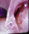
A 73-year-old female underwent open mitral valve replacement with transesophageal echocardiography (TEE) guidance. She developed upper gastrointestinal bleeding postoperatively and was found on upper endoscopy to have a bleeding site at the gastric cardia with the appearance of focal trauma and a possible puncture site. A submucosal bluish protrusion was seen throughout the esophagus with a mucosal flap at the proximal esophagus. As a unifying diagnosis, it was suspected that the intraoperative TEE probe caused a submucosal dissection with point of entry at the proximal esophagus, running the entire length of the esophagus and exiting at the gastric cardia, giving rise to a clinical upper gastrointestinal bleed. Closure of the esophageal defect was achieved using an endoclip. A CT scan showed focal pneumomediastinum along the proximal esophagus, confirming the hypothesis. We report the first case to our knowledge of iatrogenic pan-esophageal submucosal dissection, which, in this case, presented as a clinical bleed from the exit point trauma to the gastric cardia mucosa caused by a TEE probe. Endoscopic management of the gastric injury as well as the esophageal defect led to resolution of the bleeding and avoidance of mediastinitis, respectively, allowing for an excellent recovery.
Keywords: Dissection; Esophagus; Gastrointestinal bleeding; Transesophageal echocardiography; Upper endoscopy.
Copyright © 2022 by The Author(s). Published by S. Karger AG, Basel.
Figures


References
-
- Min JK, Spencer KT, Furlong KT, DeCara JM, Sugeng L, Ward RP, et al. Clinical features of complications from transesophageal echocardiography: a single-center case series of 10,000 consecutive examinations. J Am Soc Echocardiogr. 2005;18((9)):925–9. - PubMed
-
- Jovic M, Baulig W, Schneider P, Schmid ER. Esophageal dissection after transesophageal echocardiography in a patient with Barrett's esophagus and long-term systemic steroid therapy. J Cardiothorac Vasc Anesth. 2011;25((1)):150–2. - PubMed
-
- El-Chami MF, Martin RP, Lerakis S. Esophageal dissection complicating transesophageal echocardiogram − the lesson to be learned: do not force the issue. J Am Soc Echocardiogr. 2006;19((5)):579e5–7. - PubMed
-
- Stavropoulos SN, Desilets DJ, Fuchs KH, Gostout CJ, Haber G, Inoue H, et al. Per-oral endoscopic myotomy white paper summary. Gastrointest Endosc. 2014;80((1)):1–15. - PubMed
Publication types
LinkOut - more resources
Full Text Sources
Research Materials

