

Helicobacter pylori-Negative Advanced Gastric Cancer Arising from the Gastric Mucosa without Inflammation, Atrophy, or Intestinal Metaplasia
- PMID: 35949241
- PMCID: PMC9247486
- DOI: 10.1159/000524427
Helicobacter pylori-Negative Advanced Gastric Cancer Arising from the Gastric Mucosa without Inflammation, Atrophy, or Intestinal Metaplasia
Abstract
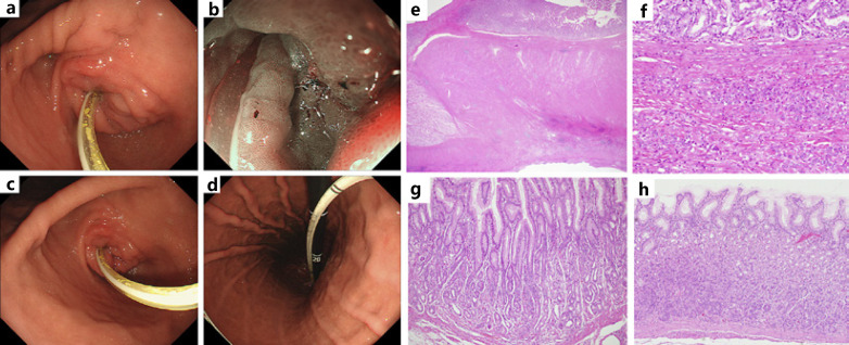
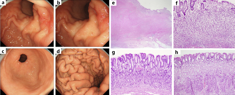
Gastric cancer is strongly associated with atrophic gastritis associated with Helicobacter pylori infection. The eradication of H. pylori has been reported to improve inflammation of the gastric mucosa, atrophy, and intestinal metaplasia and has also been shown to inhibit the development and growth of gastric cancer. Advanced gastric cancer from H. pylori-negative mucosa without inflammation, atrophy, or intestinal epithelialization is rarely found. We report on two cases of advanced gastric cancer without endoscopic evidence of gastric mucosal atrophy. One case was in the gastric angle 9 years after H. pylori eradication and the other case was in the pylorus of an uninfected stomach. Although gastric cancer is strongly associated with atrophic gastritis caused by H. pylori infection, postoperative histopathological examination of the stomach in both cases showed no inflammation, atrophy, or intestinal metaplasia. Poorly differentiated adenocarcinomas were confirmed at the microscopic level, and both cases were determined to be type 4 using the Borrmann classification. There is a body of evidence showing that H. pylori infection can cause gastric cancer and is the most prevalent infection-induced cancer in the world. The 2 cases here displayed different carcinogenesis than gastric mucosal atrophy caused by H. pylori infection. With the spread of H. pylori eradication treatments and an increasing number of uninfected patients, these case reports can contribute to future strategies for the diagnosis of gastric cancer.
Keywords: Atrophic gastritis; Carcinogenesis; Endoscopy; Gastric cancer; Helicobacter pylori infection.
Copyright © 2022 by S. Karger AG, Basel.
Conflict of interest statement
The authors declare that Hirofimi Kawamoto received advisory fees from Gadelius Medical. Other authors have no competing interests.
Figures
Similar articles
-
[The assessment of nitric oxide metabolites in gastric juice in Helicobacter pylori infected subjects in compliance with grade of inflammatory lesions in gastric mucosa].Pol Merkur Lekarski. 2008 Feb;24(140):95-100. Pol Merkur Lekarski. 2008. PMID: 18634262 Polish.
-
AGA Clinical Practice Update on the Diagnosis and Management of Atrophic Gastritis: Expert Review.Gastroenterology. 2021 Oct;161(4):1325-1332.e7. doi: 10.1053/j.gastro.2021.06.078. Epub 2021 Aug 26. Gastroenterology. 2021. PMID: 34454714 Free PMC article. Review.
-
Helicobacter pylori infection, glandular atrophy and intestinal metaplasia in superficial gastritis, gastric erosion, erosive gastritis, gastric ulcer and early gastric cancer.World J Gastroenterol. 2005 Feb 14;11(6):791-6. doi: 10.3748/wjg.v11.i6.791. World J Gastroenterol. 2005. PMID: 15682469 Free PMC article.
-
Helicobacter pylori-induced inflammation masks the underlying presence of low-grade dysplasia on gastric lesions.World J Gastroenterol. 2020 Jul 14;26(26):3834-3850. doi: 10.3748/wjg.v26.i26.3834. World J Gastroenterol. 2020. PMID: 32774061 Free PMC article.
-
Review article: exploring the link between Helicobacter pylori and gastric cancer.Aliment Pharmacol Ther. 1999 Mar;13 Suppl 1:3-11. doi: 10.1046/j.1365-2036.1999.00002.x. Aliment Pharmacol Ther. 1999. PMID: 10209681 Review.
Cited by
-
Clinicopathological and endoscopic features of Helicobacter pylori infection-negative gastric cancer in Japan: a retrospective study.Clin Endosc. 2024 Jul;57(4):486-494. doi: 10.5946/ce.2023.258. Epub 2024 Mar 22. Clin Endosc. 2024. PMID: 38514987 Free PMC article.
-
A prominent role of LncRNA H19 in H. pylori CagA induced DNA damage response and cell malignancy.Sci Rep. 2024 Jun 20;14(1):14185. doi: 10.1038/s41598-024-65221-y. Sci Rep. 2024. PMID: 38902391 Free PMC article.
References
-
- Komoto K, Haruma K, Kamada T, Tanaka S, Yoshihara M, Sumii K, et al. Helicobacter pylori infection and gastric neoplasia: correlations with histological gastritis and tumor histology. Am J Gastroenterol. 1998 Aug;93((8)):1271–6. - PubMed
-
- Shin HR, Shin A, Woo H, Fox K, Walsh N, Lo YR, et al. Prevention of infection-related cancers in the WHO Western Pacific Region. Jpn J Clin Oncol. 2016 Jan;46((1)):13–22. - PubMed
-
- Uemura N, Okamoto S, Yamamoto S, Matsumura N, Yamaguchi S, Yamakido M, et al. Helicobacter pylori infection and the development of gastric cancer. N Engl J Med. 2001 Sep;345((11)):784–9. - PubMed
-
- Sasaki A, Kitadai Y, Ito M, Sumii M, Tanaka S, Yoshihara M, et al. Helicobacter pylori infection influences tumor growth of human gastric carcinomas. Scand J Gastroenterol. 2003 Feb;38((2)):153–8. - PubMed
Publication types
LinkOut - more resources
Full Text Sources
