

Mesenchymal stem cells and their conditioned medium as potential therapeutic strategies in managing comorbid anxiety in rat sepsis induced by cecal ligation and puncture
- PMID: 35949300
- PMCID: PMC9320199
- DOI: 10.22038/IJBMS.2022.61860.13690
Mesenchymal stem cells and their conditioned medium as potential therapeutic strategies in managing comorbid anxiety in rat sepsis induced by cecal ligation and puncture
Abstract
Objectives: Sepsis-associated encephalopathy (SAE) is a common brain dysfunction following sepsis. Due to the beneficial effects of mesenchymal stem cells (MSCs) therapy on anxiety, an extreme and early manifestation of SAE, we hypothesized that MSCs-derived conditioned medium (CM) may be able to attenuate anxiety in cecal ligation and puncture (CLP)-induced sepsis.
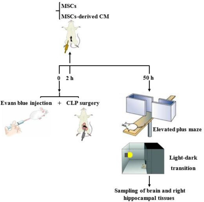
Materials and methods: Rats were assigned into 4 groups: sham, CLP, MSC, and CM. All animals, except in the sham group, underwent the CLP procedure to induce sepsis. Two hours after sepsis induction, the rats in MSC and CM groups, received 1×10⁶ MSCs and CM derived from the same number of cells, respectively. 48 hr after the treatments, anxiety-related behaviors were assessed, and brain and right hippocampal tissues were collected.
Results: MSCs and CM enhanced the percentages of open arm entries and time spent in the open arms of the elevated plus-maze and the time spent in the light side of the light-dark box. MSCs and CM decreased the Evans blue content and decreased the IL-6 and TNF-α levels in the brain tissue samples. Reductions in the expression of 5-HT2A receptors and phosphorylation of ERK1/2 and an increase in the expression of 5-HT1A receptors in the hippocampal tissue samples were observed in the MSC and CM groups.
Conclusion: MSCs and MSCs-derived CM attenuated anxiety-related behaviors to an equal extent by reducing inflammation, modifying 5-HT receptor expression changes, and inhibiting the ERK pathway. Therefore, MSCs-derived CM may be considered a promising therapy for comorbid anxiety in septic patients.
Keywords: Extracellular signal-regulated kinases; Inflammation; Mesenchymal stem cells; Sepsis; Sepsis-associated-encephalopathy; Serotonin.
Conflict of interest statement
The authors declare that they have no conflicts of interest.
Figures


Similar articles
-
Adipose-Derived Mesenchymal Stem Cells and Conditioned Medium Attenuate the Memory Retrieval Impairment During Sepsis in Rats.Mol Neurobiol. 2020 Sep;57(9):3633-3645. doi: 10.1007/s12035-020-01991-6. Epub 2020 Jun 19. Mol Neurobiol. 2020. PMID: 32562236
-
Beneficial effects of tannic acid on comorbid anxiety in cecal ligation and puncture-induced sepsis in rats and potential underlying mechanisms.Naunyn Schmiedebergs Arch Pharmacol. 2023 May;396(5):1019-1030. doi: 10.1007/s00210-022-02374-5. Epub 2023 Jan 4. Naunyn Schmiedebergs Arch Pharmacol. 2023. PMID: 36598513
-
Mesenchymal stem cell-conditioned medium prevents inflammation-induced liver and lung damage in septic mice.Int Immunopharmacol. 2024 Aug 20;137:112407. doi: 10.1016/j.intimp.2024.112407. Epub 2024 Jun 13. Int Immunopharmacol. 2024. PMID: 38875996
-
Wharton's jelly-derived mesenchymal stem cells attenuate sepsis-induced organ injury partially via cholinergic anti-inflammatory pathway activation.Am J Physiol Regul Integr Comp Physiol. 2020 Jan 1;318(1):R135-R147. doi: 10.1152/ajpregu.00098.2018. Epub 2019 Oct 9. Am J Physiol Regul Integr Comp Physiol. 2020. PMID: 31596111
-
Overexpressing TGF-β1 in mesenchymal stem cells attenuates organ dysfunction during CLP-induced septic mice by reducing macrophage-driven inflammation.Stem Cell Res Ther. 2020 Sep 3;11(1):378. doi: 10.1186/s13287-020-01894-2. Stem Cell Res Ther. 2020. PMID: 32883356 Free PMC article.
Cited by
-
Post-sepsis psychiatric disorder: Pathophysiology, prevention, and treatment.Neurol Sci. 2024 Jul;45(7):3093-3105. doi: 10.1007/s10072-024-07409-8. Epub 2024 Feb 21. Neurol Sci. 2024. PMID: 38381393 Free PMC article. Review.
-
Effects of the Co-Overexpression of the BCL and BDNF Genes on the Gamma-Aminobutyric Acid-Ergic Differentiation of Wharton's-Jelly-Derived Mesenchymal Stem Cells.Biomedicines. 2023 Jun 18;11(6):1751. doi: 10.3390/biomedicines11061751. Biomedicines. 2023. PMID: 37371846 Free PMC article.
-
MSC-derived exosomal miR-140-3p improves cognitive dysfunction in sepsis-associated encephalopathy by HMGB1 and S-lactoylglutathione metabolism.Commun Biol. 2024 May 11;7(1):562. doi: 10.1038/s42003-024-06236-z. Commun Biol. 2024. PMID: 38734709 Free PMC article.
-
The effect of adipose-derived mesenchymal stem cell transplantation on ovarian mitochondrial dysfunction in letrozole-induced polycystic ovary syndrome in rats: the role of PI3K-AKT signaling pathway.J Ovarian Res. 2024 Apr 27;17(1):91. doi: 10.1186/s13048-024-01422-3. J Ovarian Res. 2024. PMID: 38678269 Free PMC article.
-
Effects, methods and limits of the cryopreservation on mesenchymal stem cells.Stem Cell Res Ther. 2024 Sep 29;15(1):337. doi: 10.1186/s13287-024-03954-3. Stem Cell Res Ther. 2024. PMID: 39343920 Free PMC article. Review.
References
-
- Angus DC, Van der Poll T. Severe sepsis and septic shock. N Engl J Med. 2013;369:840–851. - PubMed
-
- Gofton TE, Young GB. Sepsis-associated encephalopathy. Nat Rev Neurol. 2012;8:557–566. - PubMed
-
- Dantzer R. Cytokine-induced sickness behaviour: A neuroimmune response to activation of innate immunity. Eur J Pharmacol. 2004;500:399–411. - PubMed
LinkOut - more resources
Full Text Sources
Miscellaneous