

Multiple benign fibrous histiocytomas of the mandible: A case report and review of the literature
- PMID: 35949345
- PMCID: PMC9353540
- DOI: 10.3892/etm.2022.11530
Multiple benign fibrous histiocytomas of the mandible: A case report and review of the literature
Abstract
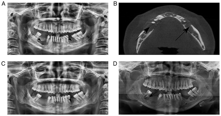
Benign fibrous histiocytoma (BFH) mostly occurs on the skin of the extremities, while it is unusual to manifest on the bone and mandibular involvement of BFH is even rarer. The present study reports a case of BFH in a 42-year-old female who had a slowly progressive swelling of the bilateral mandible and slight facial asymmetry over a period of 4 months. However, the outcome of this patient was unsatisfactory, with the first and second recurrence observed 16 and 46 months after surgery, respectively. The present case suggests that BFH has a risk of recurrence after transoral curettage. Regular follow-up is advised to detect tumor recurrence after the surgery of transoral curettage.
Keywords: benign fibrous histiocytoma; case report; clinical features; mandible; regular follow-up; surgical treatments.
Copyright: © Wang et al.
Conflict of interest statement
The authors declare that they have no competing interests.
Figures
Similar articles
-
The first total vertebral involvement of benign fibrous histiocytoma: A case report and literature review.J Bone Oncol. 2019 Dec 20;20:100274. doi: 10.1016/j.jbo.2019.100274. eCollection 2020 Feb. J Bone Oncol. 2019. PMID: 31908914 Free PMC article.
-
Benign fibrous histiocytoma: A rare case involving jaw bone.Contemp Clin Dent. 2015 Sep;6(Suppl 1):S266-8. doi: 10.4103/0976-237X.166828. Contemp Clin Dent. 2015. PMID: 26604585 Free PMC article.
-
Benign Fibrous Histiocytomas of the Oral Mucosa: Report on Three Cases and Review of the Literature.Dermatopathology (Basel). 2015 Apr 29;2(2):52-60. doi: 10.1159/000381618. eCollection 2015 Apr-Jun. Dermatopathology (Basel). 2015. PMID: 27047935 Free PMC article.
-
Benign fibrous histiocytoma of the maxilla: a case report and review of literature.Indian J Dent Res. 2014 Jan-Feb;25(1):115-8. doi: 10.4103/0970-9290.131160. Indian J Dent Res. 2014. PMID: 24748313 Review.
-
[Benign fibrous histiocytoma involving the skull: a case report and literature review].Nan Fang Yi Ke Da Xue Xue Bao. 2010 Dec;30(12):2752-5. Nan Fang Yi Ke Da Xue Xue Bao. 2010. PMID: 21177160 Review. Chinese.
Cited by
-
Literature Review, Case Presentation and Management of Non-ossifying Fibroma of Right Angle of Mandible: More Than just a Cortical Defect!Indian J Otolaryngol Head Neck Surg. 2024 Feb;76(1):1054-1061. doi: 10.1007/s12070-023-04110-8. Epub 2023 Aug 7. Indian J Otolaryngol Head Neck Surg. 2024. PMID: 38440574 Free PMC article.
References
-
- Stout AP, Lattes R. Atlas of Tumor Pathology: Tumors of the Soft Tissues. Armed Forces Institute of Pathology, Washington, DC, pp38-52, 1967.
-
- Nielsen GP, Kyriakos M. Non-ossifying fibroma/benign fibrous histiocytoma of bone. In: World Health Organization Classification of Tumours of Soft Tissue and Bone. International Agency for Research on Cancer, Lyon, 2013.
Publication types
LinkOut - more resources
Full Text Sources