. 2022 Jul 15;24(3):310.
doi: 10.3892/ol.2022.13430. eCollection 2022 Sep.

Propofol inhibits the malignant development of osteosarcoma U2OS cells via AMPK/FΟΧO1-mediated autophagy

Affiliations

Propofol inhibits the malignant development of osteosarcoma U2OS cells via AMPK/FΟΧO1-mediated autophagy

Lina Dai et al. Oncol Lett. .

Abstract

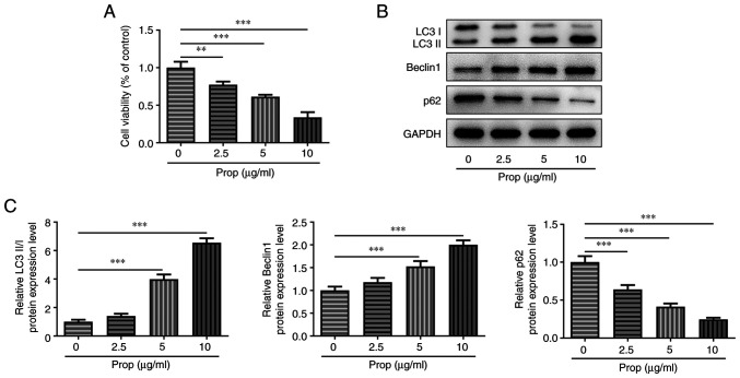
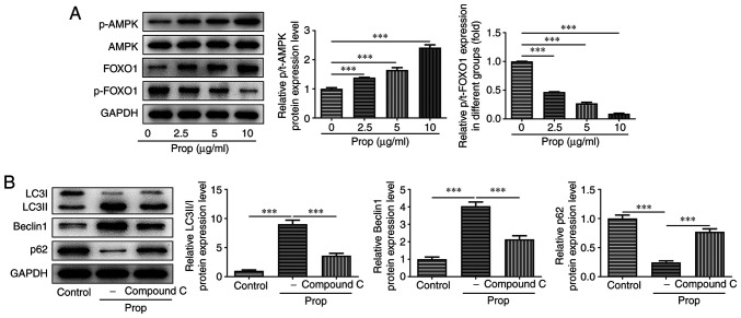
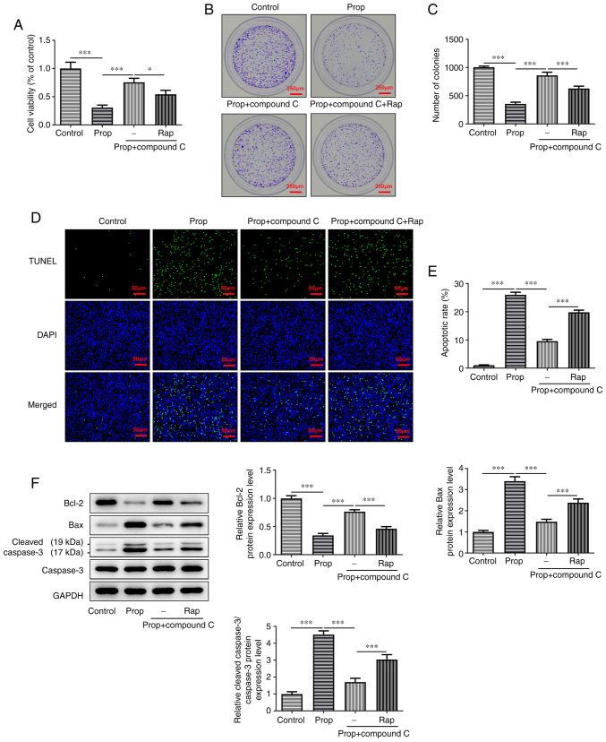
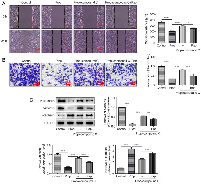
It has previously been reported that propofol regulates the development of human osteosarcoma (OS). However, the specific molecular mechanisms underlying the effect of propofol on OS remain poorly understood. Therefore, the aim of the present study was to explore the effects of propofol on OS U2OS cells and the potential underlying mechanism. The Cell Counting Kit-8 and colony formation assays were performed to assess cell viability and proliferation. Furthermore, cell apoptosis was assessed using the TUNEL assay and western blotting. Wound healing and Transwell assays were performed to evaluate OS cell migration and invasion abilities, respectively. The protein expression levels of epithelial-mesenchymal transition (EMT)-, autophagy- and adenosine monophosphate-activated protein kinase (AMPK)/FOXO1 signaling pathway-related proteins were also determined using western blotting. The results demonstrated that propofol significantly reduced the viability of OS cells and promoted autophagy in a dose-dependent manner. Moreover, cell treatment with propofol significantly enhanced the protein expression levels of phosphorylated (p)-AMPK and FOXO1, while decreasing the protein levels of p-FOXO1. Furthermore, treatment with propofol significantly suppressed cell viability, migration and invasion abilities and the EMT of OS cells, and potentially promoted cell apoptosis via inducing autophagy via the AMPK/FOXO1 signaling pathway. In summary, the present study indicated that propofol potentially had an inhibitory effect on the development of OS cells via AMPK/FOXO1-mediated autophagy. These results have therefore provided an experimental basis for further studies into the therapeutic effect of propofol on OS.

Keywords: adenosine monophosphate-activated protein kinase/FOXO1 signaling pathway; autophagy; metastasis; osteosarcoma; propofol.


Conflict of interest statement

The authors declare that they have no competing interests.

Figures

Figure 1.
Figure 1.
Prop inhibits cell viability and induces cell autophagy in U2OS cells. (A) Cell viability was detected using the Cell Counting Kit-8 assay. (B) Protein expression levels of LC3II/I, Beclin1 and p62 were determined using western blotting. (C) Semi-quantification of the related western blotting bands. Data are presented as the mean ± SD. **P<0.01 and ***P<0.001. Prop, propofol.
Figure 2.
Figure 2.
Prop induces the autophagy of U2OS cells by activating the AMPK/FOXO1 signaling pathway. (A) Protein expression levels of t-AMPK, p-AMPK, FOXO1 and p-FOXO1 were detected via western blotting. (B) Protein expression levels of LC3II/I, Beclin1 and p62 were detected via western blotting. Data are presented as the mean ± SD. ***P<0.001. Prop, propofol; AMPK, adenosine monophosphate-activated protein kinase; t, total; p, phosphorylated.
Figure 3.
Figure 3.
Prop induces adenosine monophosphate-activated protein kinase/FOXO1 pathway-mediated autophagy to inhibit cell proliferation and promote apoptosis in U2OS cells. (A) Cell viability was detected using the Cell Counting Kit-8 assay. (B) Cell proliferation was assessed using the colony formation assay. (C) Quantification of cell colony number. (D) Cell apoptosis was assessed using the TUNEL assay. Scale bar, 50 µm. (E) Quantification of the U2OS cell apoptotic rate. (F) Protein expression levels of Bcl-2, Bax, caspase-3 and cleaved caspase-3 were detected via western blotting. Data are presented as the mean ± SD. *P<0.05 and ***P<0.001. Prop, propofol; RAP, rapamycin.
Figure 4.
Figure 4.
Prop inhibits U2OS cell migration, invasion and the epithelial-mesenchymal transition via autophagy through the activation of the adenosine monophosphate-activated protein kinase/FOXO1 signaling pathway. (A) Cell migration was analyzed using the wound healing assay. (B) Cell invasion was assessed using the Transwell assay. (C) N-cadherin, Vimentin and E-cadherin protein expression levels were detected via western blotting. Data are presented as the mean ± SD. *P<0.05, **P<0.01 and ***P<0.001. Prop, propofol.

Similar articles

Cited by

References

    1. Harrison DJ, Geller DS, Gill JD, Lewis VO, Gorlick R. Current and future therapeutic approaches for osteosarcoma. Expert Rev Anticancer Ther. 2018;18:39–50. doi: 10.1080/14737140.2018.1413939. - DOI - PubMed
    1. Eaton BR, Schwarz R, Vatner R, Yeh B, Claude L, Indelicato DJ, Laack N. Osteosarcoma. Pediatr Blood Cancer. 2021;68((Suppl 2)):e28352. doi: 10.1002/pbc.28352. - DOI - PubMed
    1. Corre I, Verrecchia F, Crenn V, Redini F, Trichet V. The Osteosarcoma microenvironment: A complex but targetable ecosystem. Cells. 2020;9:976. doi: 10.3390/cells9040976. - DOI - PMC - PubMed
    1. Meazza C, Scanagatta P. Metastatic osteosarcoma: A challenging multidisciplinary treatment. Expert Rev Anticancer Ther. 2016;16:543–556. doi: 10.1586/14737140.2016.1168697. - DOI - PubMed
    1. Kager L, Tamamyan G, Bielack S. Novel insights and therapeutic interventions for pediatric osteosarcoma. Future Oncol. 2017;13:357–368. doi: 10.2217/fon-2016-0261. - DOI - PubMed