

A novel inflammation-associated prognostic signature for clear cell renal cell carcinoma
- PMID: 35949606
- PMCID: PMC9353224
- DOI: 10.3892/ol.2022.13427
A novel inflammation-associated prognostic signature for clear cell renal cell carcinoma
Abstract
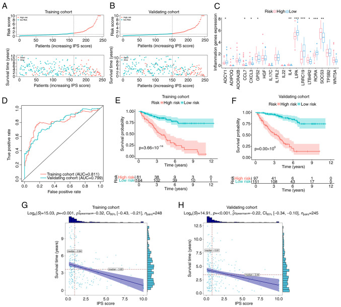
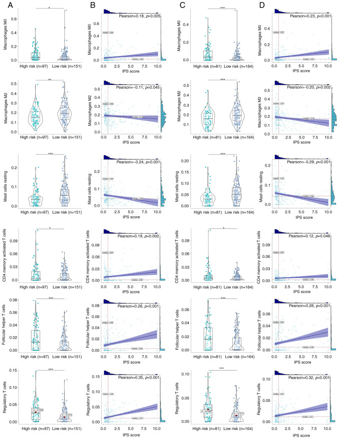
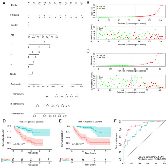
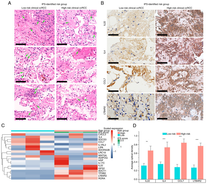
Clear cell renal cell carcinoma (ccRCC) are typically situated in a complex inflammatory and immune microenvironment, which has been reported to contribute to the unfavorable prognosis of patients with ccRCC. There would be beneficial clinical implications for elucidating the roles of its molecular characteristics in the inflammatory microenvironment. This is because it would facilitate the development of reliable biomarkers for pre-stratification prior to the designation of individualized treatment strategies. In the present study, RNA-sequencing data from 607 patients were retrospectively analyzed to elucidate the profile of inflammatory molecules. Based on this, an inflammatory prognostic signature (IPS) was developed and further validated using clinical ccRCC samples. Subsequently, the associated mechanisms in terms of the immune microenvironment and molecular pathways were then investigated. This proposed IPS was found to exhibit superior accuracy compared with the criterion of a good prognostic model for the prediction of patient prognosis from ccRCC [area under the receiver operating characteristic curve (AUC)=0.811] in addition to being an independent factor for prognostic risk stratification [hazard ratio: 11.73 (95% CI, 26.98-5.10); log-rank test, P<0.001]. Pathologically, ccRCC cells identified as high-risk according to their IPS presented with a more malignant tumor structure, including voluminous eosinophilic cytoplasm, acinar/lamellar/tubular growth patterns and atypic nuclei. High-risk ccRCC also exhibited higher infiltration levels by four types of immune cells, including T regulatory cells, but lower infiltration levels by mast cells. Pathways associated with immune-inflammation interaction, including the IL-17 pathway, were found to be upregulated in IPS-identified high-risk ccRCC. Furthermore, by combining the IPS with clinical factors, an integrated prognostic index was developed and validated for increasing the accuracy of patient risk-stratification for ccRCC (AUC=0.911). In conclusion, the complex regulatory mechanisms and molecular characteristics involved in ccRCC-inflammation interaction, coupled with their prognostic potential, were systematically elucidated in the present study. This may have important implications in furthering the understanding into the molecular mechanisms underlying this ccRCC-inflammation interaction, which can in turn be exploited for identifying high-risk patients with ccRCC prior to designing their clinical treatment strategy.
Keywords: clear cell renal cell carcinoma; immune cells; inflammation; precision medicine; prognostic signature.
Copyright: © Liu et al.
Conflict of interest statement
The authors declare that they have no competing interests.
Figures


Similar articles
-
Integrated analysis on the N6-methyladenosine-related long noncoding RNAs prognostic signature, immune checkpoints, and immune cell infiltration in clear cell renal cell carcinoma.Immun Inflamm Dis. 2021 Dec;9(4):1596-1612. doi: 10.1002/iid3.513. Epub 2021 Aug 25. Immun Inflamm Dis. 2021. PMID: 34432955 Free PMC article.
-
Transcriptome-based network analysis related to regulatory T cells infiltration identified RCN1 as a potential biomarker for prognosis in clear cell renal cell carcinoma.BioData Min. 2024 Nov 14;17(1):51. doi: 10.1186/s13040-024-00404-x. BioData Min. 2024. PMID: 39543725 Free PMC article.
-
Prognostic Value of a Long Non-coding RNA Signature in Localized Clear Cell Renal Cell Carcinoma.Eur Urol. 2018 Dec;74(6):756-763. doi: 10.1016/j.eururo.2018.07.032. Epub 2018 Aug 22. Eur Urol. 2018. PMID: 30143382
-
Updates on Immunotherapy and Immune Landscape in Renal Clear Cell Carcinoma.Cancers (Basel). 2021 Nov 22;13(22):5856. doi: 10.3390/cancers13225856. Cancers (Basel). 2021. PMID: 34831009 Free PMC article. Review.
-
Cellular milieu in clear cell renal cell carcinoma.Front Oncol. 2022 Oct 14;12:943583. doi: 10.3389/fonc.2022.943583. eCollection 2022. Front Oncol. 2022. PMID: 36313721 Free PMC article. Review.
Cited by
-
Molecular mechanisms of pancreatic cancer liver metastasis: the role of PAK2.Front Immunol. 2024 Jan 26;15:1347683. doi: 10.3389/fimmu.2024.1347683. eCollection 2024. Front Immunol. 2024. PMID: 38343537 Free PMC article.
-
Expression pattern of cancer-associated cellular senescence genes in clear cell renal cell carcinoma distinguishes tumor subclasses with clinical implications.Sci Rep. 2025 Jan 2;15(1):442. doi: 10.1038/s41598-024-84620-9. Sci Rep. 2025. PMID: 39747640 Free PMC article.
-
Unveiling efferocytosis-related signatures through the integration of single-cell analysis and machine learning: a predictive framework for prognosis and immunotherapy response in hepatocellular carcinoma.Front Immunol. 2023 Jul 27;14:1237350. doi: 10.3389/fimmu.2023.1237350. eCollection 2023. Front Immunol. 2023. PMID: 37575252 Free PMC article.
-
Revealing the role of regulatory T cells in the tumor microenvironment of lung adenocarcinoma: a novel prognostic and immunotherapeutic signature.Front Immunol. 2023 Aug 21;14:1244144. doi: 10.3389/fimmu.2023.1244144. eCollection 2023. Front Immunol. 2023. PMID: 37671160 Free PMC article.
-
GADD45B regulates the carcinogenesis process of chronic atrophic gastritis and the metabolic pathways of gastric cancer.Front Endocrinol (Lausanne). 2023 Aug 7;14:1224832. doi: 10.3389/fendo.2023.1224832. eCollection 2023. Front Endocrinol (Lausanne). 2023. PMID: 37608794 Free PMC article.
References
-
- Che Z, Fan J, Zhou Z, Li Q, Ma Z, Hu Z, Wu Y, Jin Y, Su Y, Liang P, Li H. Activation-induced cytidine deaminase expression facilitates the malignant phenotype and epithelial-to-mesenchymal transition in clear cell renal cell carcinoma. DNA Cell Biol. 2020;39:1299–1312. doi: 10.1089/dna.2019.5119. - DOI - PubMed
LinkOut - more resources
Full Text Sources