

T2-Sampling Perfection With Application-Optimized Contrasts by Using Flip Angle Evolution (SPACE) Protocol MRI: A Safe, Minimally Invasive Screening Tool for Spinal CSF Leak Causing Spontaneous Intracranial Hypotension
- PMID: 35949747
- PMCID: PMC9357252
- DOI: 10.7759/cureus.26626
T2-Sampling Perfection With Application-Optimized Contrasts by Using Flip Angle Evolution (SPACE) Protocol MRI: A Safe, Minimally Invasive Screening Tool for Spinal CSF Leak Causing Spontaneous Intracranial Hypotension
Abstract
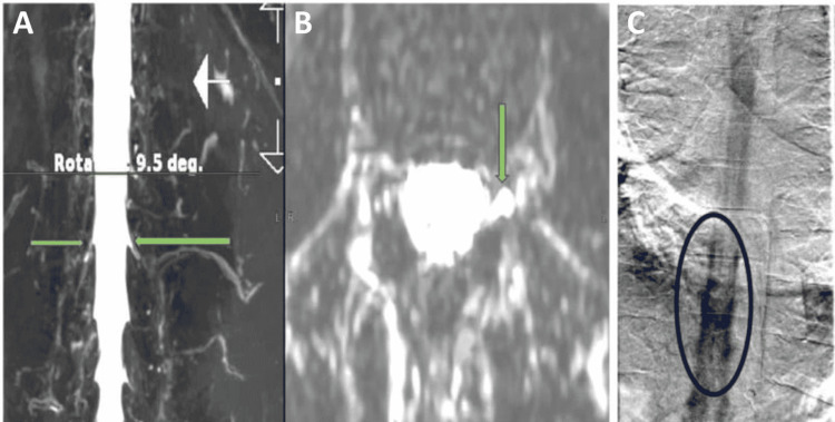
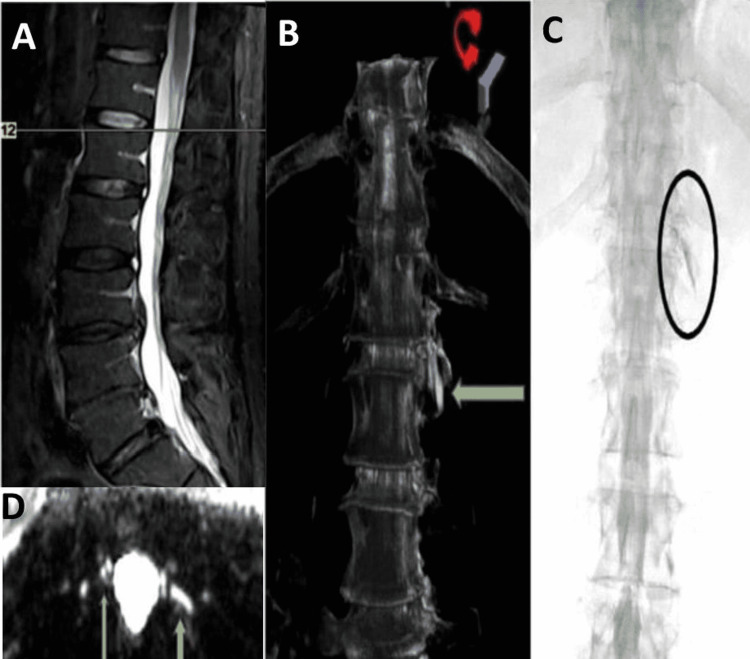
Spontaneous intracranial hypotension (SIH) due to a spinal cerebrospinal fluid (CSF) leak is secondary cause of headache with potentially devastating consequences. Its diagnosis is complicated owing to the lack of a reasonable, minimally invasive screening test. This results in many patients remaining undiagnosed for years after the headache onset. Current testing approaches are either overly invasive, namely the CSF infusion protocol or both invasive and insensitive viz. lumbar puncture (LP) with an opening pressure (OP) or computed tomography myelogram (CTM). These diagnostic methods are frequently employed in a clinical setting since they require access to the thecal space; they unfortunately have a dearth of sensitivity. CTM will not document a leak if it is intermittent or very slow and in the setting of a spinal CSF leak, the OP on LP may be high, low, or normal. A potential remedy for this state is the T2-sampling perfection with application-optimized contrasts by using flip angle evolution (SPACE) protocol spinal magnetic resonance imaging (MRI). We present two cases that demonstrate its potential value as a screening tool. It is well known for its high sensitivity for identifying spinal pathology and is minimally invasive, making it a good choice for a screening modality when diagnosing possible SIH cases.
Keywords: ds myelogram; screening tool; spinal csf leak; spontaneous intracranial hypotension (sih); t2-space protocol mri.
Copyright © 2022, Daripa et al.
Conflict of interest statement
The authors have declared that no competing interests exist.
Figures
Similar articles
-
Assessing Spinal Cerebrospinal Fluid Leaks in Spontaneous Intracranial Hypotension With a Scoring System Based on Brain Magnetic Resonance Imaging Findings.JAMA Neurol. 2019 May 1;76(5):580-587. doi: 10.1001/jamaneurol.2018.4921. JAMA Neurol. 2019. PMID: 30776059 Free PMC article.
-
Spontaneous Intracranial Hypotension - A Dilemma.Neurol India. 2021 Nov-Dec;69(Supplement):S456-S462. doi: 10.4103/0028-3886.332255. Neurol India. 2021. PMID: 35103002 Review.
-
Clinical Presentation, Investigation Findings, and Treatment Outcomes of Spontaneous Intracranial Hypotension Syndrome: A Systematic Review and Meta-analysis.JAMA Neurol. 2021 Mar 1;78(3):329-337. doi: 10.1001/jamaneurol.2020.4799. JAMA Neurol. 2021. PMID: 33393980 Free PMC article.
-
Cerebrospinal fluid outflow resistance as a diagnostic marker of spontaneous cerebrospinal fluid leakage.J Neurosurg Spine. 2017 Aug;27(2):227-234. doi: 10.3171/2017.1.SPINE16548. Epub 2017 Jun 2. J Neurosurg Spine. 2017. PMID: 28574328
-
Patterns of cerebrospinal fluid (CSF) distribution in patients with spontaneous intracranial hypotension: Assessed with magnetic resonance myelography.J Chin Med Assoc. 2017 Feb;80(2):109-116. doi: 10.1016/j.jcma.2016.02.013. Epub 2016 Oct 12. J Chin Med Assoc. 2017. PMID: 27743810
References
Publication types
LinkOut - more resources
Full Text Sources
Miscellaneous