. 2022 Jul 12;17(3):133.
doi: 10.3892/mco.2022.2566. eCollection 2022 Sep.

Overexpression of Annexin A1 is associated with the formation of capillaries in infantile hemangioma

Affiliations

Overexpression of Annexin A1 is associated with the formation of capillaries in infantile hemangioma

Xinyuan Pan et al. Mol Clin Oncol. .

Abstract

Infantile hemangioma is a common benign tumor in infants. However, the molecular mechanism that controls the proliferation and differentiation of hemangioma is not well understood. Annexin A1 (ANX A1) is a phospholipid-binding protein involved in a variety of biological processes, including inflammation, cell proliferation and apoptosis. To explore the significance of ANX A1 in the process of proliferation or differentiation of hemangioma, proliferating and involuting hemangioma tissues were collected to detect the expression of ANX A1 using immunohistochemistry and western blotting. Normal skin tissues were used as the negative control. The results revealed that ANX A1 was upregulated in the proliferative phase of hemangioma, and its expression was decreased when the hemangioma entered the involuting phase. Additionally, in the proliferative phase, the strongest staining of ANX A1 was observed in newly born capillaries, and the staining of ANX A1 became weaker in enlarged vessels, indicating that ANX A1 plays an important role in promoting the formation of capillaries. The expression of hypoxia-inducible factor (HIF)-1α was positively associated with the expression trend of ANX A1, suggesting that the overexpression of ANX A1 may be associated with the increase of HIF-1α. In summary, the results of the present study revealed that the expression of ANX A1 was increased in proliferating hemangioma tissue, and that high expression of ANX A1 may be closely associated with the formation of capillaries in infantile hemangioma.

Keywords: Annexin A1; capillary; extracellular signal-regulated kinase 1/2; hemangioma; hypoxia-inducible factor-1α; proliferation.


Conflict of interest statement

The authors declare that they have no competing interests.

Figures
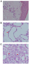
Figure 1
Figure 1
Histopathological analysis of hemangioma tissues. (A) Normal skin, (B) involuting hemangioma and (C) proliferative hemangioma tissue staining by hematoxylin and eosin. The red arrow in B indicates apoptotic bodies in endothelial cells. The red arrows in C indicate the formation of small capillaries. Scale bar, 20 µM.
Figure 2
Figure 2
Detection of ANX A1 expression using western blot analysis. Detection of ANX A1 expression in Cont (normal skin tissue), IH and PH by western blot analysis. Tubulin was used as an internal control and the expression of ANX A1 protein was quantified using densitometric analysis. ANX A1 was upregulated in PH and IH compared with Cont. Each data point represents the mean ± SD. *P<0.01 vs. Cont; #P<0.01 vs. IH. n=15 for each group. ANX A1, Annexin A1; Cont, control; IH, involuting hemangioma; PH, proliferative hemangioma.
Figure 3
Figure 3
Detection of ANX A1 using immunohistochemical staining. Immunohistochemical staining of ANX A1 expression in (A) normal skin, (B) involuting hemangioma and (C) proliferative hemangioma. ANX A1 was strongly expressed in both proliferative hemangioma and involuting hemangioma compared with normal skin tissue. Red arrows in C indicate a strong positive reaction of ANX A1 in the newly born capillaries. Scale bar, 20 µM. ANX A1, Annexin A1.
Figure 4
Figure 4
Detection of HIF-1α expression using western blot analysis. HIF-1α expression was detected in Cont (normal skin tissue), IH and PH by western blot analysis. Tubulin was used as an internal control. HIF-1α was upregulated in PH and IH compared with Cont. Each data point represents the mean ± SD. *P<0.01 vs. Cont; #P<0.01 vs. IH. n=15 for each group. HIF-1α, hypoxia-inducible factor-1α; Cont, control; IH, involuting hemangioma; PH, proliferative hemangioma.
Figure 5
Figure 5
Detection of the localization of HIF-1α and ANX A1 expression in hemangioma tissues. Double-label immunofluorescence of whole-mount sections showing the co-localization of HIF-1α (green fluorescence) with ANX A1 (red fluorescence). Cell nuclei were stained with DAPI (blue fluorescence). The composite images reveal cells co-labeled with HIF-1α and ANX A1. The arrows indicate cells co-labeled with HIF-1α and ANX A1. Scale bar, 20 µM. HIF-1α, hypoxia-inducible factor-1α; ANX A1, Annexin A1.
Figure 6
Figure 6
Phosphorylation of ERK1/2 evaluated using western blot analysis. The phosphorylation level of ERK1/2 in the IH and PH tissues was significantly higher than that in the Cont group, and the phosphorylation of ERK1/2 in IH was lower than that in PH. Each data point represents the mean ± SD. *P<0.01 vs. Cont; #P<0.01 vs. IH. n=15 for each group. Cont, control; IH, involuting hemangioma; PH, proliferative hemangioma; p-, phosphorylated.

Similar articles

Cited by

References

    1. Kanada KN, Merin MR, Munden A, Friedlander SF. A prospective study of cutaneous fifindings in newborns in the United States: Correlation with race, ethnicity, and gestational status using updated classifification and nomenclature. J Pediatr. 2012;161:240–245. doi: 10.1016/j.jpeds.2012.02.052. - DOI - PubMed
    1. Kilcline C, Frieden IJ. Infantile hemangiomas: How common are they? A systematic review of the medical literature. Pediatr Dermatol. 2008;25:168–173. doi: 10.1111/j.1525-1470.2008.00626.x. - DOI - PubMed
    1. Couto RA, Maclellan RA, Zurakowski D, Greene AK. Infantile hemangioma: Clinical assessment of the involuting phase and implications for management. Plast Reconstr Surg. 2012;130:619–624. doi: 10.1097/PRS.0b013e31825dc129. - DOI - PubMed
    1. Nguyen HP, Pickrell BB, Wright TS. Beta-blockers as therapy for infantile hemangiomas. Semin Plast Surg. 2014;28:87–90. doi: 10.1055/s-0034-1376259. - DOI - PMC - PubMed
    1. Priya C, Varshini C, Biswakumar B. Case report: A rare case of infantile hemangioma, treated in a private clinic as out patient. Prim Health Care. 2019;9(321)