Case Reports
. 2022 Jul 15;17(3):136.
doi: 10.3892/mco.2022.2569. eCollection 2022 Sep.

Application of PD-L1 blockade in refractory histiocytic sarcoma: A case report

Affiliations
Case Reports

Application of PD-L1 blockade in refractory histiocytic sarcoma: A case report

Osamu Imataki et al. Mol Clin Oncol. .

Abstract

Histiocytic sarcoma (HS) is a rare hematological malignancy, which exhibits morphological and immunophenotypic features of histiocytes. A standard therapy for HS has not yet been established due to its rareness; therefore, disease control is not always possible. A multimodal treatment strategy has been suggested for HS. The present study reported on a case of a 43-year-old female patient who complained of left femoral pain, which was caused by left femoral bone mass. A biopsy of their left femoral bone tumor revealed that the patient had HS. Their sarcoma was localized in the femoral bone and was not considered to be curable, due to local infiltration of the bone tumor beyond the periosteum. The patient then underwent two types of HS-specific chemotherapy; however, both regimens were ineffective. As a result, they underwent radiation therapy at the sites of progressive disease. Because the HS cells of the patient expressed PD-L1, they were treated with nivolumab (240 mg/body, biweekly) for residual diseases in the right occipital bone, multiple lung nodules, intrapelvic right lymph node and primary site. Nivolumab treatment resulted in a complete response at all sites, with the exception of the primary site, which was confirmed by 18F-fluorodeoxyglucose-positron emission tomography/computed tomography. The patient received additional nivolumab treatment as consolidation therapy for 1 year. In addition, residual disease of the femoral head was completely resected. The surgically resected refractory tumor revealed the tumor cells no longer pathologically expressed PD-L1 . In conclusion, for refractory and recurrent HS in which surgical resection is not appropriate, treatment with immune-checkpoint inhibitors, such as nivolumab, may be considered an optional but promising immunotherapy if the tumor histologically expresses PD-L1. The present study detected one of the refractory mechanisms of ICI treatment.

Keywords: 18F-FDG-PET; PD-L1; histiocytic sarcoma; immune-checkpoint inhibitor; nivolumab; resistance mechanism.


Conflict of interest statement

The authors declare that they have no competing interests.

Figures

Figure 1
Figure 1
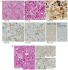
Pathological feature of HS. Pathology was used to diagnose the patient's bone biopsy. (A) HS tumor infiltrates cortical bone and is diffusely involved in the bone marrow. Histologic findings in the patient's first excisional biopsy H&E stain revealed atypical histiocytic proliferation with scattered large, hyperchromatic cells (indicated with asterisks), as well as small lymphocytes and eosinophils in the background. The PD-L1 stain (clone 28-8) was found to be positive in approximately 75% of the atypical cells. (B) The tumor cells were found to be positive for CD68 and CD163. Lysozyme stain was variable in its positivity in atypical cells. The tumor tissue was infiltrated by CD4+ T cells (indicated with small arrowheads). H&E, hematoxylin and eosin; HS, histiocytic sarcoma.
Figure 2
Figure 2
Imaging diagnosis of left femoral HS. (A) 18F-FDG-PET/CT study. The femoral lesion, which was diagnosed as HS, only 18F-FDG-PET/CT could detect. Left leg MRI was difficult to draw the tumor status is active or necrotic. (B) The patient recurred with new lesions; right occipital bone, multiple lung nodules, and intrapelvic right lymph node, and primary site; left femoral bone (left panel). (C) Imaging follow-up after radiation and nivolumab salvage therapy for recurrent HS. Following radiation therapy, FDG-PET/CT revealed a decrease in FDG accumulation in the right occipital bone and intrapelvic lymph node (right panel). Only after nivolumab therapy did the accumulation of multiple lung nodules disappeared completely and the left femoral head lesion diminished (right panel). 18F-FDG-PET/CT, 18F-fluorodeoxyglucose-positron emission tomography/computed tomography; HS, histiocytic sarcoma.
Figure 3
Figure 3
Pathological findings of relapsed and refractory primary lesions. After surgical resection of residual primary disease on the patient's left femoral head. (A) Tumor cells were shown as CD68 positive cells in the tissue (upper panels). CD4+ T cells infiltrated into interstitial tissues, however, PD-L1 (clone 28-8) was no longer positive on the tumor cells (0%) (lower panels). (B) Surrounding the alive tumor cells, many HS tumor cells still remain positive for PD-L1 antigen (right upper panel) and become apoptotic (left panel, asterisk) by the immunity of CD4+ T cells (left panel, shown by small arrowheads) (right lower panel). H&E, hematoxylin and eosin; HS, histiocytic sarcoma.

Similar articles

Cited by

References

    1. Swerdlow SH, Campo E, Harris NL, Jaffe ES, Pileri SA, Stein H, Thiele J. WHO classification of tumours of haematopoietic and lymphoid tissues. Revised Fourth Edition World Health Organization classification of tumours. IARC, Lyon, 2017.
    1. Swerdlow SH, Campo E, Pileri SA, Harris NL, Stein H, Siebert R, Advani R, Ghielmini M, Salles GA, Zelenetz AD, Jaffe ES. The 2016 revision of the world health organization classification of lymphoid neoplasms. Blood. 2016;127:2375–2390. doi: 10.1182/blood-2016-01-643569. - DOI - PMC - PubMed
    1. Emile JF, Abla O, Fraitag S, Horne A, Haroche J, Donadieu J, Requena-Caballero L, Jordan MB, Abdel-Wahab O, Allen CE, et al. Revised classification of histiocytoses and neoplasms of the macrophage-dendritic cell lineages. Blood. 2016;127:2672–2681. doi: 10.1182/blood-2016-01-690636. - DOI - PMC - PubMed
    1. Hung YP, Qian X. Histiocytic sarcoma. Arch Pathol Lab Med. 2020;144:650–654. doi: 10.5858/arpa.2018-0349-RS. - DOI - PubMed
    1. Gatalica Z, Bilalovic N, Palazzo JP, Bender RP, Swensen J, Millis SZ, Vranic S, Von Hoff D, Arceci RJ. Disseminated histiocytoses biomarkers beyond BRAFV600E: Frequent expression of PD-L1. Oncotarget. 2015;6:19819–19825. doi: 10.18632/oncotarget.4378. - DOI - PMC - PubMed

Publication types