

Effect of acquisition techniques, latest kernels, and advanced monoenergetic post-processing for stent visualization with third-generation dual-source CT
- PMID: 35950281
- PMCID: PMC9634938
- DOI: 10.5152/dir.2022.21107
Effect of acquisition techniques, latest kernels, and advanced monoenergetic post-processing for stent visualization with third-generation dual-source CT
Abstract
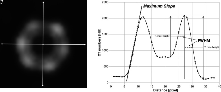
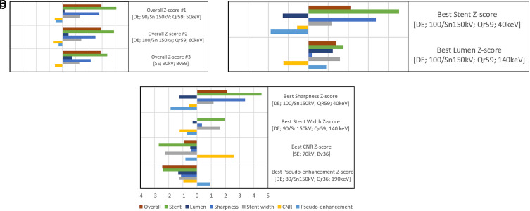
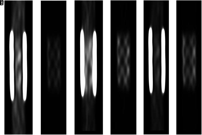
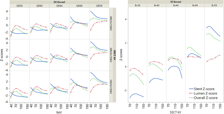
PURPOSE The purpose of this study is to systematically evaluate the effect of tube voltage, current kernels, and monoenergetic post-processing on stent visualization. METHODS A 6 mm chrome-cobalt peripheral stent was placed in a dedicated phantom and scanned with the available tube voltage settings of a third-generation dual-source scanner in single-energy (SE) and dual-energy (DE) mode. Images were reconstructed using the latest convolution kernels and monoenergetic reconstructions (40-190 keV) for DE. The sharpness of stent struts (S), struts width (SW), contrast-to-noise-ratios (CNR), and pseudoenhancement (PE) between the vessel with and without stent were analyzed using an in-house built automatic analysis tool. Measurements were standardized through calculated z-scores. Z-scores were combined for stent (SQ), luminal (LQ), and overall depiction quality (OQ) by adding S and SW, CNR and SW and PE, and S and SW and CNR and PE. Two readers rated overall stent depiction on a 5-point Likert-scale. Agreement was calculated using linear-weighted kappa. Correlations were calculated using Spearman correlation coefficient. RESULTS Maximum values of S and CNR were 169.1 HU/pixel for [DE; 100/ Sn 150 kV; Qr59; 40 keV] and 50.0 for [SE; 70 kV; Bv36]. Minimum values of SW and PE were 2.615 mm for [DE; 80 to 90/ Sn 150 kV; Qr59; 140 to 190 keV] and 0.12 HU for [DE; 80/ Sn 150 kV; Qr36; 190 keV]. Best combined z-scores of SQ, LQ, and OQ were 4.53 for [DE; 100/ Sn 150 kV; Qr 59; 40 keV], 1.23 for [DE; 100/ Sn 150 kV; Qr59; 140 keV] and 2.95 for [DE; 90/ Sn 150 kV; Qr59; 50 keV]. Best OQ of SE was ranked third with 2.89 for [SE; 90 kV; Bv59]. Subjective agreement was excellent (kappa=0.86; P < .001) and correlated well with OQ (rs=0.94, P < .001). CONCLUSION Combining DE computed tomography (CT) acquisition with the latest kernels and monoenergetic post-processing allows for improved stent visualization as compared with SECT. The best overall results were obtained for monoenergetic reconstructions with 50 keV from DECT 90/Sn 150 kV acquisitions using kernel Qr59.
Conflict of interest statement
Figures
Similar articles
-
Impact of an advanced image-based monoenergetic reconstruction algorithm on coronary stent visualization using third generation dual-source dual-energy CT: a phantom study.Eur Radiol. 2016 Jun;26(6):1871-8. doi: 10.1007/s00330-015-3997-4. Epub 2015 Sep 15. Eur Radiol. 2016. PMID: 26373752
-
Advanced virtual monoenergetic computed tomography of hyperattenuating and hypoattenuating liver lesions: ex-vivo and patient experience in various body sizes.Invest Radiol. 2015 Oct;50(10):695-702. doi: 10.1097/RLI.0000000000000171. Invest Radiol. 2015. PMID: 26002623
-
Assessment of an advanced image-based technique to calculate virtual monoenergetic computed tomographic images from a dual-energy examination to improve contrast-to-noise ratio in examinations using iodinated contrast media.Invest Radiol. 2014 Sep;49(9):586-92. doi: 10.1097/RLI.0000000000000060. Invest Radiol. 2014. PMID: 24710203
-
Maximizing Iodine Contrast-to-Noise Ratios in Abdominal CT Imaging through Use of Energy Domain Noise Reduction and Virtual Monoenergetic Dual-Energy CT.Radiology. 2015 Aug;276(2):562-70. doi: 10.1148/radiol.2015140857. Epub 2015 Apr 10. Radiology. 2015. PMID: 25860839 Free PMC article.
-
Assessment of the hepatic veins in poor contrast conditions using dual energy CT: evaluation of a novel monoenergetic extrapolation software algorithm.Rofo. 2014 Jun;186(6):591-7. doi: 10.1055/s-0034-1366423. Epub 2014 Apr 22. Rofo. 2014. PMID: 24756426
Cited by
-
Diagnostic Value of DECT-Based Collagen Mapping for Assessing the Distal Tibiofibular Syndesmosis in Patients with Acute Trauma.Diagnostics (Basel). 2023 Feb 1;13(3):533. doi: 10.3390/diagnostics13030533. Diagnostics (Basel). 2023. PMID: 36766638 Free PMC article.
-
Determining the Optimal Energy Level of Virtual Monoenergetic Images in Dual-Source CT for Diagnosis of Bowel Obstruction and Colitis.Diagnostics (Basel). 2023 Nov 21;13(23):3491. doi: 10.3390/diagnostics13233491. Diagnostics (Basel). 2023. PMID: 38066732 Free PMC article.
-
Diagnostic value of DECT-based colored collagen maps for the assessment of cruciate ligaments in patients with acute trauma.Eur Radiol. 2023 Sep;33(9):6339-6350. doi: 10.1007/s00330-023-09558-4. Epub 2023 Mar 31. Eur Radiol. 2023. PMID: 37000215 Free PMC article.
References
MeSH terms
LinkOut - more resources
Full Text Sources
Medical
