Common Supraventricular and Ventricular Arrhythmias in Children
- PMID: 35950741
- PMCID: PMC9524439
- DOI: 10.5152/TurkArchPediatr.2022.22099
Common Supraventricular and Ventricular Arrhythmias in Children
Abstract
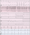
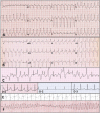
The most common pediatric arrhythmias are tachycardias, and the most common type is supraventricular tachycardia, originating from or above the atrioventricular node and HIS bundle. Ventricular tachycardias are less common but more dangerous. Supraventricular tachycardias usually cause a narrow complex tachycardia unless there is a basal bundle branch block or rate-dependent aberration. A wide QRS tachycardia should be treated as ventricular tachycardias unless proven to be an supraventricular tachycardia with aberration. Diagnosis of both tachyarrhythmia types depends mainly on 12-lead electrocardiography. The most common supraventricular tachycardia type in newborns and infants is atrioventricular reentry tachycardia, related to manifest or concealed accessory pathways and in adolescent atrioventricular nodal reentry tachycardia, whereas focal atrial tachycardias consist of 10%-15% of supraventricular tachycardias during all ages. Supraventricular tachycardias have a low risk of morbidity, and ablation therapy is successful in most types with success rates over 90%. Ventricular tachycardias can be monomorphic or polymorphic, nonsustained or sustained, and can cause more hemodynamic instability than supraventricular tachycardias, requiring more close monitoring and urgent therapies. If hemodynamically unstable, synchronized cardioversion must be performed. Polymorphic ventricular tachycardias are very dangerous and often associated with primary ion channel defects (channelopathies), which can cause sudden cardiac death.
Figures


Similar articles
-
[Catheter ablation in supraventricular tachycardia].Z Kardiol. 1996;85 Suppl 6:45-60. Z Kardiol. 1996. PMID: 9064982 Review. German.
-
What an anesthesiologist should know about pediatric arrhythmias.Paediatr Anaesth. 2024 Dec;34(12):1187-1199. doi: 10.1111/pan.14980. Epub 2024 Aug 15. Paediatr Anaesth. 2024. PMID: 39148245 Review.
-
Using the right drug: a treatment algorithm for regular supraventricular tachycardias.Eur Heart J. 1997 May;18 Suppl C:C27-32. doi: 10.1093/eurheartj/18.suppl_c.27. Eur Heart J. 1997. PMID: 9152672 Review.
-
Importance of the Activation Sequence of the His or Right Bundle for Diagnosis of Complex Tachycardia Circuits.Circ Arrhythm Electrophysiol. 2021 Oct;14(10):e009194. doi: 10.1161/CIRCEP.120.009194. Epub 2021 Oct 4. Circ Arrhythm Electrophysiol. 2021. PMID: 34601885 Review.
-
Clinical Features and Sites of Ablation for Patients With Incessant Supraventricular Tachycardia From Concealed Nodofascicular and Nodoventricular Tachycardias.JACC Clin Electrophysiol. 2017 Dec 26;3(13):1547-1556. doi: 10.1016/j.jacep.2017.07.015. Epub 2017 Nov 6. JACC Clin Electrophysiol. 2017. PMID: 29759837
Cited by
-
Malignant Ventricular Arrhythmia With Apical Biventricular Noncompaction.Cureus. 2024 Nov 9;16(11):e73323. doi: 10.7759/cureus.73323. eCollection 2024 Nov. Cureus. 2024. PMID: 39659346 Free PMC article.
-
Evaluating antiarrhythmic drugs for managing infants with supraventricular tachycardia; a review.Am J Cardiovasc Dis. 2024 Jun 15;14(3):144-152. doi: 10.62347/ZTXC5809. eCollection 2024. Am J Cardiovasc Dis. 2024. PMID: 39021523 Free PMC article. Review.
-
Incidence and trend of cardiac events among children and young adults exposed to psychopharmacological treatment (2006-2018): A nationwide register-based study.Br J Clin Pharmacol. 2025 Mar;91(3):817-828. doi: 10.1111/bcp.16321. Epub 2024 Oct 24. Br J Clin Pharmacol. 2025. PMID: 39448545 Free PMC article.
-
Neurological Consequences of Cardiac Arrhythmias: Relationship Between Stroke, Cognitive Decline, and Heart Rhythm Disorders.Cureus. 2024 Mar 29;16(3):e57159. doi: 10.7759/cureus.57159. eCollection 2024 Mar. Cureus. 2024. PMID: 38681361 Free PMC article. Review.
-
The Effects of Pediatric Acute Lymphoblastic Leukemia Treatment on Cardiac Repolarization.Children (Basel). 2024 Sep 24;11(10):1158. doi: 10.3390/children11101158. Children (Basel). 2024. PMID: 39457123 Free PMC article.
References
-
- Cannon BC, Snyder CS. Disorders of cardiac rhythm and conduction. In: Allen HD, Driscoll DJ, Shaddy RE, Feltes TF.eds. Moss and Adams Heart Disease in Infants, Children and Adolescents Including the Fetus and Young Adult. Philadelphia, PA; 2013:441 472.
LinkOut - more resources
Full Text Sources
