

Cytocompatibility and bioactive potential of AH Plus Bioceramic Sealer: An in vitro study
- PMID: 35950780
- PMCID: PMC9541143
- DOI: 10.1111/iej.13805
Cytocompatibility and bioactive potential of AH Plus Bioceramic Sealer: An in vitro study
Abstract
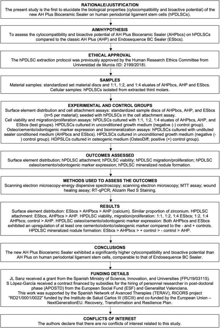
Aim: To assess the cytocompatibility and bioactive potential of the new calcium silicate cement-based sealer AH Plus Bioceramic Sealer (AHPbcs) on human periodontal ligament stem cells (hPDLSCs) compared with the epoxy resin-based sealer AH Plus (AHP) and the calcium silicate cement-based sealer Endosequence BC Sealer (ESbcs).
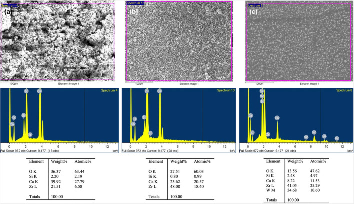
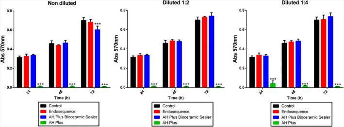
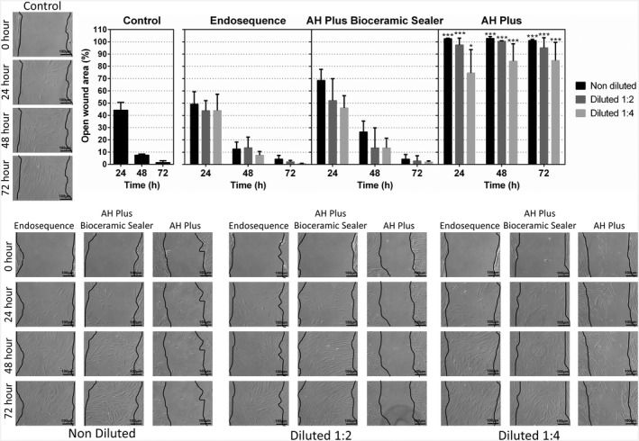
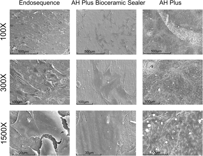
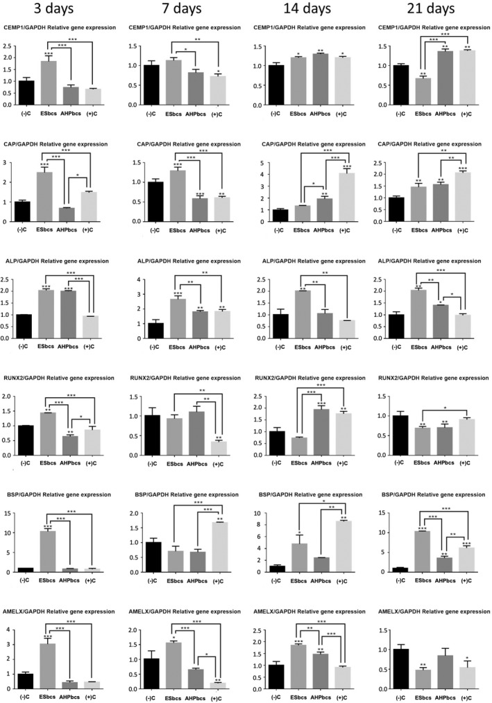
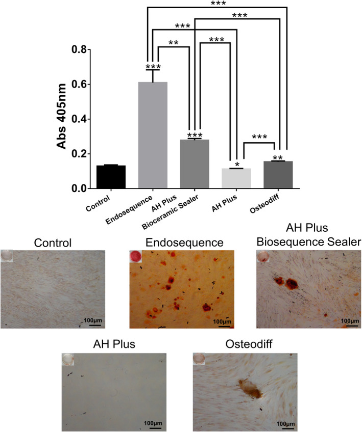
Methodology: Standardized sample discs and 1:1, 1:2 and 1:4 eluates of the tested materials were prepared. The following assays were performed: surface element distribution via SEM-EDX, cell attachment and morphology via SEM, cell viability via a MTT assay, cell migration/proliferation via a wound-healing assay, osteo/cemento/odontogenic marker expression via RT-qPCR and cell mineralized nodule formation via Alizarin Red S staining. HPDLSCs were isolated from extracted third molars. Comparisons were made with hPDLSCs cultured in unconditioned (negative control) or osteogenic (positive control) culture media. Statistical significance was established at p < .05.
Results: A higher peak of Ca2 + was detected from ESbcs compared with AHPbcs and AHP in SEM-EDX. Both AHPbcs and ESbcs showed significantly positive results in the cytocompatibility assays (cell viability, migration/proliferation, attachment and morphology) compared with a negative control group, whilst AHP showed significant negative results. Both AHPbcs and ESbcs exhibited an upregulation of at least one osteo/odonto/cementogenic marker compared with the negative and positive control groups. Both ESbcs and AHPbcs showed a significantly higher calcified nodule formation than the negative and positive control groups, indicative of their biomineralization potential and were also significantly higher than AHP group.
Conclusion: AH Plus Bioceramic Sealer exhibited a significantly higher cytocompatibility and bioactive potential than AH Plus and a similar cytocompatibility to that of Endosequence BC Sealer. Endosequence BC Sealer exhibited a significantly higher mineralization potential than the other tested sealers. The results from this in vitro study act as supporting evidence for the use of AH Plus Bioceramic Sealer in root canal treatment.
Keywords: AH Plus Bioceramic Sealer; AH Plus sealer; Endosequence BC sealer; bioactivity; biocompatibility.
© 2022 The Authors. International Endodontic Journal published by John Wiley & Sons Ltd on behalf of British Endodontic Society.
Conflict of interest statement
The authors declare no conflicts of interest related to this study.
Figures
References
-
- Aminoshariae, A. & Kulild, J.C. (2020) The impact of sealer extrusion on endodontic outcome: a systematic review with meta‐analysis. Australian Endodontic Journal, 46, 123–129. - PubMed
-
- Arzate, H. , Zeichner‐David, M. & Mercado‐Celis, G. (2015) Cementum proteins: role in cementogenesis, biomineralization, periodontium formation and regeneration. Periodontology 2000, 67, 211–233. - PubMed
-
- Bakir, E.P. , Yildirim, Z.S. , Bakir, Ş. & Ketani, A. (2022) Are resin‐containing pulp capping materials as reliable as traditional ones in terms of local and systemic biological effects? Dental Materials Journal, 41, 78–86. - PubMed
-
- Bartold, P.M. & Gronthos, S. (2017) Standardization of criteria defining periodontal ligament stem cells. Journal of Dental Research, 96, 487–490. - PubMed
MeSH terms
Substances
Grants and funding
LinkOut - more resources
Full Text Sources
Miscellaneous
