. 2022 Aug 31;10(4):e0166122.
doi: 10.1128/spectrum.01661-22. Epub 2022 Aug 11.

Amyloid Aggregates Are Localized to the Nonadherent Detached Fraction of Aging Streptococcus mutans Biofilms

Affiliations

Amyloid Aggregates Are Localized to the Nonadherent Detached Fraction of Aging Streptococcus mutans Biofilms

Elena Yarmola et al. Microbiol Spectr. .

Abstract

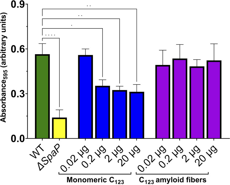
The number of bacterial species recognized to utilize purposeful amyloid aggregation within biofilms continues to grow. The oral pathogen Streptococcus mutans produces several amyloidogenic proteins, including adhesins P1 (also known as AgI/II, PAc) and WapA, whose truncation products, namely, AgII and AgA, respectively, represent the amyloidogenic moieties. Amyloids demonstrate common biophysical properties, including recognition by Thioflavin T (ThT) and Congo red (CR) dyes that bind to the cross β-sheet quaternary structure of amyloid aggregates. Previously, we observed amyloid formation to occur only after 60 h or more of S. mutans biofilm growth. Here, we extend those findings to investigate where amyloid is detected within 1- and 5-day-old biofilms, including within tightly adherent compared with those in nonadherent fractions. CR birefringence and ThT uptake demonstrated amyloid within nonadherent material removed from 5-day-old cultures but not within 1-day-old or adherent samples. These experiments were done in conjunction with confocal microscopy and immunofluorescence staining with AgII- and AgA-reactive antibodies, including monoclonal reagents shown to discriminate between monomeric protein and amyloid aggregates. These results also localized amyloid primarily to the nonadherent fraction of biofilms. Lastly, we show that the C-terminal region of P1 loses adhesive function following amyloidogenesis and is no longer able to competitively inhibit binding of S. mutans to its physiologic substrate, salivary agglutinin. Taken together, our results provide new evidence that amyloid aggregation negatively impacts the functional activity of a widely studied S. mutans adhesin and are consistent with a model in which amyloidogenesis of adhesive proteins facilitates the detachment of aging biofilms. IMPORTANCE Streptococcus mutans is a keystone pathogen and causative agent of human dental caries, commonly known as tooth decay, the most prevalent infectious disease in the world. Like many pathogens, S. mutans causes disease in biofilms, which for dental decay begins with bacterial attachment to the salivary pellicle coating the tooth surface. Some strains of S. mutans are also associated with bacterial endocarditis. Amyloid aggregation was initially thought to represent only a consequence of protein mal-folding, but now, many microorganisms are known to produce functional amyloids with biofilm environments. In this study, we learned that amyloid formation diminishes the activity of a known S. mutans adhesin and that amyloid is found within the nonadherent fraction of older biofilms. This finding suggests that the transition from adhesin monomer to amyloid facilitates biofilm detachment. Knowing where and when S. mutans produces amyloid will help in developing therapeutic strategies to control tooth decay and other biofilm-related diseases.

Keywords: Streptococcus mutans; adhesins; amyloid; biofilms.


Conflict of interest statement

The authors declare no conflict of interest.

Figures

FIG 1
FIG 1
Schematic representation of P1(AgI/II) and relevant domains. (A) Schematic representation of relevant domains identified within the primary sequence of adhesin P1. A1-3, series of three alanine-rich tandem repeats; V, variable region where most sequence differences among strains are clustered; P1-3, series of three proline-rich tandem repeats; C1, C2, C3, beta-rich domains each of which adopts a DE-variant IgG fold; LPXTG, Sortase A cleavage motif. (B) Schematic representation of the tertiary structure of P1. The color scheme and indicated domains from the N to C terminus matches those shown in A. The protein folds into an unusual tertiary structure whereby the alanine-rich and proline-rich repeat regions interact to form an extended helical stalk. AgII, comprised largely of the C1 through C3 domains, not only is contained as an integral part of the cell-surface localized adhesin but also is liberated as an independent fragment capable of interacting with the globular head of the cell-surface localized adhesin.
FIG 2
FIG 2
Evaluation of S. mutans macrocolony morphology. S. mutans wild-type UA159; a ΔgtfBCD mutant lacking glycosyltransferases B, C, and D; a ΔspaP/wapA/smu_63c mutant lacking amyloidogenic proteins P1, WapA, and Smu_63C; and a ΔsrtA mutant lacking the Sortase A transpeptidase were spotted onto Todd-Hewitt plus yeast extract (THYE) agar plates containing Thioflavin S, with or without the amyloid inhibitor epigallocatechin gallate (EGCG). A halo-like morphology was observed for the ΔspaP/wapA/smu_63c mutant strain lacking known amyloidogenic proteins, for the ΔsrtA mutant that cannot link substrate proteins to the cell surface, and for all four strains when grown in the presence of EGCG, suggesting that amyloid can influence S. mutans biofilm architecture.
FIG 3
FIG 3
Evaluation of Congo red birefringence in adherent and nonadherent fractions of S. mutans biofilms. (A) Images of Congo red (CR)-stained adherent (left) and nonadherent (right) fractions from 1-day-old (top) or 5-day-old (bottom) biofilms viewed with and without crossed polarizers (“crossed” and “uncrossed,” respectively). (B) Images of CR-stained curli-negative (left) and curli-positive strains of Escherichia coli grown on YESCA agar and included here as negative and positive controls for CR birefringence. Scale bar: 115 μm (all panels).
FIG 4
FIG 4
Thioflavin T fluorescence assay of adherent and nonadherent fractions of S. mutans biofilms before and after filtration. (A) ThT fluorescence assay of samples of adherent (Ad) or nonadherent (nonAd) material from 1- and 5-day-old S. mutans biofilms before and after filtration through a 0.2-μm filter. The experiment was performed in triplicate, with statistical analysis by one-way ANOVA (****, P < 0.0001). (B) Detection of residual cells or cell wall material in each sample by dot blot. Serial 3-fold dilutions were spotted onto the filter and probed with S. mutans serotype c-specific typing antiserum (CDC). A suspension of S. mutans from an overnight planktonic culture (beginning at ~3 × 107 CFU), and growth media alone, served as positive and negative controls for the antiserum, respectively.
FIG 5
FIG 5
Evaluation of reactivity of anti-P1 monoclonal antibodies with monomeric, amyloid mat, and purified fibril forms of C123 by dot blot. The indicated amount of the total protein of monomeric C123, amyloid mats induced by mechanical agitation, or purified amyloid fibrils derived by proteinase K treatment of amyloid mats were spotted onto the filter and probed with the indicated anti-P1 MAb.
FIG 6
FIG 6
Confocal microscopy and immunostaining of adherent and nonadherent fractions of S. mutans biofilms. S. mutans (UA159::Pldh-gfp) biofilms were grown for 1 or 5 days and then immunostained with the indicated antibody. Adherent fractions were stained directly on the slide on which the biofilm was grown. Nonadherent fractions were transferred to a tube and stained separately. Goat anti-mouse or anti-rabbit secondary reagents were conjugated to Alexa Fluor 594 (red). (A) Maximum intensity projections. Scale bar: 20 μm. (B) Volume snapshots corresponding to images shown in A.
FIG 6
FIG 6
Confocal microscopy and immunostaining of adherent and nonadherent fractions of S. mutans biofilms. S. mutans (UA159::Pldh-gfp) biofilms were grown for 1 or 5 days and then immunostained with the indicated antibody. Adherent fractions were stained directly on the slide on which the biofilm was grown. Nonadherent fractions were transferred to a tube and stained separately. Goat anti-mouse or anti-rabbit secondary reagents were conjugated to Alexa Fluor 594 (red). (A) Maximum intensity projections. Scale bar: 20 μm. (B) Volume snapshots corresponding to images shown in A.
FIG 7
FIG 7
Competitive inhibition of S. mutans binding to immobilized salivary agglutinin by C123 monomers compared with that by C123 amyloid fibrils. Microtiter plate wells were coated with human salivary agglutinin and then incubated with the indicated amount of C123 monomers or purified amyloid fibrils. Following washing, ~3.5 × 108 CFU of S. mutans was added to the wells and incubated for 3 h, cell attachment was assayed by staining with crystal violet, and measurement of absorbance at 595 nm was conducted. Wild-type S. mutans in the absence of an added inhibitor and a ΔspaP mutant lacking the gene encoding adhesin P1 were included as positive and negative controls, respectively. The experiment was performed in triplicate, with statistical analysis by one-way ANOVA. ns, not significant; *, P < 0.05; **, P < 0.01; ***, P < 0.001; ****, P < 0.0001.
FIG 8
FIG 8
Working model for amyloid-induced detachment of S. mutans mature biofilms. Initially, monomeric cell surface-anchored adhesins are available for binding to their substrates, allowing for initial colonization and biofilm development. As the biofilm grows and matures, amyloidogenic moieties associated with the cell surface or in the extracellular matrix undergo amyloid aggregation, which can either act as a scaffold for the development of a robust adherent biofilm layer (outcome 1) or can disrupt adhesive activity and promote biofilm detachment (outcome 2). Created with BioRender.com.

References

    1. Paula AJ, Hwang G, Koo H. 2020. Dynamics of bacterial population growth in biofilms resemble spatial and structural aspects of urbanization. Nat Commun 11:1354. doi:10.1038/s41467-020-15165-4. - DOI - PMC - PubMed
    1. Orazi G, Jean-Pierre F, O’Toole GA. 2020. Pseudomonas aeruginosa PA14 enhances the efficacy of norfloxacin against Staphylococcus aureus newman biofilms. J Bacteriol 202:e00159-20. doi:10.1128/JB.00159-20. - DOI - PMC - PubMed
    1. Bridier A, Piard JC, Pandin C, Labarthe S, Dubois-Brissonnet F, Briandet R. 2017. Spatial organization plasticity as an adaptive driver of surface microbial communities. Front Microbiol 8:1364. doi:10.3389/fmicb.2017.01364. - DOI - PMC - PubMed
    1. Donlan RM. 2002. Biofilms: microbial life on surfaces. Emerg Infect Dis 8:881–890. doi:10.3201/eid0809.020063. - DOI - PMC - PubMed
    1. Flemming HC, Wingender J, Szewzyk U, Steinberg P, Rice SA, Kjelleberg S. 2016. Biofilms: an emergent form of bacterial life. Nat Rev Microbiol 14:563–575. doi:10.1038/nrmicro.2016.94. - DOI - PubMed

Publication types