. 2022 Sep 14;96(17):e0055522.
doi: 10.1128/jvi.00555-22. Epub 2022 Aug 11.

Dominant Negative Mutants of Human Immunodeficiency Virus Type 1 Viral Infectivity Factor (Vif) Disrupt Core-Binding Factor Beta-Vif Interaction

Affiliations

Dominant Negative Mutants of Human Immunodeficiency Virus Type 1 Viral Infectivity Factor (Vif) Disrupt Core-Binding Factor Beta-Vif Interaction

Sizhu Duan et al. J Virol. .

Abstract

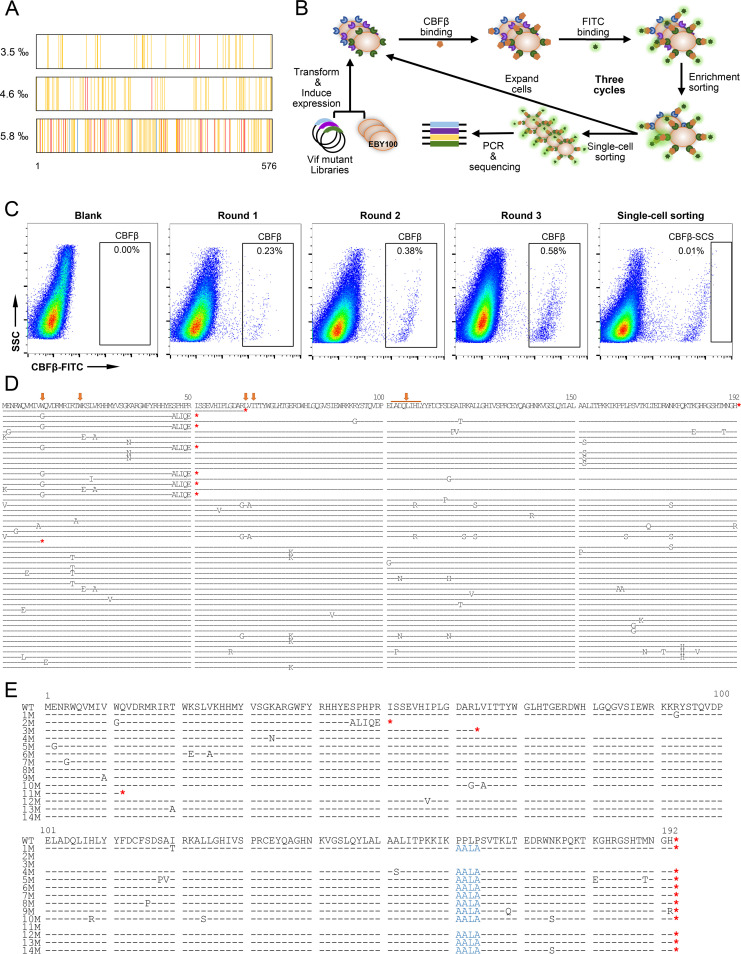
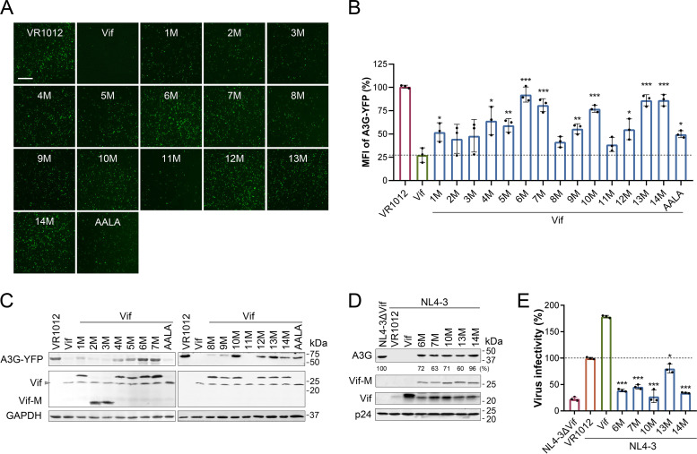
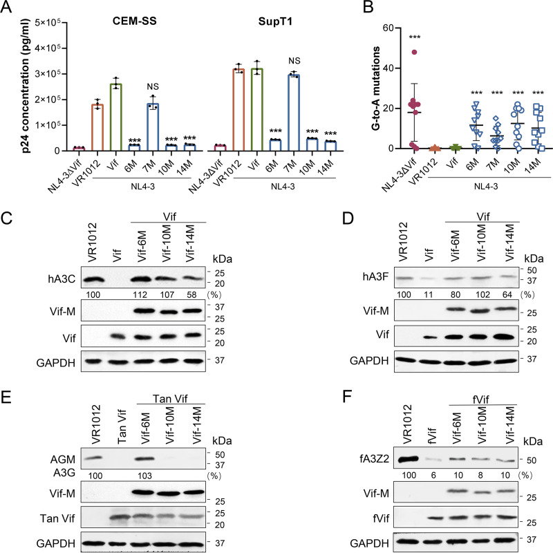
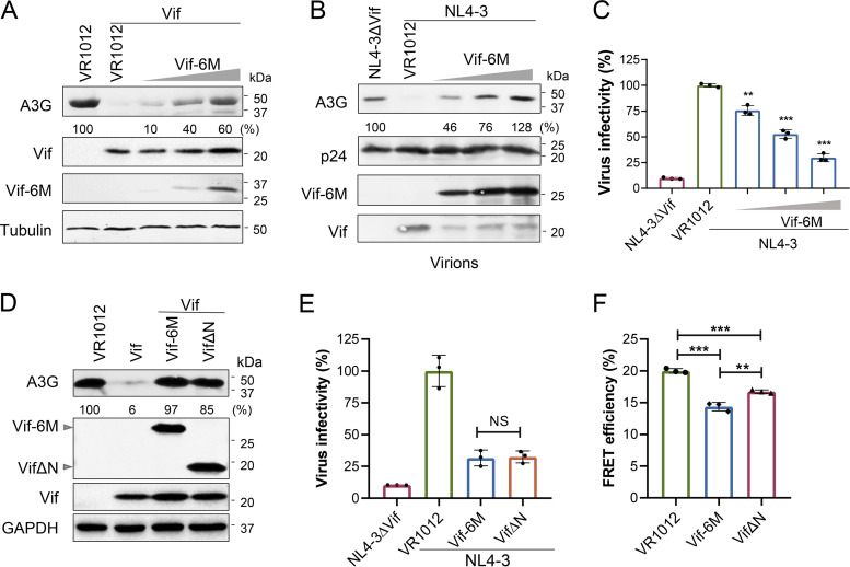
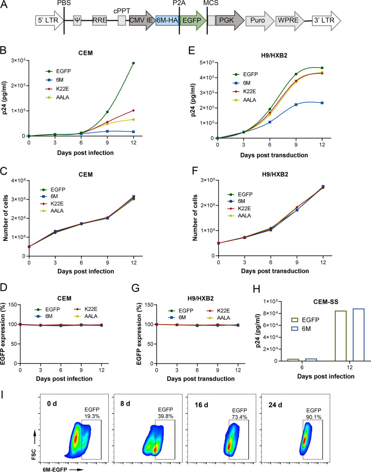
Apolipoprotein B mRNA-editing catalytic polypeptide-like 3 family members (APOBEC3s) are host restriction factors that inhibit viral replication. Viral infectivity factor (Vif), a human immunodeficiency virus type 1 (HIV-1) accessory protein, mediates the degradation of APOBEC3s by forming the Vif-E3 complex, in which core-binding factor beta (CBFβ) is an essential molecular chaperone. Here, we screened nonfunctional Vif mutants with high affinity for CBFβ to inhibit HIV-1 in a dominant negative manner. We applied the yeast surface display technology to express Vif random mutant libraries, and mutants showing high CBFβ affinity were screened using flow cytometry. Most of the screened Vif mutants containing random mutations of different frequencies were able to rescue APOBEC3G (A3G). In the subsequent screening, three of the mutants restricted HIV-1, recovered G-to-A hypermutation, and rescued APOBEC3s. Among them, Vif-6M showed a cross-protection effect toward APOBEC3C, APOBEC3F, and African green monkey A3G. Stable expression of Vif-6M in T lymphocytes inhibited the viral replication in newly HIV-1-infected cells and the chronically infected cell line H9/HXB2. Furthermore, the expression of Vif-6M provided a survival advantage to T lymphocytes infected with HIV-1. These results suggest that dominant negative Vif mutants acting on the Vif-CBFβ target potently restrict HIV-1. IMPORTANCE Antiviral therapy cannot eliminate HIV and exhibits disadvantages such as drug resistance and toxicity. Therefore, novel strategies for inhibiting viral replication in patients with HIV are urgently needed. APOBEC3s in host cells are able to inhibit viral replication but are antagonized by HIV-1 Vif-mediated degradation. Therefore, we screened nonfunctional Vif mutants with high affinity for CBFβ to compete with the wild-type Vif (wtVif) as a potential strategy to assist with HIV-1 treatment. Most screened mutants rescued the expression of A3G in the presence of wtVif, especially Vif-6M, which could protect various APOBEC3s and improve the incorporation of A3G into HIV-1 particles. Transduction of Vif-6M into T lymphocytes inhibited the replication of the newly infected virus and the chronically infected virus. These data suggest that Vif mutants targeting the Vif-CBFβ interaction may be promising in the development of a new AIDS therapeutic strategy.

Keywords: apolipoprotein B mRNA-editing catalytic polypeptide-like 3 family member (APOBEC3); core-binding factor beta (CBFβ); dominant negative mutant; human immunodeficiency virus (HIV); viral infectivity factor (Vif).


Conflict of interest statement

The authors declare no conflict of interest.

Figures

FIG 1
FIG 1
Screening of Vif mutants with higher affinity for CBFβ by yeast surface display technology. (A) Sequencing analysis of the distribution of mutation sites of the Vif mutant libraries. A total of 25 clones from each of 3 Vif mutant libraries with different mutation frequencies (0.35%, 0.46%, 0.58%) were sequenced. The nucleotide mutation sites are highlighted (one mutation: orange, two mutations: red, three mutations: blue). (B) Identification strategy based on yeast surface cDNA display. The Vif mutant library was displayed on the surface of yeast cells, incubated with CBFβ protein, and stained with FITC antibody. Cells binding to CBFβ were identified by fluorescence-activated cell sorting (FACS)-based screening. (C) Yeast clones displaying Vif protein mutants with high affinity for CBFβ were enriched by three rounds of FACS. Single cells were sorted into 96-well PCR plates, and Vif mutants were sequenced after nested PCR. (D) Sequences of 50 Vif mutants aligned to wild-type Vif (wtVif), amplified from single cells with higher CBFβ affinity. The red asterisks indicate the termination codon in each sequence. The reported key sites in Vif for interaction with CBFβ are marked with orange arrows. (E) Fourteen sequences were selected for further identification. The PPLP motif was mutated to AALA (shown in blue) based on the sequences of the sorted Vif mutants. The red asterisks indicate the termination codon in each sequence.
FIG 2
FIG 2
Vif mutants dominantly interfered with the function of wtVif. (A) A3G-YFP levels detected under a fluorescence microscope. 293T cells were cotransfected with A3G-YFP and wtVif alone or with each Vif mutant. Scale bar = 100 μm. (B) Mean fluorescence intensity (MFI) of A3G-YFP detected by flow cytometry. 293T cells were cotransfected with A3G-YFP and wtVif alone or with each Vif mutant for 48 h. Data were pooled from three independent experiments; mean ± SD. The significance of differences between each group and the Vif group was assessed by Student’s t test. (C) Western blot analysis of A3G recovered by Vif mutants (Vif-M). GAPDH was detected as a loading control. (D) Western blot analysis of A3G packaged into HIV-1 virions in the presence of Vif mutants. A3G-cmyc, pNL4-3, and wtVif alone or with each Vif mutant were cotransfected into 293T cells. Cells transfected with pNL4-3ΔVif were included as a control. After 48 h, viral particles were obtained by ultracentrifugation. p24 was detected as a loading control, and expression of A3G was normalized with p24. Protein expression levels were quantified using Quantity One software. (E) Infectivity of viruses determined by TZM-bl cells. Data were pooled from triplicate experiments; mean ± SD. The significance of differences between each group and the NL4-3 group was assessed by Student’s t test.
FIG 3
FIG 3
Vif mutants rescued the anti-HIV-1 activity of A3G and the level of other APOBEC3s. (A) Replication of viruses packaged in the presence of Vif mutants. A3G, pNL4-3, and wtVif or each Vif mutant or empty vector (VR1012) were cotransfected into 293T cells. Cells transfected with pNL4-3ΔVif were included as a control. After 48 h, cell supernatants containing viruses were collected by centrifugation. CEM-SS and SupT1 cells were infected with equal amounts of virus (4 ng RT). After 48 h, the concentration of produced viruses was detected using a Lenti RT activity ELISA kit. (B) G-to-A mutations induced by A3G in the presence of Vif mutants. At 48 h after viral infection, genomic DNA of CEM-SS cells was extracted. An 885-bp fragment of pol was amplified by nested PCR and ligated into the pGEM-T-easy cloning vector for sequencing (mean ± SD; n = 10). The values on the y axis represent the number of G-to-A mutants in each sequence. The significance of difference between each group and the NL4-3 group was assessed by Student’s t test. (C and D) Western blot analysis of hA3C and hA3F rescued by Vif mutants by interfering with wtVif. 293T cells were cotransfected with hA3C-FLAG (C) or hA3F-cmyc (D), Vif-HA, and Vif mutants. (E) Western blot analysis of AGM A3G saved by Vif mutants from SIVagmTan Vif (Tan Vif)-mediated degradation. 293T cells transfected with AGM A3G-HA, Tan Vif-cmyc, and Vif mutants were cultured for 48 h. (F) Western blot analysis of the effect of Vif mutants on feline immunodeficiency virus Vif (fVif). 293T cells transfected with feline A3Z2-HA (fA3Z2-HA), fVif-cmyc, and Vif mutants were cultured for 48 h. In Western blot analysis, GAPDH was detected as a loading control, and APOBEC3 expression was normalized with GAPDH.
FIG 4
FIG 4
Vif-6M influenced assembly of Vif and E3 complex by competitively binding CBFβ. (A) The ability of Vif-6M to rescue A3G when CBFβ was overexpressed. 293T cells were cotransfected with CBFβ, A3G, wtVif, and Vif-6M and then cultured for 48 h. In Western blot analysis, GAPDH was detected as a loading control, and A3G expression was normalized with GAPDH. (B) The affinity of Vif-6M for CBFβ detected by YSD. Vif or Vif-6M was displayed on the surface of EBY100 cells; the cells were incubated with CBFβ protein for 4 h, stained with FITC-antibody, and monitored by flow cytometry. (C) Coimmunoprecipitation analysis of the affinity of wtVif or Vif-6M with CBFβ. 293T cells were cotransfected with CBFβ-FLAG and Vif-HA or HA-Vif-6M-cmyc. At 48 h posttransfection, cell lysates were prepared and immunoprecipitated with anti-HA antibody-conjugated agarose beads. Expression of CBFβ in the cells and its interaction with Vif or Vif-6M were detected by Western blotting. The CBFβ level was normalized with Vif and Vif-6M. (D) Coimmunoprecipitation analysis of the ability of Vif-6M to block the interaction of Vif-CBFβ. 293T cells were cotransfected with CBFβ-FLAG and Vif-HA with Vif-6M-cmyc or VR1012. Expression of CBFβ in cells and its interaction with Vif were detected by Western blotting. The CBFβ level was normalized with Vif. (E) Western blot analysis of A3G degradation mediated by wtVif and Vif with K22E and V25A mutations. 293T cells were transfected with A3G, wtVif or Vif22E25A for 48 h. GAPDH was detected as a loading control, and the expression of A3G was normalized with GAPDH. Protein expression levels were quantified using Quantity One. (F) FRET efficiency of wtVif, Vif-6M, Vif-K22E, or Vif-AALA to CBFβ. 293T cells were cotransfected with CBFβ-CFP and Vif-YFP or Vif mutant-YFP and then cultured for 48 h. Samples were excited at λmaxCFP (440 nm), and fluorescence emission was detected by scanning fluorometry. FRET efficiency was calculated as described in Materials and Methods. Three independent experiments; mean ± SD. The significance of difference between each group and the Vif group was assessed by Student’s t test. (G) The inhibitory effect of wtVif, Vif-6M, Vif-K22E, or Vif-AALA on Vif-CBFβ interaction. 293T cells were cotransfected with Vif-YFP, CBFβ-CFP, and Vif, Vif-6M, Vif-K22E, Vif-AALA, or VR1012 and then cultured for 48 h. Three independent experiments; mean ± SD. The significance of difference between each group and the VR1012 group was assessed by Student’s t test. (H) Coimmunoprecipitation analysis of the interaction between Vif-6M and members of the E3-complex. 293T cells were cotransfected with Cul5-cmyc, A3G-cmyc, CBFβ-cmyc, EB-cmyc, EC-cmyc, and Vif-HA or HA-Vif-6M-cmyc. Expression of proteins in the cells and their interaction with Vif or Vif-6M were detected by Western blotting. (I and J) Western blot analysis of the stability of wtVif and Vif-6M. The 293T cells transfected with Vif-6M (I) or wtVif (J) were treated with 100 μg/mL cycloheximide (CHX) at the indicated time. GAPDH was detected as a loading control, and expression of Vif or Vif-6M was normalized with GAPDH.
FIG 5
FIG 5
Vif-6M protected A3G from HIV-1 Vif-mediated degradation as efficiently as VifΔN in a dose-dependent manner. (A) The protection of Vif-6M on A3G in the presence of wtVif. 293T cells were cotransfected with A3G-cmyc and Vif-HA alone or with Vif-6M (200, 400, or 800 ng). After 48 h, the cells were harvested and analyzed by Western blotting. (B) Western blot analysis of A3G restored by Vif-6M in the presence of NL4-3. Tubulin and p24 were detected as loading controls. A3G expression was normalized with tubulin or p24. (C) Vif-6M inhibited the infection of viruses in a dose-dependent manner. 293T cells were cotransfected with A3G, pNL4-3, and Vif-6M (200, 400, or 800 ng). Cells transfected with pNL4-3ΔVif were included as a control. After 48 h, the cell supernatant containing virus was collected by centrifugation. Viral infectivity was determined using TZM-bl cells. Triplicate experiments; mean ± SD. The significance of difference between each group and the NL4-3 group was assessed by Student’s t test. (D) Western blot analysis of the recovery of A3G by Vif-6M or VifΔN. 293T cells were cotransfected by A3G, wtVif, and Vif-6M or VifΔN and then cultured for 48 h. GAPDH was detected as a loading control, and the expression of A3G was normalized with GAPDH. (E) The ability of Vif-6M and VifΔN to inhibit viral infection. A3G, pNL4-3, and VR1012, Vif-6M or VifΔN were cotransfected into 293T cells. Cells transfected with pNL4-3ΔVif were included as a control. After 48 h, the infectivity was detected by TZM-bl cells. Triplicate experiments; mean ± SD. (F) The FRET efficiency of Vif-CBFβ with the expression of Vif-6M or VifΔN. 293T cells were cotransfected with Vif-YFP, CBFβ-CFP, and Vif-6M or VifΔN and then cultured for 48 h. Samples were excited at λmaxCFP (440 nm), and the fluorescence emission was detected by scanning fluorometry. Three independent experiments; mean ± SD.
FIG 6
FIG 6
Transduction of Vif-6M inhibited long-term HIV-1 replication in T lymphocytes. (A) Schematic representation of the HIV-1-based 6M-HA-EGFP-pLVX-puro lentiviral vector in the proviral genome form. A P2A peptide cleavage site is added after the C-terminal HA-tag of the Vif mutants. (B) Vif-6M transduction into CEM cells inhibited HIV-1 replication. Vif-6M, EGFP, Vif-K22E, and Vif-AALA were transduced into CEM cells. After infection with HIV-1 NL4-3, the virus production was monitored at the indicated time points using a p24 ELISA kit. (C) Growth curve of CEM cells over time. (D) EGFP expression was detected by flow cytometry. (E) Vif-6M transduction into H9/HXB2 cells inhibited HIV-1 replication. Similarly, Vif-6M, EGFP, Vif-K22E, and Vif-AALA were transduced into H9/HXB2 cells. Virus production was monitored at the indicated time points using a p24 ELISA kit. (F) Growth curve of H9/HXB2 cells over time. (G) EGFP expression was detected by flow cytometry. (H) The effect on HIV-1 replication in CEM-SS cells after transduction of Vif-6M. Virus production was monitored at the indicated time points using a p24 ELISA kit. (I) Survival advantage of CEM-6M cells. The CEM cells stably expressing Vif-6M (20%) were mixed with CEM cells (80%) and then infected by NL4-3. The proportion of CEM-6M cells was monitored by flow cytometry for 24 days.

Similar articles

Cited by

References

    1. Li G, De Clercq E. 2016. HIV genome-wide protein associations: a review of 30 years of research. Microbiol Mol Biol Rev 80:679–731. 10.1128/MMBR.00065-15. - DOI - PMC - PubMed
    1. Gabuzda DH, Lawrence K, Langhoff E, Terwilliger E, Dorfman T, Haseltine WA, Sodroski J. 1992. Role of Vif in replication of human immunodeficiency virus type 1 in CD4+ T lymphocytes. J Virol 66:6489–6495. 10.1128/JVI.66.11.6489-6495.1992. - DOI - PMC - PubMed
    1. Sheehy AM, Gaddis NC, Choi JD, Malim MH. 2002. Isolation of a human gene that inhibits HIV-1 infection and is suppressed by the viral Vif protein. Nature 418:646–650. 10.1038/nature00939. - DOI - PubMed
    1. Alce TM, Popik W. 2004. APOBEC3G is incorporated into virus-like particles by a direct interaction with HIV-1 Gag nucleocapsid protein. J Biol Chem 279:34083–34086. 10.1074/jbc.C400235200. - DOI - PubMed
    1. Khan MA, Goila-Gaur R, Opi S, Miyagi E, Takeuchi H, Kao S, Strebel K. 2007. Analysis of the contribution of cellular and viral RNA to the packaging of APOBEC3G into HIV-1 virions. Retrovirology 4:48. 10.1186/1742-4690-4-48. - DOI - PMC - PubMed

Publication types

Substances