. 2022 Aug 15;149(16):dev200499.
doi: 10.1242/dev.200499. Epub 2022 Aug 22.

PFN4 is required for manchette development and acrosome biogenesis during mouse spermiogenesis

Affiliations

PFN4 is required for manchette development and acrosome biogenesis during mouse spermiogenesis

Naila Umer et al. Development. .

Abstract

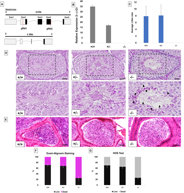
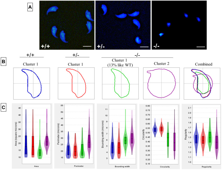
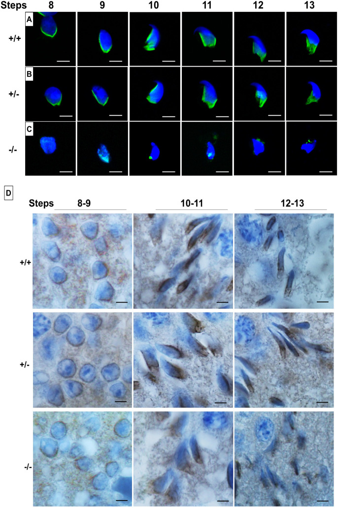
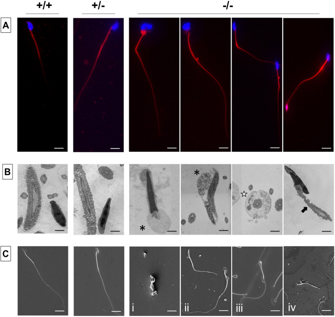
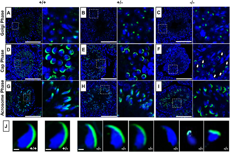
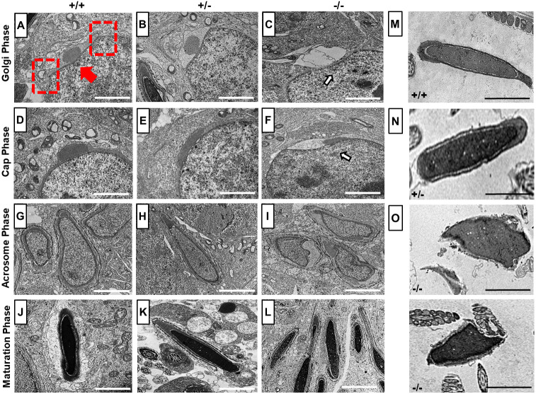
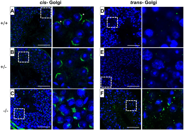
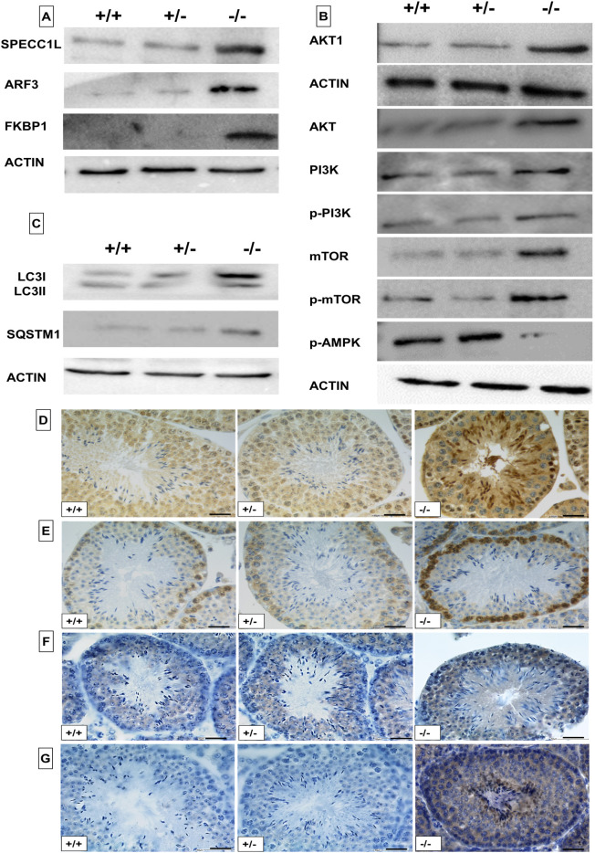
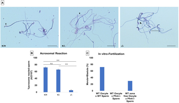
Profilin 4 (Pfn4) is expressed during spermiogenesis and localizes to the acrosome-acroplaxome-manchette complex. Here, we generated PFN4-deficient mice, with sperm displaying severe impairment in manchette formation. Interestingly, HOOK1 staining suggests that the perinuclear ring is established; however, ARL3 staining is disrupted, suggesting that lack of PFN4 does not interfere with the formation of the perinuclear ring and initial localization of HOOK1, but impedes microtubular organization of the manchette. Furthermore, amorphous head shape and flagellar defects were detected, resulting in reduced sperm motility. Disrupted cis- and trans-Golgi networks and aberrant production of proacrosomal vesicles caused impaired acrosome biogenesis. Proteomic analysis showed that the proteins ARF3, SPECC1L and FKBP1, which are involved in Golgi membrane trafficking and PI3K/AKT pathway, are more abundant in Pfn4-/- testes. Levels of PI3K, AKT and mTOR were elevated, whereas AMPK level was reduced, consistent with inhibition of autophagy. This seems to result in blockage of autophagic flux, which could explain the failure in acrosome formation. In vitro fertilization demonstrated that PFN4-deficient sperm is capable of fertilizing zona-free oocytes, suggesting a potential treatment for PFN4-related human infertility.

Keywords: Cis-Golgi; In vitro fertilization; Trans-Golgi; Acrosome biogenesis; Infertility; Manchette; Morula; PI3K/AKT signaling; Profilin 4; Spermiogenesis.


Conflict of interest statement

Competing interests The authors declare no competing or financial interests.

Figures

Fig. 1.
Fig. 1.
Generation and characterization of PFN4-deficient mice (Pfn4Δa). (A) Schematic of the Pfn4 locus and established PFN4-deficient mouse line. Red lines mark positions of the guide RNAs used for gene editing. (B) Validation of PFN4-deficient mice (Pfn4Δa). qRT-PCR was performed to check the relative expression of Pfn4 mRNA in murine testis of WT, Pfn4+/− and Pfn4−/− mice for the Pfn4Δa mutation. (C) Mating statistics of WT, Pfn4+/− and Pfn4−/− male mice (n=9/genotype). (D) PAS staining on WT, Pfn4+/− and Pfn4−/− testes sections. Dashed boxes indicate the areas shown at higher magnification below. Arrows indicate abnormal head morphology. (E) Hematoxylin and Eosin staining on WT, Pfn4+/− and Pfn4−/− cauda epididymis sections. (F) Quantification of E&N staining (n=3 biological replicates/genotype) of mature WT, Pfn4+/− and Pfn4−/− sperm isolated from cauda epididymis. (G) Hypo-osmotic swelling (HOS) test (n=3 biological replicates/genotype) of mature WT, Pfn4+/− and Pfn4−/− sperm isolated from cauda epididymis. Error bars represent mean±s.d. Scale bars: 10 μm.
Fig. 2.
Fig. 2.
Sperm head morphology analysis. Sperm head morphology analysis on WT, Pfn4+/− and Pfn4−/− sperm cells (n=3 biological replicates/genotype). In total, 901 nuclei were analyzed: 309 for WT (cluster 1), 276 for Pfn4+/− (cluster 1) and 541 for Pfn4−/− (86 sperm nuclei in cluster 1 and 455 nuclei in cluster 2). (A) Sperm cells stained with DAPI. (B) Head morphology of WT, Pfn4+/− and Pfn4−/− sperm cells. (C) Area, perimeter, regularity, bounding width, and circularity of WT, Pfn4+/− and Pfn4−/− sperm cells. Bonferroni's corrected P-values were calculated using the non-parametric Mann–Whitney–Wilcoxon test. Error bars show the mean±s.d. Box boundaries indicate consensus nucleus (overall examination of the population). Horizontal lines indicate mean of the respective parameter and error bars indicate coefficient of variability, standard error and standard deviation per parameter.
Fig. 3.
Fig. 3.
Manchette staining using α-tubulin and HOOK1 antibodies. (A-C) Immunofluorescence staining for the manchette using an α-tubulin antibody (green) on a germ cell population isolated from WT (A), Pfn4+/− (B) and Pfn4−/− (C) testes (n=3/genotype). Nuclei were stained with DAPI (blue). (D) IHC using an anti-HOOK1 antibody on WT, Pfn4+/− and Pfn4−/− testes sections. Scale bars: 20 µm.
Fig. 4.
Fig. 4.
Flagellar staining and ultrastructural analysis. (A-C) Flagellar staining and ultrastructural analysis using MitoRed (A), TEM (B) and SEM (C) of WT, Pfn4+/− and Pfn4−/− sperm isolated from the cauda epididymis (n=3 biological replicates/genotype). Black asterisks indicate cytoplasmic droplets in the midpiece, white star indicates a defective fibrous sheath, and black arrow indicates mitochondrial structural defects. Scale bars: 10 μm.
Fig. 5.
Fig. 5.
Acrosome analysis using PNA-FITC fluorescence staining. (A-I) Immunofluorescence staining for acrosome biogenesis on testes sections and mature sperm of WT, Pfn4+/− and Pfn4−/− mice (n=3 biological replicates/genotype). In the Golgi phase, the proacrosomal granule (green) was labeled by PNA-FITC in WT (A), Pfn4+/− (B) and Pfn4−/− (C) round spermatozoa. In the cap phase, acrosomal caps (green) were stained in WT (D), Pfn4+/− (E) and Pfn4−/− (F) round spermatozoa (white arrows show abnormal cap structures). In the acrosomal phase, PNA-FITC-labeled the acrosomal area in WT (G), Pfn4+/− (H) and Pfn4−/− (I) elongated spermatids. Dashed boxes indicate the areas enlarged to the right. (J) Immunofluorescence staining using PNA-FITC (green) on epididymal sperm cells of WT, Pfn4+/− and Pfn4−/− mice (n=3/genotype) to show the variety of Pfn4−/− sperm such as malformed acrosome and abnormal head morphology. Scale bars: 20 μm.
Fig. 6.
Fig. 6.
TEM analysis. (A-L) Ultrastructural analysis using TEM on testes sections of WT, Pfn4+/− and Pfn4−/− mice (n=3 biological replicates/genotype). (A-C) Golgi phase spermatozoa of WT (A), Pfn4+/− (B) Pfn4−/− (C) mice (arrow indicates deformed developing acrosome). Red arrow indicates the acroplaxome, red dashed rectangles indicate the marginal ring. (D-F) Cap phase spermatozoa of WT (D), Pfn4+/− (E) and Pfn4−/− (F) mice (arrow indicates abnormal cap-like structures). (G-I) Acrosomal phase spermatids of WT (G), Pfn4+/− (H) and Pfn4−/− (I) mice. (J-L) Maturation phase spermatids of WT (J), Pfn4+/− (K) and Pfn4−/− (L) mice. (M-O) Ultrastructural analysis of cauda epididymal sperms of WT (M), Pfn4+/− (N) and Pfn4−/− (O) mice. Scale bars: 2 μm.
Fig. 7.
Fig. 7.
Fluorescence staining of the Golgi network. Cis- and trans-Golgi immunofluorescence staining using GM130 (green) and TGN46 (green) antibodies and nuclei stained with DAPI (blue) on WT, Pfn4+/− and Pfn4−/− (n=3/genotype) testes sections. (A-C) WT (A), Pfn4+/− (B) and Pfn4−/− (C) spermatozoa stained for the cis-Golgi compartment (asterisks). (D-F) WT (D), Pfn4+/− (E) and Pfn4−/− (F) spermatozoa stained for the trans-Golgi network. Dashed boxes indicate the areas enlarged to the right. Scale bars: 50 μm.
Fig. 8.
Fig. 8.
Analysis of autophagy regulators. Western blot analysis and immunohistochemical staining on WT, Pfn4+/− and Pfn4−/− mice (n=3/genotype). (A) Western blot using SPECC1L, ARF3 and FKBP1 antibodies on protein extracts from WT, Pfn4+/− and Pfn4−/− testes. (B) Western blot using AKT, AKT1, PI3K, p-PI3K, mTOR, p-mTOR and p-AMPK antibodies on protein extracts from WT, Pfn4+/− and Pfn4−/− testes. (C) Western blot using LC3I/II and SQSTM1 antibodies on protein extracts from WT, Pfn4+/− and Pfn4−/− testes. (D-G) IHC using LC3I/II (D), SQSTM1 (E), AKT1 (F) and ARF3 (G) antibodies on WT, Pfn4+/− and Pfn4−/− testes sections. Scale bars: 50 μm.
Fig. 9.
Fig. 9.
Acrosomal reaction and in vitro fertilization. Calcium ionophore-induced acrosomal reaction in WT, Pfn4+/− and Pfn4−/− sperm cells (n=3/genotype). (A) Coomassie-stained sperm cells of WT, Pfn4+/− and Pfn4−/−. Black arrows indicate that a successful acrosomal reaction took place. Black arrowhead indicates a crescent-shaped acrosome on a sperm head, indicating that the acrosomal reaction did not take place. Scale bars: 20 μm. (B) Percentage of acrosomal-reacted sperm WT, Pfn4+/− and Pfn4−/− (n=3 biological replicates/genotype). ***P<0.0005, one-tailed, paired Student's t-test. P-value relates to acrosomal-reacted WT and Pfn4+/− sperm compared with Pfn4−/− sperm. (C) Percentage of oocytes developing into blastocysts after fertilization of oocytes with WT and Pfn4−/− sperm and zona-free oocytes with Pfn4−/− sperm (n=1/genotype).

References

    1. Alkanderi, S., Molinari, E., Shaheen, R., Elmaghloob, Y., Stephen, L. A., Sammut, V., Ramsbottom, S. A., Srivastava, S., Cairns, G., Edwards, N.et al. (2018). ARL3 mutations cause Joubert syndrome by disrupting ciliary protein composition. Am. J. Hum. Genet. 103, 612-620. 10.1016/j.ajhg.2018.08.015 - DOI - PMC - PubMed
    1. Behnen, M., Murk, K., Kursula, P., Cappallo-Obermann, H., Rothkegel, M., Kierszenbaum, A. L. and Kirchhoff, C. (2009). Testis-expressed profilins 3 and 4 show distinct functional characteristics and localize in the acroplaxome-manchette complex in spermatids. BMC Cell Biol. 10, 34. 10.1186/1471-2121-10-34 - DOI - PMC - PubMed
    1. Calvi, A., Wong, A. S. W., Wright, G., Wong, E. S. M., Loo, T. H., Stewart, C. L. and Burke, B. (2015). SUN4 is essential for nuclear remodeling during mammalian spermiogenesis. Dev. Biol. 407, 321-330. 10.1016/j.ydbio.2015.09.010 - DOI - PubMed
    1. Chapin, R. E., Wine, R. N., Harris, M. W., Borchers, C. H. and Haseman, J. K. (2001). Structure and control of a cell-cell adhesion complex associated with spermiation in rat seminiferous epithelium. J. Androl. 22, 1030-1052. 10.1002/j.1939-4640.2001.tb03444.x - DOI - PubMed
    1. Chen, C., Bao, H., Lin, W., Chen, X., Huang, Y., Wang, H., Yang, Y., Liu, J., Lv, X. and Teng, L. (2022). ASF1b is a novel prognostic predictor associated with cell cycle signaling pathway in gastric cancer. J. Cancer 13, 1985-2000. 10.7150/jca.69544 - DOI - PMC - PubMed

Publication types