

Microglia-dependent remodeling of neuronal circuits
- PMID: 35950924
- PMCID: PMC9826178
- DOI: 10.1111/jnc.15689
Microglia-dependent remodeling of neuronal circuits
Abstract
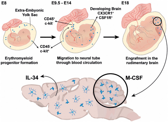
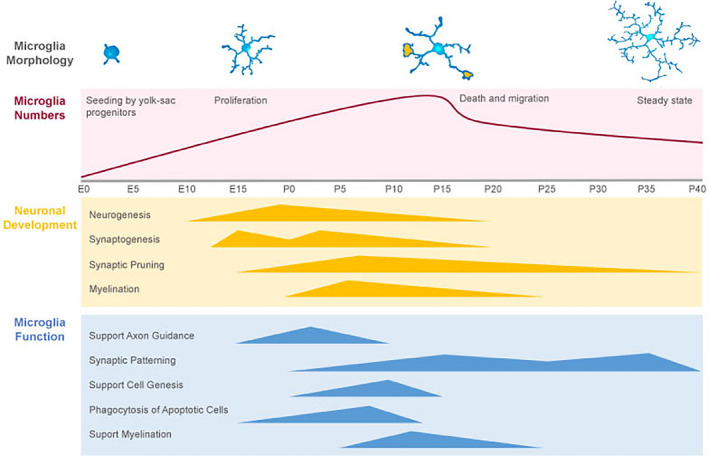
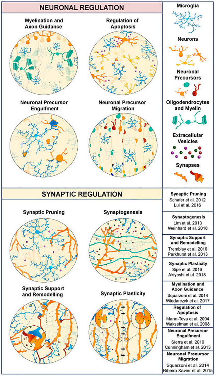
Microglia are tissue-resident macrophages responsible for the surveillance, neuronal support, and immune defense of the brain parenchyma. Recently, the role played by microglia in the formation and function of neuronal circuits has garnered substantial attention. During development, microglia have been shown to engulf neuronal precursors and participate in pruning mechanisms while, in the mature brain, they influence synaptic signaling, provide trophic support and shape synaptic plasticity. Recently, studies have unveiled different microglial characteristics associated with specific brain regions. This emerging view suggests that the maturation and function of distinct neuronal circuits may be potentially associated with the molecular identity microglia adopts across the brain. Here, we review and summarize the known role of these cells in the thalamus, hippocampus, cortex, and cerebellum. We focus on in vivo studies to highlight the characteristics of microglia that may be important in the remodeling of these neuronal circuits and in relation to neurodevelopmental and neuropsychiatric disorders.
Keywords: brain wiring; cerebellum; cortex; hippocampus; microglia; neurodevelopment; thalamus.
© 2022 The Authors. Journal of Neurochemistry published by John Wiley & Sons Ltd on behalf of International Society for Neurochemistry.
Conflict of interest statement
The authors have no conflicts of interest to declare that are relevant to the content of this article.
Figures
References
-
- Aavani, T. , Rana, S. A. , Hawkes, R. , & Pittman, Q. J. (2015). Maternal immune activation produces cerebellar hyperplasia and alterations in motor and social behaviors in male and female mice. Cerebellum, 14, 491–505. - PubMed
-
- Arnò, B. , Grassivaro, F. , Rossi, C. , Bergamaschi, A. , Castiglioni, V. , Furlan, R. , Greter, M. , Favaro, R. , Comi, G. , Becher, B. , Martino, G. , & Muzio, L. (2014). Neural progenitor cells orchestrate microglia migration and positioning into the developing cortex. Nature Communications, 5, 5611. - PubMed
-
- Ashwell, K. (1990). Microglia and cell death in the developing mouse cerebellum. Brain Research Developmental Brain Research, 55, 219–230. - PubMed
-
- Askew, K. , Li, K. , Olmos‐Alonso, A. , Garcia‐Moreno, F. , Liang, Y. , Richardson, P. , Tipton, T. , Chapman, M. A. , Riecken, K. , Beccari, S. , Sierra, A. , Molnár, Z. , Cragg, M. S. , Garaschuk, O. , Perry, V. H. , & Gomez‐Nicola, D. (2017). Coupled proliferation and apoptosis maintain the rapid turnover of microglia in the adult brain. Cell Reports, 18, 391–405. - PMC - PubMed
Publication types
MeSH terms
LinkOut - more resources
Full Text Sources
