. 2022 Aug 12;377(6607):eabm4096.
doi: 10.1126/science.abm4096. Epub 2022 Aug 12.

Prokaryotic innate immunity through pattern recognition of conserved viral proteins

Affiliations

Prokaryotic innate immunity through pattern recognition of conserved viral proteins

Linyi Alex Gao et al. Science. .

Abstract

Many organisms have evolved specialized immune pattern-recognition receptors, including nucleotide-binding oligomerization domain-like receptors (NLRs) of the STAND superfamily that are ubiquitous in plants, animals, and fungi. Although the roles of NLRs in eukaryotic immunity are well established, it is unknown whether prokaryotes use similar defense mechanisms. Here, we show that antiviral STAND (Avs) homologs in bacteria and archaea detect hallmark viral proteins, triggering Avs tetramerization and the activation of diverse N-terminal effector domains, including DNA endonucleases, to abrogate infection. Cryo-electron microscopy reveals that Avs sensor domains recognize conserved folds, active-site residues, and enzyme ligands, allowing a single Avs receptor to detect a wide variety of viruses. These findings extend the paradigm of pattern recognition of pathogen-specific proteins across all three domains of life.


Conflict of interest statement

Competing interests: F.Z. is a scientific advisor and cofounder of Editas Medicine, Beam Therapeutics, Pairwise Plants, Arbor Biotechnologies, and Sherlock Biosciences.

Figures

Fig. 1.
Fig. 1.
Prokaryotic STAND NTPases recognize phage terminase and portal proteins. (A) Maximum likelihood tree of the ATPase domain of selected NLR-like STAND NTPases in four model organisms across kingdoms of life. (B) Domain architectures of representative NLR-like genes in (A). LRR, leucine-rich repeat; TPR, tetratricopeptide repeat; WD40, WD40 repeat; ankyrin, ankyrin repeat; BIR, baculoviral inhibitor of apoptosis repeat; PYD, pyrin domain; FIIND, function to find domain; CARD, caspase activation and recruitment domain; RX-CC, potato virus X resistance protein coiled-coil domain; PLP, patatin-like phospholipase; TIR, toll/interleukin-1 receptor homology domain. (C) Schematic of genetic screening approach to identify phage-encoded activators of Avs proteins that induce cell death. (D) Genetic screen results for phage-encoded activators. (E) Quantification of the phage DNA band intensity in a Southern blot of DNA isolated from phage-infected E. coli. (F) Photographs of E. coli co-transformation assays with Avs genes and phage activators identified in (D).
Fig. 2.
Fig. 2.
Avs proteins are pattern-recognition receptors for the terminase and portal of diverse tailed phages. (A) Schematic of plasmid depletion assay. (B) Heatmaps of plasmid depletion for the terminase and portal proteins of representative phages spanning nine major tailed phage families. The native Avs promoter was retained for all homologs except for those outside of the Enterobacteriaceae family (EpAvs1 and CcAvs4). Terminases and portals were induced with 0.002% arabinose. Horizontal black bars indicate groups of terminase proteins with at least 20% pairwise sequence identity. Asterisks indicate prophages. S. flava, Sphingopyxis flava R11H; D. archaeon, Desulfurococcales archaeon ex4484_217_2; E. coli-1, Escherichia coli NCTC9020; E. coli-2, Escherichia coli M885. (C) Pairwise amino acid sequence identity between the core folds of the terminases and portals in (B), excluding non-conserved regions. (D) Activity of four Avs proteins against the human herpesvirus 8 (HHV-8) terminase and portal.
Fig. 3.
Fig. 3.
SeAvs3 and EcAvs4 are phage-activated DNA endonucleases. (A) Domain architecture of SeAvs3 and EcAvs4. (B) Alignment of Avs D-QxK nuclease motifs with characterized Cap4 and Mrr representatives. (C, D) Agarose gel analysis of SeAvs3 nuclease function in vitro with a linear dsDNA substrate and (E) cofactor requirements. (F, G) Agarose gel analysis of EcAvs4 nuclease function in vitro with a linear dsDNA substrate and (H) cofactor requirements.
Fig. 4.
Fig. 4.
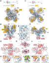
Cryo-EM structures of SeAvs3 and EcAvs4 in complex with their cognate triggers. (A, B) Structure of the SeAvs3-terminase complex. (C, D) Structure of the EcAvs4-portal complex. (E, F) ATP molecule in the STAND ATPase active site of EcAvs4 and SeAvs3. Cryo-EM density is shown as a transparent surface. (G) SeAvs3 Cap4-like nuclease effector domain. (H, I) Active sites for the inward and outward facing protomers of the SeAvs3 Cap4-like nuclease. (J) Equivalent view of the active site of HindIII bound to target DNA with two divalent metal ions (PDB 3A4K). (K) Electrostatic surface potential for the SeAvs3 Cap4-like nuclease and the EcAvs4 Mrr-like nuclease. Active sites are indicated by purple circles. Ideal B-form DNA is modeled on both surfaces based on the crystal structure of HindIII bound to its target (PDB 3A4K). (L) EcAvs4 Mrr-like nuclease effector domain. (M, N) Active sites for the inward and outward facing protomers of the EcAvs4 Mrr-like nuclease. TPR, tetratricopeptide repeat.
Fig. 5.
Fig. 5.
Structural basis for viral fold recognition by SeAvs3 and EcAvs4. (A) The interface between SeAvs3 and the PhiV-1 terminase. An SeAvs3 surface view is shown in transparency. SeAvs3 is colored from N to C terminus according to the key. (B) AlphaFold or crystal structures of different terminases modeled into SeAvs3. The ATPase and nuclease domains were individually aligned to the PhiV-1 terminase domains. (C, D) Recognition of the PhiV-1 terminase ATPase and nuclease active sites by the SeAvs3 TPR domain. (E) Sequence logos for terminase ATPase Walker A motifs and terminase nuclease active sites. A total of 11,000 terminase sequences were clustered at 30% sequence identity, and motifs were extracted from clusters containing terminases targeted or not targeted by SeAvs3 according to Fig. 2B (see also fig. S21). (F) Plasmid depletion assay for SeAvs3 co-expressed in E. coli with a terminase ATPase or nuclease domain harboring active site mutations. (G) The interface between EcAvs4 and the PhiV-1 portal. An EcAvs4 surface view is shown in transparency. EcAvs4 is colored from N to C terminus according to the key. (H) β-sheet augmentation between EcAvs4 and the portal clip domain. (I) Comparison of the EcAvs4-bound state of the PhiV-1 portal, the cryo-EM structure of the highly homologous T7 portal in its native virion, and AlphaFold models of diverse portals. A top view of the assembled dodecamer of the T7 portal is also shown. TPR, tetratricopeptide repeat; CTD, C-terminal domain.
Fig. 6.
Fig. 6.
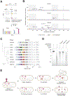
Taxonomic distribution and domain architectures of Avs families. (A) Distribution of avs genes across phyla. The values above the bars indicate the number and percentage of genomes containing each gene. (B) Number of bacterial and archaeal phyla (minimum 100 sequenced isolates) with at least one detected instance of an avs gene. (C) Kernel density plots of the length distribution of Avs proteins, excluding the N-terminal domain. The red lines indicate medians. ****p < 0.0001 (Mann-Whitney). Maximum likelihood tree of representatives of the ATPase + C-terminal domain of (D) Avs2 terminase sensors (n = 1,255) and (E) Avs4 portal sensors (n = 1,089) clustered at 95% sequence identity. See fig. S24 for the trees for Avs1 and Avs3. Stars on the outer ring indicate homologs investigated experimentally in this study. MBL, metallo-β-lactamase; REase, restriction endonuclease; TIR, Toll/interleukin-1 receptor homology domain; SIR2, sirtuin; CMP, cytidine monophosphate; HTH, helix-turn-helix. (F) Anti-phage defense activity of a chimeric Avs4 with transmembrane N-terminal helices from Sulfurospirillum sp. replacing the nuclease domain of EcAvs4.
Fig. 7:
Fig. 7:
Phage-encoded genes inhibit Avs activity. (A) Schematic of a pooled screen in E. coli for phage early genes that rescue Avs-mediated toxicity. (B) Deep sequencing readout of anti-defense candidate genes co-expressed with SeAvs3, EcAvs4, or KpAvs4. (C) A hypervariable early gene locus within a closely related set of Autographiviridae phages contains abundant anti-defense genes. The tree was constructed from a concatenated alignment of conserved proteins present in all ten phages. Colors represent groups of proteins clustered at 40% sequence identity at 70% coverage. (D) In vitro reconstitution of anti-SeAvs3 activity by three anti-defense candidates. (E) Schematic of the mechanism of Avs proteins as anti-phage pattern recognition receptors.

Comment in

References

    1. Makarova KS, Wolf YI, Snir S, Koonin EV, Defense islands in bacterial and archaeal genomes and prediction of novel defense systems. J. Bacteriol 193, 6039–6056 (2011). - PMC - PubMed
    1. Koonin EV, Makarova KS, Wolf YI, Evolutionary Genomics of Defense Systems in Archaea and Bacteria. Annu. Rev. Microbiol 71, 233–261 (2017). - PMC - PubMed
    1. Doron S, Melamed S, Ofir G, Leavitt A, Lopatina A, Keren M, Amitai G, Sorek R, Systematic discovery of antiphage defense systems in the microbial pangenome. Science. 359 (2018), doi:10.1126/science.aar4120. - DOI - PMC - PubMed
    1. Gao L, Altae-Tran H, Böhning F, Makarova KS, Segel M, Schmid-Burgk JL, Koob J, Wolf YI, Koonin EV, Zhang F, Diverse enzymatic activities mediate antiviral immunity in prokaryotes. Science. 369, 1077–1084 (2020). - PMC - PubMed
    1. Hampton HG, Watson BNJ, Fineran PC, The arms race between bacteria and their phage foes. Nature. 577, 327–336 (2020). - PubMed

MeSH terms