Multicenter Study
. 2022 Nov 21;145(11):3859-3871.
doi: 10.1093/brain/awac224.

Interpretable surface-based detection of focal cortical dysplasias: a Multi-centre Epilepsy Lesion Detection study

Hannah Spitzer  1 Mathilde Ripart  2 Kirstie Whitaker  3 Felice D'Arco  4 Kshitij Mankad  4 Andrew A Chen  5   6 Antonio Napolitano  7 Luca De Palma  8 Alessandro De Benedictis  9 Stephen Foldes  10 Zachary Humphreys  10 Kai Zhang  11 Wenhan Hu  11 Jiajie Mo  11 Marcus Likeman  12 Shirin Davies  13   14 Christopher Güttler  15 Matteo Lenge  16 Nathan T Cohen  17 Yingying Tang  18   19 Shan Wang  19   20 Aswin Chari  2   4 Martin Tisdall  2   4 Nuria Bargallo  21   22 Estefanía Conde-Blanco  23 Jose Carlos Pariente  23 Saül Pascual-Diaz  23 Ignacio Delgado-Martínez  24 Carmen Pérez-Enríquez  25 Ilaria Lagorio  26 Eugenio Abela  27 Nandini Mullatti  28 Jonathan O'Muircheartaigh  28   29 Katy Vecchiato  29   30 Yawu Liu  31 Maria Eugenia Caligiuri  32 Ben Sinclair  33 Lucy Vivash  33   34 Anna Willard  33 Jothy Kandasamy  35 Ailsa McLellan  35 Drahoslav Sokol  35 Mira Semmelroch  36 Ane G Kloster  37 Giske Opheim  37   38 Letícia Ribeiro  39   40 Clarissa Yasuda  39   40 Camilla Rossi-Espagnet  41 Khalid Hamandi  13   42 Anna Tietze  15 Carmen Barba  16 Renzo Guerrini  16 William Davis Gaillard  17 Xiaozhen You  17 Irene Wang  19 Sofía González-Ortiz  43   44 Mariasavina Severino  26 Pasquale Striano  26   45 Domenico Tortora  26 Reetta Kälviäinen  31   46 Antonio Gambardella  47 Angelo Labate  48 Patricia Desmond  49 Elaine Lui  49 Terence O'Brien  33   50 Jay Shetty  35 Graeme Jackson  51   52 John S Duncan  53 Gavin P Winston  53   54 Lars H Pinborg  37   55 Fernando Cendes  39   40 Fabian J Theis  1   56 Russell T Shinohara  57 J Helen Cross  2   58 Torsten Baldeweg  2   4 Sophie Adler  2 Konrad Wagstyl  2   59
Affiliations
Multicenter Study

Interpretable surface-based detection of focal cortical dysplasias: a Multi-centre Epilepsy Lesion Detection study

Hannah Spitzer et al. Brain. .

Abstract

One outstanding challenge for machine learning in diagnostic biomedical imaging is algorithm interpretability. A key application is the identification of subtle epileptogenic focal cortical dysplasias (FCDs) from structural MRI. FCDs are difficult to visualize on structural MRI but are often amenable to surgical resection. We aimed to develop an open-source, interpretable, surface-based machine-learning algorithm to automatically identify FCDs on heterogeneous structural MRI data from epilepsy surgery centres worldwide. The Multi-centre Epilepsy Lesion Detection (MELD) Project collated and harmonized a retrospective MRI cohort of 1015 participants, 618 patients with focal FCD-related epilepsy and 397 controls, from 22 epilepsy centres worldwide. We created a neural network for FCD detection based on 33 surface-based features. The network was trained and cross-validated on 50% of the total cohort and tested on the remaining 50% as well as on 2 independent test sites. Multidimensional feature analysis and integrated gradient saliencies were used to interrogate network performance. Our pipeline outputs individual patient reports, which identify the location of predicted lesions, alongside their imaging features and relative saliency to the classifier. On a restricted 'gold-standard' subcohort of seizure-free patients with FCD type IIB who had T1 and fluid-attenuated inversion recovery MRI data, the MELD FCD surface-based algorithm had a sensitivity of 85%. Across the entire withheld test cohort the sensitivity was 59% and specificity was 54%. After including a border zone around lesions, to account for uncertainty around the borders of manually delineated lesion masks, the sensitivity was 67%. This multicentre, multinational study with open access protocols and code has developed a robust and interpretable machine-learning algorithm for automated detection of focal cortical dysplasias, giving physicians greater confidence in the identification of subtle MRI lesions in individuals with epilepsy.

Keywords: epilepsy; focal cortical dysplasia; machine learning; structural MRI.


Figures

Figure 1
Figure 1
MELD processing pipeline. (A) Local sites extract surface-based morphological features from structural T1 and FLAIR MRI, along with manually delineated lesion masks. These were coregistered to a symmetric template surface and anonymized data matrices are shared with the MELD team. (B) Central preprocessing: the MELD team carried out outlier detection and data harmonization to minimize interscanner feature differences. (C) Morphological features underwent intrasubject, interhemispheric and intersubject normalization. (D) The full cohort was randomly subdivided 50:50 into training/validation cohorts and withheld test cohort. To avoid overfitting, all optimization experiments were carried out on the training/validation cohort prior to final testing on the test cohort and new site cohorts. (E) The neural network classifier was trained to identify lesional vertices from MRI features. Vertex-wise predictions were collected into connected clusters. (F) Classifier predictions mapped to cortical surfaces, lesional features and their relative saliency were plotted; lesional features across the cohort were analysed.
Figure 2
Figure 2
Non-linear 2D UMAP embedding of lesional T1 features. (A) Manual lesion masks of patients (black) compared to equivalent cortex on healthy controls (grey). Lesions differ from control cortex and exhibit different patterns of structural abnormality. (B) Data-driven clustering of UMAP embedding reveals three distinct groups of lesions. Colour-associated pie charts describe the proportion of each histopathological subtype present in each group. (C) Patient lesions coloured by intra- and intersubject normalized features. Group 1 is predominantly FCD IIA and IIB, along with unoperated patients. It is characterized by increased intrinsic curvature, increased cortical thickness, decreased grey–white matter contrast, bottom of sulcus and increased FLAIR in the white matter. Group 2 is characterized by increased intrinsic curvature, decreased grey–white matter contrast and decreased intracortical FLAIR. It contains proportionally more FCD I and III lesions. Group 3 largely overlaps healthy control clusters. Lesional features in this cluster are more heterogeneous and less extreme.
Figure 3
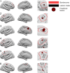
Figure 3
Neural network predictions. Classifier predictions for six patients are displayed. Patients 1–4 are examples where the classifier has correctly identified the lesion. In Patient 4 there is an additional cluster in the left insula. Patient 5 is an example where the classifier detects an area in the border zone. Patient 6 is an example of where the neural network has not identified the lesion. An additional cluster is detected in the right post-central gyrus. Left column = lateral view, middle column = medial view, right column = enlarged view around lesion mask. Black = lesion mask; red = border zone; burgundy = classifier-predicted clusters.
Figure 4
Figure 4
UMAP embedding of classifier predictions. (A) Data-driven clustering of UMAP embedding of lesional T1 features reveals three distinct groups of lesions. (B) True positive and false positive clusters derived from the neural network superimposed on A. Feature values in true positive and false positive clusters are similar to either group 1 or 2. Clusters are not similar to healthy cortex or group 3. (C) Predicted clusters overlapping lesion masks from group 3 lesions are superimposed. The feature values in the predicted clusters are similar to group 1 or 2, i.e. the network has identified vertices exhibiting characteristically abnormal MRI features in FCD.
Figure 5
Figure 5
Individual patient reports. Example classifier predictions with saliency scores for ‘Patient 1’ (an example with FLAIR data) and ‘Patient 2’ (without FLAIR data). (A) Classifier predictions (dark red) and manual lesion mask (black line) visualized on brain surfaces (only lesional hemisphere is shown). Classifier predictions (dark red) visualized on T1 volume. (B) Z-scored mean feature values within predicted lesions coloured with Integrated Gradients saliency scores. Positive saliency scores indicate feature values driving the classifier’s ‘lesion’ prediction. Negative scores indicate feature values that are inconsistent with the prediction. (C) Lesional cortex highlighted on the patients’ MRI scans exhibit salient features automatically identified by the classifier.

References

    1. Bien CG, Szinay M, Wagner J, Clusmann H, Becker AJ, Urbach H. Characteristics and surgical outcomes of patients with refractory magnetic resonance imaging-negative epilepsies. Arch Neurol. 2009;66(12):1491–1499. - PubMed
    1. McGonigal A, Bartolomei F, Régis J, et al. . Stereoelectroencephalography in presurgical assessment of MRI-negative epilepsy. Brain. 2007;130(Pt 12):3169–3183. - PubMed
    1. Colombo N, Tassi L, Deleo F, et al. . Focal cortical dysplasia type IIa and IIb: MRI aspects in 118 cases proven by histopathology. Neuroradiology. 2012;54(10):1065–1077. - PubMed
    1. Irene Wang Z, Alexopoulos AV, Jones SE, Jaisani Z, Najm IM, Prayson RA. The pathology of magnetic-resonance-imaging-negative epilepsy. Mod Pathol. 2013;26(8):1051–1058. - PubMed
    1. Téllez-Zenteno JF, Hernández Ronquillo L, Moien-Afshari F, Wiebe S. Surgical outcomes in lesional and non-lesional epilepsy: A systematic review and meta-analysis. Epilepsy Res. 2010;89(2-3):310–318. - PubMed

Publication types

MeSH terms