. 2022 Sep 14;42(37):7060-7076.
doi: 10.1523/JNEUROSCI.0071-22.2022. Epub 2022 Aug 11.

Crossed Corticostriatal Projections in the Macaque Brain

Affiliations

Crossed Corticostriatal Projections in the Macaque Brain

Elena Borra et al. J Neurosci. .

Abstract

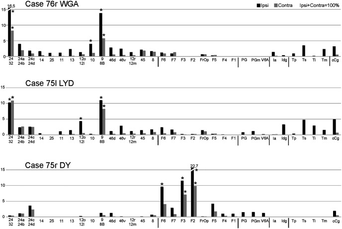
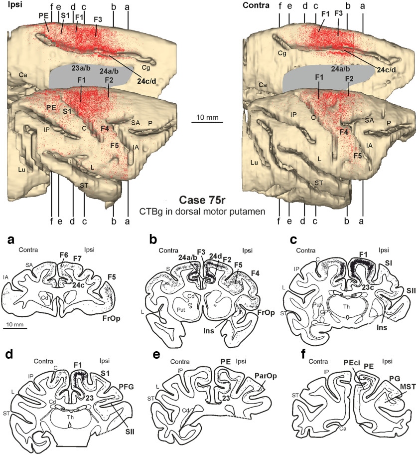
In nonhuman primates, major input to the striatum originates from ipsilateral cortex and thalamus. The striatum is a target also of crossed corticostriatal (CSt) projections from the contralateral hemisphere, which have been so far somewhat neglected. In the present study, based on neural tracer injections in different parts of the striatum in macaques of either sex, we analyzed and compared qualitatively and quantitatively the distribution of labeled CSt cells in the two hemispheres. The results showed that crossed CSt projections to the caudate and the putamen can be relatively robust (up to 30% of total labeled cells). The origin of the direct and the crossed CSt projections was not symmetrical as the crossed ones originated almost exclusively from motor, prefrontal, and cingulate areas and not from parietal and temporal areas. Furthermore, there were several cases in which the contribution of contralateral areas tended to equal that of the ipsilateral ones. The present study is the first detailed description of this anatomic pathway of the macaque brain and provides the substrate for bilateral distribution of motor, motivational, and cognitive signals for reinforcement learning and selection of actions or action sequences, and for learning compensatory motor strategies after cortical stroke.SIGNIFICANCE STATEMENT In nonhuman primates the striatum is a target of projections originating from the contralateral hemisphere (crossed CSt projections), which have been so far poorly investigated. The present study analyzed qualitatively and quantitatively in the macaque brain the origin of the crossed CSt projections compared with those originating from the ipsilateral hemisphere. The results showed that crossed CSt projections originate mostly from frontal and rostral cingulate areas and in some cases their contribution tended to equal that from ipsilateral areas. These projections could provide the substrate for bilateral distribution of motor, motivational, and cognitive signals for reinforcement learning and action selection, and for learning compensatory motor strategies after cortical stroke.

Keywords: basal ganglia; cingulate cortex; frontal cortex; interhemispheric transfer; monkey; striatum.


Figures

Figure 1.
Figure 1.
Location of the injection sites shown in drawings of coronal sections and in brightfield (for WGA and LYD injections) and epifluorescence (for CTBg, DY, and FB injections) photomicrographs. In the drawings, all injection sites except for WGA are depicted as a black zone corresponding to the core, surrounded by a gray zone, corresponding to the halo. WGA injection sites are depicted as a gray zone because of the poor definition of the core versus the halo. Calibration bars shown for the section drawing and the photomicrograph of Case 75l LYD apply to all section drawings and photomicrographs, respectively. C, Central sulcus; Cd, caudate nucleus; Cg, cingulate sulcus; Cla, claustrum; GP, globus pallidus; IA, inferior arcuate sulcus; L, lateral sulcus; Put, putamen; S, spur of the arcuate sulcus; SA, superior arcuate sulcus; ST, superior temporal sulcus.
Figure 2.
Figure 2.
Percentage of areal distribution of the total retrograde labeling observed in the ipsilateral hemisphere (black) compared with that of the total retrograde labeling observed in the contralateral hemisphere (gray) in all the cases of the present study. In each graph, areas are ordered based on the amount of ipsilateral labeling (only areas with ipsilateral labeling >1%). Superior parietal (SPL) areas and inferior parietal (IPL) areas are grouped. The results of the statistical analysis are reported in which the frequency distributions of the labeled CSt cells per area in the ipsilateral and the contralateral hemisphere were compared (Pearson chi-square test for independence). cCg, Caudal cingulate cortex (areas 23, 31, 29, 30); DF, degree of freedom; Ia, agranular insula; Idg, disgranular insula.
Figure 3.
Figure 3.
Distribution of the retrograde labeling observed after injection of WGA in the head of the caudate in Case 76r. The labeling is shown in dorsolateral and medial views of the 3D reconstructions of the injected (ipsilateral) and the contralateral hemisphere and in drawings of coronal sections. For the sake of comparison, in this and in the subsequent figures, all the 3D reconstructions are shown as a right hemisphere with the injected hemisphere on the left and all drawings with the injected hemisphere on the right. a–f, Sections are shown in a rostral to caudal order. The levels at which the sections were taken are shown on the 3D reconstructions of both hemispheres. Each dot corresponds to one labeled neuron. C, Central sulcus; Ca, calcarine fissure; Cd, caudate nucleus; Cg, cingulate sulcus; GP, globus pallidus; IA, inferior arcuate sulcus; Ins, insula; IP, intraparietal sulcus; L, lateral sulcus; LO, lateral orbital sulcus; Lu, lunate sulcus; MO, medial orbital sulcus; Opt, occipito-temporo-parietal area; P, principal sulcus; Put, putamen; SA, superior arcuate sulcus; ST, superior temporal sulcus; Th, thalamus.
Figure 4.
Figure 4.
Distribution of the retrograde labeling observed after injection of LYD in the head of the caudate in Case 75l. The labeling is shown in dorsolateral and medial views of the 3D reconstructions of the injected (ipsilateral) and contralateral hemispheres and in drawings of coronal sections. C, Central sulcus; Ca, calcarine fissure; Cd, caudate nucleus; Cg, cingulate sulcus; GP, globus pallidus; IA, inferior arcuate sulcus; Ins, insula; IP, intraparietal sulcus; L, lateral sulcus; LO, lateral orbital sulcus; Lu, lunate sulcus; MO, medial orbital sulcus; P, principal sulcus; ParOp, parietal operculum; Put, putamen; SA, superior arcuate sulcus; ST, superior temporal sulcus; Th, thalamus. Conventions are defined in the legend to Figure 3.
Figure 5.
Figure 5.
Percentage distribution of the total (ipsi plus contra) retrograde labeling in ipsilateral (black) and contralateral (gray) areas observed after the tracer injections in the caudate. The asterisks indicate the five most labeled areas. cCg, Caudal cingulate cortex (areas 23, 31, 29, 30); Ia, agranular insula; Idg, disgranular insula.
Figure 6.
Figure 6.
Distribution of the retrograde labeling observed after injection of DY in the caudate body in Case 75r. The labeling is shown in dorsolateral and medial views of the 3D reconstructions of the injected (ipsilateral) and contralateral hemisphere and in drawings of coronal sections. AIP, anterior intraparietal area; C, Central sulcus; Ca, calcarine fissure; Cd, caudate nucleus; Cg, cingulate sulcus; IA, inferior arcuate sulcus; IP, intraparietal sulcus; L, lateral sulcus; LO, lateral orbital sulcus; Lu, lunate sulcus; MST, medial superior temporal area; MO, medial orbital sulcus; P, principal sulcus; PO, parieto occipital sulcus; Put, putamen; S, spur of the arcuate sulcus; SA, superior arcuate sulcus; ST, superior temporal sulcus. Conventions as in Figure 3.
Figure 7.
Figure 7.
Distribution of the retrograde labeling observed after injection of WGA in the precommissural putamen in Case 77r. The labeling is shown in dorsolateral and medial views of the 3D reconstructions of the injected (ipsilateral) and contralateral hemisphere and in drawings of coronal sections. AIP, anterior intraparietal area; C, central sulcus; Ca, calcarine fissure; Cd, caudate nucleus; Cg, cingulate sulcus; GP, globus pallidus; IA, inferior arcuate sulcus; Ins, insula; IP, intraparietal sulcus; L, lateral sulcus; LO, lateral orbital sulcus; Lu, lunate sulcus; MO, medial orbital sulcus; P, principal sulcus; ParOp, parietal operculum; Put, putamen; SA, superior arcuate sulcus; S, spur of the arcuate sulcus; ST, superior temporal sulcus; Th, thalamus. Conventions as in Figure 3.
Figure 8.
Figure 8.
Percentage distribution of the total (ipsi plus contra) retrograde labeling in the ipsilateral (black) and contralateral (gray) areas observed after the tracer injections in the putamen. The asterisks indicate the five most labeled areas. AIP, anterior intraparietal area; cCg, caudal cingulate cortex (areas 23, 31, 29, 30); Ia, agranular insula; Idg, disgranular insula; ParOp, parietal operculum.
Figure 9.
Figure 9.
Distribution of the retrograde labeling observed after injection of CTBg in the dorsal part of the motor putamen in Case 75r. The labeling is shown in dorsolateral and medial views of the 3D reconstructions of the injected (ipsilateral) and contralateral hemisphere and in drawings of coronal sections. C, central sulcus; Ca, calcarine fissure; Cd, caudate nucleus; Cg, cingulate sulcus; GP, globus pallidus; IA, inferior arcuate sulcus; Ins, insula; IP, intraparietal sulcus; L, lateral sulcus; LO, lateral orbital sulcus; Lu, lunate sulcus; MO, medial orbital sulcus; MST, medial superior temporal area; P, principal sulcus; ParOp, parietal operculum; PO, parieto occipital sulcus; Put, putamen; S, spur of the arcuate sulcus; SA, superior arcuate sulcus; ST, superior temporal sulcus; Th, thalamus. Conventions as in Figure 3.
Figure 10.
Figure 10.
Distribution of the retrograde labeling observed after injection of CTBg in Case 77l, DY in Case 71r, and FB in 71l in the midventral motor putamen, shown in dorsolateral and medial views of the 3D reconstructions of the injected (ipsilateral) and contralateral hemispheres. On the 3D reconstructions of both hemispheres of all cases are indicated the levels at which the sections, shown in Figure 11, were taken. C, Central sulcus; Cg, cingulate sulcus; IA, inferior arcuate sulcus; IP, intraparietal sulcus; L, lateral sulcus; Lu, lunate sulcus; P, principal sulcus; SA, superior arcuate sulcus; ST, superior temporal sulcus. Conventions as in Figure 3.
Figure 11.
Figure 11.
Distribution of the retrograde labeling observed after injection of CTBg in Case 77l, DY in Case 71r, and FB in 71l in the midventral motor putamen, shown in drawings of coronal sections taken at levels indicated in Figure 10. AIP, anterior intraparietal area; C, central sulcus; Cd, caudate nucleus; Cg, cingulate sulcus; GP, globus pallidus; IA, inferior arcuate sulcus; Ins, insula; IP, intraparietal sulcus; L, lateral sulcus; ParOp, parietal operculum; Put, putamen; S, spur of the arcuate sulcus; SA, superior arcuate sulcus; ST, superior temporal sulcus; Th, thalamus. Conventions as in Figure 3.
Figure 12.
Figure 12.
Contralaterality [contralateral cells in area X/(ipsi + contra cells in area X)] of CSt projections from rostral cingulate and prefrontal (A), rostral premotor (B), and caudal premotor and primary motor (C) areas shown in relation to the richness (percentage of ipsilateral plus contralateral CSt cells) of the labeling. Only areas in which the total (ipsi plus contra) labeling was >1% are considered in the graphs. Values from injections in the caudate are shown with dots, those from injections in rostral and dorsal motor putamen with diamonds, and those from injections in midventral motor putamen with stars. Different colors identify different areas.

References

    1. Alexander GE, DeLong MR, Strick PL (1986) Parallel organization of functionally segregated circuits linking basal ganglia and cortex. Annu Rev Neurosci 9:357–381. 10.1146/annurev.ne.09.030186.002041 - DOI - PubMed
    1. Alexander GE, DeLong MR (1985) Microstimulation of the primate neostriatum: II. Somatotopic organization of striatal microexcitable zones and their relation to neuronal response properties. J Neurophysiol 53:1417–1430. 10.1152/jn.1985.53.6.1417 - DOI - PubMed
    1. Averbeck BB, Lehman J, Jacobson M, Haber SN (2014) Estimates of projection overlap and zones of convergence within frontal-striatal circuits. J Neurosci 34:9497–9505. 10.1523/JNEUROSCI.5806-12.2014 - DOI - PMC - PubMed
    1. Averbeck B, O'Doherty JP (2022) Reinforcement-learning in fronto-striatal circuits. Neuropsychopharmacology 47:147–162. 10.1038/s41386-021-01108-0 - DOI - PMC - PubMed
    1. Balbinot G, Schuch CP (2019) Compensatory relearning following stroke: cellular and plasticity mechanisms in rodents. Front Neurosci 12:1023. 10.3389/fnins.2018.01023 - DOI - PMC - PubMed

Publication types