. 2022 Aug 11;13(1):4708.
doi: 10.1038/s41467-022-32434-6.

Megahertz pulse trains enable multi-hit serial femtosecond crystallography experiments at X-ray free electron lasers

Susannah Holmes  1   2 Henry J Kirkwood  3 Richard Bean  3 Klaus Giewekemeyer  3 Andrew V Martin  4 Marjan Hadian-Jazi  1   3   5 Max O Wiedorn  6 Dominik Oberthür  6 Hugh Marman  1   2 Luigi Adriano  7 Nasser Al-Qudami  3 Saša Bajt  6   8 Imrich Barák  9 Sadia Bari  7 Johan Bielecki  3 Sandor Brockhauser  3 Mathew A Coleman  10 Francisco Cruz-Mazo  11   12 Cyril Danilevski  3 Katerina Dörner  3 Alfonso M Gañán-Calvo  11 Rita Graceffa  3 Hans Fanghor  3   13   14 Michael Heymann  15 Matthias Frank  10 Alexander Kaukher  3 Yoonhee Kim  3 Bostjan Kobe  16 Juraj Knoška  6   17 Torsten Laurus  7 Romain Letrun  3 Luis Maia  3 Marc Messerschmidt  18 Markus Metz  6 Thomas Michelat  3 Grant Mills  3 Serguei Molodtsov  3   19   20 Diana C F Monteiro  8   21 Andrew J Morgan  6   22 Astrid Münnich  3 Gisel E Peña Murillo  6 Gianpietro Previtali  3 Adam Round  3 Tokushi Sato  3   7 Robin Schubert  3 Joachim Schulz  3 Megan Shelby  10 Carolin Seuring  6   8 Jonas A Sellberg  23 Marcin Sikorski  3 Alessandro Silenzi  3 Stephan Stern  3 Jola Sztuk-Dambietz  3 Janusz Szuba  3 Martin Trebbin  19   24 Patrick Vagovic  3 Thomas Ve  25 Britta Weinhausen  3 Krzysztof Wrona  3 Paul Lourdu Xavier  3   6   13 Chen Xu  3 Oleksandr Yefanov  6 Keith A Nugent  26 Henry N Chapman  6   8   17 Adrian P Mancuso  3   2 Anton Barty  6 Brian Abbey  27   28 Connie Darmanin  29   30
Affiliations

Megahertz pulse trains enable multi-hit serial femtosecond crystallography experiments at X-ray free electron lasers

Susannah Holmes et al. Nat Commun. .

Abstract

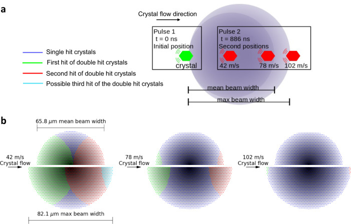
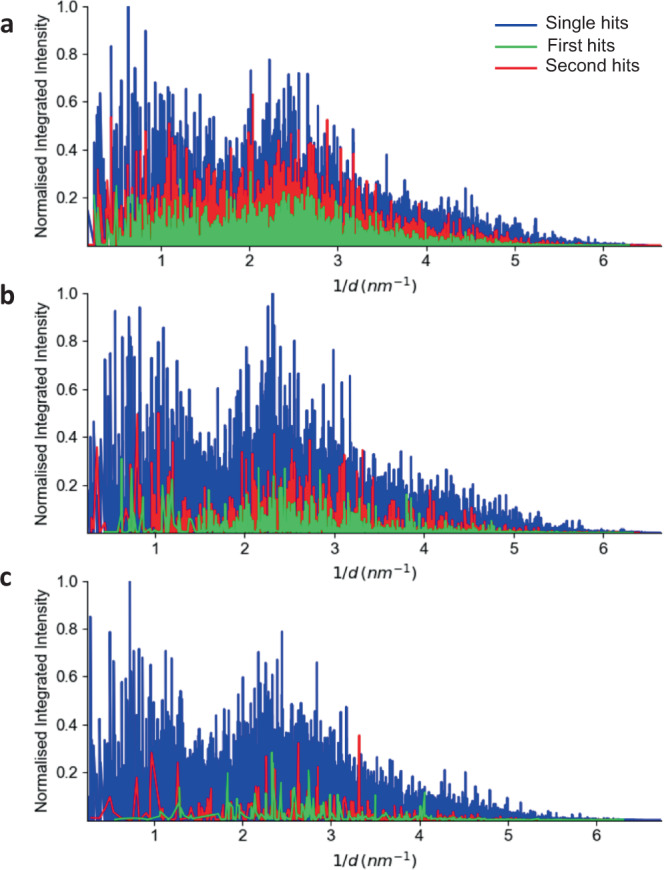
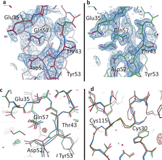
The European X-ray Free Electron Laser (XFEL) and Linac Coherent Light Source (LCLS) II are extremely intense sources of X-rays capable of generating Serial Femtosecond Crystallography (SFX) data at megahertz (MHz) repetition rates. Previous work has shown that it is possible to use consecutive X-ray pulses to collect diffraction patterns from individual crystals. Here, we exploit the MHz pulse structure of the European XFEL to obtain two complete datasets from the same lysozyme crystal, first hit and the second hit, before it exits the beam. The two datasets, separated by <1 µs, yield up to 2.1 Å resolution structures. Comparisons between the two structures reveal no indications of radiation damage or significant changes within the active site, consistent with the calculated dose estimates. This demonstrates MHz SFX can be used as a tool for tracking sub-microsecond structural changes in individual single crystals, a technique we refer to as multi-hit SFX.


Conflict of interest statement

The authors declare no competing interest.

Figures

Fig. 1
Fig. 1. Frequency of diffraction patterns as a function of the relative change in crystal orientation.
The change in crystal orientation was characterized by the reciprocal space vector a, between consecutive diffraction measurements (separated in time by 886 ns) within the X-ray pulse train. The liquid jet speed was 42 m/s. An increase in frequency above 0.04 for consecutive images with a change in orientation of less than 5 degrees, indicated by the region shaded in red, can be observed and were classified as double hit crystals.
Fig. 2
Fig. 2. Model for how multi-hits occur for a single crystal.
a Schematic diagram (not to scale) illustrating the minimum distances travelled by an 8 µm crystal for the three different jet speeds overlaid with the average beam Full Width (FW, dark purple shaded region) and the maximum beam FW (light purple shaded region). The green crystal depicts the initial position, and the red crystal illustrates how far the crystal travels after the first hit for 42 m/s, 78 m/s, and 102 m/s jet speeds. b Schematic representation of the crystal path through the X-ray beam for each of the three jet speeds for the mean beam FW (upper half) and maximum beam FW (lower half) as indicated on the first image. The mean FW is consistent for all jet speeds. The beam profile (shaded grey) is overlaid with the regions that the crystal travels through for the single hits (blue) to occur as well as the first (green) and second (red) hits of the double hit crystal. For the 42 m/s jet speed it also shows a possibility of the crystals being hit a third time (aqua) if the crystal and beam conditions were optimal. Note, for 42 m/s and 78 m/s, regions where no hits occur are possible.
Fig. 3
Fig. 3. Characteristic beam profile.
a A histogram showing the X-ray beam profile. The beam profile was modelled using a Lorentzian distribution with a Full Width Half Maximum (FWHM) = 2γ (50% of beam) and Full Width (FW) = 7.04γ (82.4% of beam). b The Lorentzian distribution used to determine the FWHM and FW from 6773 YAG images. The furthermost outlier, minimum, Q1 (25th percentile), mean, median, Q3 (75th percentile), and maximum have been indicated. To obtain the FWHM and FW for each YAG image, a 7.5% noise threshold was applied to the image combined with a 3 × 3 median filter to account for the noise.
Fig. 4
Fig. 4. Normalized integrated intensity plots.
Integrated intensities were extracted from the data, normalized and plotted against 1/d (where d is the lattice spacing) for the a 42 m/s jet data; b 78 m/s jet data; and c 102 m/s jet data. Blue represents data for single hit crystals only; green represents the first hit of the double hit crystal; red represents the second hit of the double hit crystal. A threshold of I/sig(I) > 2 was applied to the analysis.
Fig. 5
Fig. 5. Lysozyme structural maps showing the active site pocket.
The electron density map with the omit map displayed for the active site region of lysozyme in a the first hit structure (7TUM) and b the second hit structure (6WEC). c Shows the first (red) and second (green) hit structures superimposed and overlaid with the difference electron density (DED) map for the active site and d shows the DED maps for a representative di-sulphide bond Cys115-Cys30. No differences density is detected between the first and second hit structures. The 2Fo-Fc map at 1σ is shown in blue and difference maps at 3σ are shown in green (positive) and red (negative) density.
Fig. 6
Fig. 6. Parameters for optimizing the collection of double-hit data at the European XFEL.
a European XFEL repetition rate of 1.1 MHz during this experiment and (b) European XFEL repetition rate of 4.5 MHz. The green shaded area indicates parameter combinations that will result in double-hits that allow the second hit to occur within the horizontal FWHM of the beam; the blue shaded area indicates parameter combinations that will result in double-hits that allow a second hit to occur within in the tail region of the beam, and the grey shaded area indicates parameter combinations that will result in only single hits. This analysis is independent of crystal size (i.e., crystal centre-to-crystal centre hits).

References

    1. Wiedorn MO, et al. Megahertz serial crystallography. Nat. Commun. 2018;9:4025. doi: 10.1038/s41467-018-06156-7. - DOI - PMC - PubMed
    1. Chapman HN, et al. Femtosecond X-ray protein nanocrystallography. Nature. 2011;470:73–77. doi: 10.1038/nature09750. - DOI - PMC - PubMed
    1. Suga M, et al. Native structure of photosystem II at 1.95 angstrom resolution viewed by femtosecond X-ray pulses. Nature. 2015;517:99–103. doi: 10.1038/nature13991. - DOI - PubMed
    1. Boutet S, et al. High-resolution protein structure determination by serial femtosecond crystallography. Science. 2012;337:362–364. doi: 10.1126/science.1217737. - DOI - PMC - PubMed
    1. Gati C, et al. Atomic structure of granulin determined from native nanocrystalline granulovirus using an X-ray free-electron laser. Proc. Natl Acad. Sci. USA. 2017;114:2247–2252. doi: 10.1073/pnas.1609243114. - DOI - PMC - PubMed

Publication types