. 2022 Sep;28(9):1797-1801.
doi: 10.1038/s41591-022-01925-w. Epub 2022 Aug 11.

Plasma p-tau231 and p-tau217 as state markers of amyloid-β pathology in preclinical Alzheimer's disease

Affiliations

Plasma p-tau231 and p-tau217 as state markers of amyloid-β pathology in preclinical Alzheimer's disease

Marta Milà-Alomà et al. Nat Med. 2022 Sep.

Erratum in

Abstract

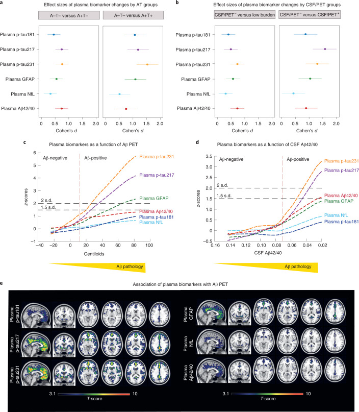
Blood biomarkers indicating elevated amyloid-β (Aβ) pathology in preclinical Alzheimer's disease are needed to facilitate the initial screening process of participants in disease-modifying trials. Previous biofluid data suggest that phosphorylated tau231 (p-tau231) could indicate incipient Aβ pathology, but a comprehensive comparison with other putative blood biomarkers is lacking. In the ALFA+ cohort, all tested plasma biomarkers (p-tau181, p-tau217, p-tau231, GFAP, NfL and Aβ42/40) were significantly changed in preclinical Alzheimer's disease. However, plasma p-tau231 reached abnormal levels with the lowest Aβ burden. Plasma p-tau231 and p-tau217 had the strongest association with Aβ positron emission tomography (PET) retention in early accumulating regions and associated with longitudinal increases in Aβ PET uptake in individuals without overt Aβ pathology at baseline. In summary, plasma p-tau231 and p-tau217 better capture the earliest cerebral Aβ changes, before overt Aβ plaque pathology is present, and are promising blood biomarkers to enrich a preclinical population for Alzheimer's disease clinical trials.


Conflict of interest statement

E.V. is a co-founder of ADx NeuroSciences. T.A.D. is an employee and shareholder of Eli Lilly and Company. J.L.M. is currently a full-time employee of Lundbeck and has previously served as a consultant or on advisory boards for the following for-profit companies, or has given lectures in symposia sponsored by the following for-profit companies: Roche Diagnostics, Genentech, Novartis, Lundbeck, Oryzon, Biogen, Lilly, Janssen, Green Valley, MSD, Eisai, Alector, BioCross, GE Healthcare and ProMIS Neurosciences. J.L.D. has served as a consultant for Genotix Biotechnologies Inc., Gates Ventures, Karuna Therapeutics, AlzPath Inc. and Cognito Therapeutics, Inc. J.L.D. received research support from ADx Neurosciences, Roche Diagnostics and Eli Lilly and Company in the past 2 years. H.Z. has served on scientific advisory boards for Eisai, Denali, Roche Diagnostics, Wave, Samumed, Siemens Healthineers, Pinteon Therapeutics, Nervgen, AZTherapies and CogRx, has given lectures in symposia sponsored by Cellectricon, Fujirebio, Alzecure and Biogen, and is a co-founder of Brain Biomarker Solutions in Gothenburg AB (BBS). J.D.G. has received speaker’s fees from Philips and Biogen and research support from GE Healthcare, Roche and Roche Diagnostics. M.S.C. has served as a consultant and on advisory boards for Roche Diagnostics International Ltd, and has given lectures in symposia sponsored by Roche Diagnostics, S.L.U. and Roche Farma, S.A. K.B. has served as a consultant, on advisory boards or at data monitoring committees for Abcam, Axon, BioArctic, Biogen, JOMDD/Shimadzu, Julius Clinical, Lilly, MagQu, Novartis, Ono Pharma, Roche Diagnostics and Siemens Healthineers, and is a co-founder of BBS in Gothenburg. The remaining authors declare no competing interests.

Figures

Fig. 1
Fig. 1. Plasma biomarkers and Aβ pathology.
a,b, Effect sizes of plasma biomarker levels change by AT groups (a; n = 397; n = 249 A−T−, n = 104 A+T−, n = 31 A+T+, n = 13 A−T+) and by CSF/PET groups (b; n = 339; n = 224 CSF/PET Aβ negative, n = 89 low burden, n = 26 CSF/PET Aβ positive). Individuals with a low burden of Aβ pathology were defined as CSF Aβ42/40 <0.071 and Aβ PET <30 Centiloids. The effect size of group differences was estimated by calculating Cohen’s d, in which the dependent variable was the residual of log(transformed) plasma biomarkers regressed on age and sex. The error bars denote the 95% CIs. c,d, The graphs represent the z-score changes of each plasma biomarker using the mean and the s.d. of that plasma biomarker in the group of participants with CSF Aβ42/40 >0.1 as a reference. The resulting z-scores are shown as a function of Aβ PET Centiloids (c) or CSF Aβ42/40 (d) using a robust local weighted regression method. The vertical dashed lines depict the Aβ PET 12 Centiloids (c) and CSF Aβ42/40 positivity cut-off (d). The horizontal dashed lines depict the abnormality threshold held at 1.5 and 2 s.d. above the mean. The horizontal axis direction of CSF Aβ42/40 (d) was inverted. e, Association of plasma biomarkers with Aβ PET at the voxel level. Associations were tested using voxel-wise, univariate, independent, linear regression models with age and sex as covariates. All plasma biomarkers showed a significant association with Aβ deposition in orbitofrontal and precuneus. These associations were stronger with plasma p-tau231 and p-tau217 and also extended to the insula and striatum. Statistical significance was set at P < 0.001 uncorrected for multiple comparisons with a cluster size of k > 100 voxels. All tests were one sided but contrasts in both directions were tested. No significant associations were found in the opposite direction. Statistical maps were resliced to 0.5 mm3 (cubic) for visualization purposes.
Extended Data Fig. 1
Extended Data Fig. 1. Plasma biomarkers by CSF/PET Aβ groups.
Violin plots comparing plasma biomarkers between CSF/PET Aβ groups (n = 339; n = 224 CSF/PET Aβ-negative, n = 89 Low burden, n = 26 CSF/PET Aβ-positive). Individuals with a low burden of Aβ pathology were defined as CSF Aβ42/40 < 0.071 and Aβ PET Centiloid < 30. The box plots depict the median (horizontal bar), interquartile range (IQR, hinges), and 1.5 × IQR (whiskers). Group comparisons were computed with a one-way ANCOVA adjusting for age and sex, followed by Tukey-corrected post hoc pairwise comparisons. The percentage (%) of change in mean levels of plasma biomarkers in the low burden group compared to the CSF/PET Aβ-negative group is shown.
Extended Data Fig. 2
Extended Data Fig. 2. ROC curves for the discrimination of Aβ PET Centiloid 12 or 30.
ROC analysis was performed to test the accuracy of plasma biomarkers (A), and plasma biomarkers in combination with a base risk factors model (age, sex and APOE ε4 status) (B), to discriminate participants with Aβ PET Centiloid ≥ 12 from those with Aβ PET Centiloid < 12. The same analyses were performed on both plasma biomarkers alone (C) or in combination with a base risk factors model (D) to discriminate participants with Aβ PET Centiloid ≥ 30 from those with Aβ PET Centiloid < 30.
Extended Data Fig. 3
Extended Data Fig. 3. ROC curves for the discrimination of Aβ status (CSF Aβ42/40).
ROC analysis was performed to test the accuracy of plasma biomarkers (A), and plasma biomarkers in combination with a base risk factors model (age, sex and APOE ε4 status) (B), to discriminate between Aβ status as defined by CSF Aβ42/40.
Extended Data Fig. 4
Extended Data Fig. 4. Association of plasma biomarkers with longitudinal change in cognition by Aβ status (CSF Aβ42/40).
Scatter plots representing the associations of each of the plasma biomarkers with annualized change in PACC scores. Each point depicts the value of the plasma biomarker of an individual and the solid lines indicate the regression line for each of the groups. The dashed line indicates the regression line of the whole sample. The error bands denote the 95% CIs. The standardized regression coefficients (β) and P values are shown and were computed using a linear regression with the annualized change in PACC scores as the dependent variable, adjusting by age, sex and years of education. All tests were two-sided. Annualized change in PACC scores was computed as the subtraction of PACC scores at visit 2 minus those at visit 1 divided by the time difference between the two visits in years. At the nominal level, there was a significant interaction between CSF Aβ status (as defined by CSF Aβ42/40) and plasma p-tau231. Thus, we performed a stratified analysis by CSF Aβ status for this biomarker, and we found a significant association of plasma p-tau231 with PACC score longitudinal changes in the CSF Aβ-positive group (β = –0.27, P = 0.023) but not in the Aβ-negative group (β = 0.054; P = 0.51). See Supplementary Table 8 for the detailed analyses, including FDR correction for multiple testing.
Extended Data Fig. 5
Extended Data Fig. 5. Association of plasma biomarkers with longitudinal change in Aβ deposition in individuals with Aβ PET Centiloids < 30.
In order to assess whether plasma biomarkers are associated with longitudinal Aβ aggregation in the earliest stages of the Alzheimer’s continuum, we conducted a sensitivity analysis in those individuals with Aβ PET Centiloid < 30. Plasma p-tau231 and p-tau217 were significantly associated with Aβ PET Centiloid increases. Scatter plots represent the associations of each of the plasma biomarkers with annualized change in Aβ PET Centiloids in individuals with Aβ PET Centiloids < 30. Each point depicts the value of the plasma biomarker of an individual and the solid lines indicate the regression line. The error bands denote the 95% CIs. The standardized regression coefficients (β) and P values are shown and were computed using a linear regression with the annualized change in Aβ PET Centiloids as the dependent variable, adjusting by age and sex. All tests were two-sided. Annualized change in Aβ PET Centiloids was computed as the subtraction of Centiloid values at visit 2 minus those at visit 1 divided by the time difference between the two visits in years. See Supplementary Table 10 for the detailed analyses, including FDR correction for multiple testing.

References

    1. Palmqvist S, et al. Discriminative accuracy of plasma Phospho-tau217 for Alzheimer disease vs other neurodegenerative disorders. JAMA. 2020;324:772. doi: 10.1001/jama.2020.12134. - DOI - PMC - PubMed
    1. Ashton NJ, et al. Plasma p-tau231: a new biomarker for incipient Alzheimer’s disease pathology. Acta Neuropathol. 2021;141:709–724. doi: 10.1007/s00401-021-02275-6. - DOI - PMC - PubMed
    1. Karikari TK, et al. Blood phosphorylated tau 181 as a biomarker for Alzheimer’s disease: a diagnostic performance and prediction modelling study using data from four prospective cohorts. Lancet Neurol. 2020;19:422–433. doi: 10.1016/S1474-4422(20)30071-5. - DOI - PubMed
    1. Lantero Rodriguez J, et al. Plasma p-tau181 accurately predicts Alzheimer’s disease pathology at least 8 years prior to post-mortem and improves the clinical characterisation of cognitive decline. Acta Neuropathol. 2020;140:267–278. doi: 10.1007/s00401-020-02195-x. - DOI - PMC - PubMed
    1. Jack CR, et al. NIA-AA Research Framework: toward a biological definition of Alzheimer’s disease. Alzheimers Dement. 2018;14:535–562. doi: 10.1016/j.jalz.2018.02.018. - DOI - PMC - PubMed

Publication types