Review
. 2022 Jul 31;12(15):1946.
doi: 10.3390/ani12151946.

Structural and Metabolic Changes in Bone

Affiliations
Review

Structural and Metabolic Changes in Bone

Agata Wawrzyniak et al. Animals (Basel). .

Abstract

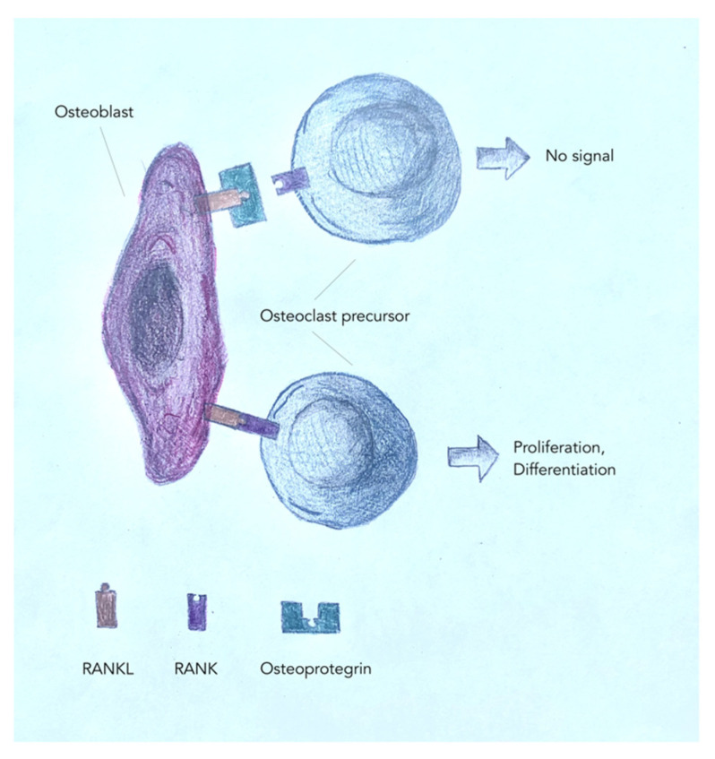
As an essential component of the skeleton, bone tissue provides solid support for the body and protects vital organs. Bone tissue is a reservoir of calcium, phosphate, and other ions that can be released or stored in a controlled manner to provide constant concentration in body fluids. Normally, bone development or osteogenesis occurs through two ossification processes (intra-articular and intra-chondral), but the first produces woven bone, which is quickly replaced by stronger lamellar bone. Contrary to commonly held misconceptions, bone is a relatively dynamic organ that undergoes significant turnover compared to other organs in the body. Bone metabolism is a dynamic process that involves simultaneous bone formation and resorption, controlled by numerous factors. Bone metabolism comprises the key actions. Skeletal mass, structure, and quality are accrued and maintained throughout life, and the anabolic and catabolic actions are mostly balanced due to the tight regulation of the activity of osteoblasts and osteoclasts. This activity is also provided by circulating hormones and cytokines. Bone tissue remodeling processes are regulated by various biologically active substances secreted by bone tissue cells, namely RANK, RANKL, MMP-1, MMP-9, or type 1 collagen. Bone-derived factors (BDF) influence bone function and metabolism, and pathophysiological conditions lead to bone dysfunction. This work aims to analyze and evaluate the current literature on various local and systemic factors or immune system interactions that can affect bone metabolism and its impairments.

Keywords: bone; bone metabolism disorders; bone morphogenetic proteins; glucocorticoids; obesity; osteoporosis.


Conflict of interest statement

The authors declare no conflict of interest.

Figures

Figure 1
Figure 1
Histological structure of normal bone tissue. (A) A cross-section of a properly structured bone showing the compact bone. The photograph of the area of an unstained section of compact bone. Visible osteons with concentric lamellae arranged around central canals (Haversian canal). Externally visible periosteum (P), cancellous bone covered by endosteum (E). Ground bone; scale bar: 2 mm. (B) A cross-section of a properly structured bone showing compact bone. Because the osteons are located very close together, compact bone is stronger and harder than spongy bone. Haversian canals contain blood vessels and supply developing bone with essential nutrients. Small trabeculae create a highly porous medullary bone. They provide support, offering considerable strength without significantly increasing the weight of the bone. H & E section; scale bar: 100 µm. (C) Osteons are structural and functional units, characteristic of compact bone. An osteon with concentric lamellae (L) surrounding a central canal (CC) is shown. The interstitial lamellae of the osteon are also visible; these are remnants of old osteons; ground bone; scale bar: 10 µm. (D) Photomicrograph of osteocytes and their associated processes. Through these long processes, cell-to-cell communication takes place and substances needed by the cells are transferred. Ground bone; scale bar: 5 µm. (E) Longitudinal section of the compact bone. Visible Haversian canal (H) connected to the transverse perforating canal (Volkmann) (V). Osteocytes embedded with lamellae (L). Ground bone; scale bar: 200 µm. (F) Histological structure of spongy bone. This structure is characteristic of bone marrow. Bone trabeculae surround the bone marrow along with blood vessels, and this arrangement strengthens the spongy bone, making it stronger. H & E section; scale bar: 50 µm.
Figure 2
Figure 2
Mechanisms of osteoblast/osteoclast regulation.
Figure 3
Figure 3
Histological structure of altered bone tissue. (A) Paget’s disease is a bone disorder. Histologically, the bone appears mosaic in appearance. It has thick cement lines that demarcate the disorderly lamellar bone. Such extensive trabeculae and lack of organization of the cortex are the cause of the loss of resistance to strain and therefore greatly increase the susceptibility to fracture. H & E section; scale bar: 200 µm. (B) In osteoporotic bone, there is an increased amount of adipose tissue appearing in the intertrabecular area. Therefore, for the same volume, the bone has much less density but is more frangible. Increased medial adiposity, associated with intratrabecular connectivity, emphasizes the functional connection between bone and marrow. Thin trabeculae are separated from each other. H & E section; scale bar: 100 µm. (C) Longitudinal section of the cancellous bone of the obese rat. Visible network of trabeculae separated by intercellular spaces containing islands and clusters of haematopoietic cells admixed with adipocytes. H & E section; scale bar: 500 µm.

References

    1. Raggatt L.J., Partridge N.C. Cellular and Molecular Mechanisms of Bone Remodeling. J. Biol. Chem. 2010;285:25103–25108. doi: 10.1074/jbc.R109.041087. - DOI - PMC - PubMed
    1. Florencio-Silva R., da Silva Sasso G.R., Sasso-Cerri E., Simões M.J., Cerri P.S. Biology of Bone Tissue: Structure, Function, and Factors That Influence Bone Cells. BioMed Res. Int. 2015;2015:421746. doi: 10.1155/2015/421746. - DOI - PMC - PubMed
    1. Mizokami A., Kawakubo-Yasukochi T., Hirata M. Osteocalcin and its endocrine functions. Biochem. Pharmacol. 2017;132:1–8. doi: 10.1016/j.bcp.2017.02.001. - DOI - PubMed
    1. Avtandilashvili M., Tolmachev S.Y. Modeling the Skeleton Weight of an Adult Caucasian Man. Health Phys. 2019;117:149–155. doi: 10.1097/HP.0000000000000881. - DOI - PubMed
    1. Boskey A.L., Posner A.S. Bone structure, composition, and mineralization. Orthop. Clin. N. Am. 1984;15:597–612. doi: 10.1016/S0030-5898(20)31258-X. - DOI - PubMed