. 2022 Jul 27;11(15):2306.
doi: 10.3390/cells11152306.

Hyperoxaluria Induces Endothelial Dysfunction in Preglomerular Arteries: Involvement of Oxidative Stress

Affiliations

Hyperoxaluria Induces Endothelial Dysfunction in Preglomerular Arteries: Involvement of Oxidative Stress

Javier Saenz-Medina et al. Cells. .

Abstract

Urolithiasis is a worldwide problem and a risk factor for kidney injury. Oxidative stress-associated renal endothelial dysfunction secondary to urolithiasis could be a key pathogenic factor, similar to obesity and diabetes-related nephropathy. The aim of the present study was to characterize urolithiasis-related endothelial dysfunction in a hyperoxaluria rat model of renal lithiasis.

Experimental approach: Endothelial dysfunction was assessed in preglomerular arteries isolated from control rats and in which 0.75% ethylene glycol was administered in drinking water. Renal interlobar arteries were mounted in microvascular myographs for functional studies; superoxide generation was measured by chemiluminescence and mRNA and protein expression by RT-PCR and immunofluorescence, respectively. Selective inhibitors were used to study the influence of the different ROS sources, xanthine oxidase, COX-2, Nox1, Nox2 and Nox4. Inflammatory vascular response was also studied by measuring the RNAm expression of NF-κB, MCP-1 and TNFα by RT-PCR.

Results: Endothelium-dependent vasodilator responses were impaired in the preglomerular arteries of the hyperoxaluric group along with higher superoxide generation in the renal cortex and vascular inflammation developed by MCP-1 and promoted by NF-κB. The xanthine oxidase inhibitor allopurinol restored the endothelial relaxations and returned superoxide generation to basal values. Nox1 and Nox2 mRNA were up-regulated in arteries from the hyperoxaluric group, and Nox1 and Nox2 selective inhibitors also restored the impaired vasodilator responses and normalized NADPH oxidase-dependent higher superoxide values of renal cortex from the hyperoxaluric group.

Conclusions: The current data support that hyperoxaluria induces oxidative stress-mediated endothelial dysfunction and inflammatory response in renal preglomerular arteries which is promoted by the xanthine oxidase, Nox1 and Nox2 pathways.

Keywords: endothelial dysfunction; oxidative stress; urolithiasis.


Conflict of interest statement

The authors declare no conflict of interest.

Figures
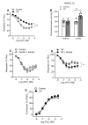
Figure 1
Figure 1
Impaired endothelium-dependent relaxations of the renal preglomerular arteries are associated with higher basal O2 generation in the renal cortex in hyperoxaluric rats (OX) and ameliorated by the SOD mimetic tempol. (A) Comparative endothelium-dependent relaxations of intrarenal arteries levels in control and OX rats. Results are given as a percentage of the phenylephrine (Phe)-induced contraction. Data are means ± SEM of 4 animals (1–2 arteries from each rat). Statistical differences were calculated with unpaired t-test *** p < 0.001. (B) Basal ROS generation in kidney cortex and renal interlobar arteries measured by lucigenin-enhanced chemiluminescence. Results are given as counts per minute (cpm) per mg of tissue. Bars represent mean ± SEM of 5 animals. Statistical differences were calculated with unpaired Student’s t-test *** p < 0.001 versus basal levels. (C,D) Effect of tempol on the relaxant responses elicited by ACh in rat renal interlobar arteries in control (C) and OX groups (D). Statistical differences were calculated by unpaired Student’s t-test ** p < 0.01. (E) Constrictor responses to Phe in renal interlobar arteries from control and hyperoxaluric rats (OX). Results are expressed as absolute values (Nm−1). Data are shown as the mean ± SEM of 7–8 arteries (4 animals).
Figure 2
Figure 2
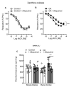
COX-2 inhibition improved renal endothelial relaxations in both control and OX rats and reduced elevated O2 levels in the renal cortex of the OX group, with no differences in COX-2 mRNA expression between the two groups. (A,B) Comparative effects of NS398 on endothelial relaxations of rat intrarenal arteries in the Control (A) and OX groups (B). Results are given as a percent of the phenylephrine (Phe) contraction as the mean ± SEM of 7–8 arteries (4 animals). Statistical differences were calculated by paired t-test ** p < 0.01. (C) Effect of NS398 on basal ROS generation in the renal arteries and kidney cortex of control and hyperoxaluric rats, measured by lucenin-enhanced chemiluminescence. Results are expressed as counts per minute (cpm) per mg of tissue. Bars represented mean ± SEM of 5–10 animals. Statistical differences were calculated with t-test *** p < 0.001 versus basal levels. (D) Comparative COX-2 mRNA expression in the renal arteries of the control group (n = 6) and OX group (n = 8), performed by RT-PCR. The delta–delta CT method was used to determine the fold change gene relative expression in the arteries that were treated with water (control group) and with 0.75% EG (OX group). The values represent the mean ±  SD. No statistical differences were detected.
Figure 3
Figure 3
Xanthine oxidase inhibition reduced elevated O2 levels in the renal cortex from the OX group and improved impaired endothelial relaxations in the OX group. (A,B) Effect of the xanthine oxidase inhibitor allopurinol on the endothelial relaxations of rat intrarenal arteries in the control (A) and OX groups (B). Results are given as percentage of the phenylephrine (Phe) contraction as the mean ± SEM of 4 animals (1–2 arteries per animal). Statistical differences were calculated by paired Student’s t-test *** p < 0.001. (C) Effect of allopurinol on basal ROS generation in the kidney cortex and renal interlobar arteries measured by lucigenin-enhanced chemiluminescence. Results are expressed as counts per minute (cpm) per mg of tissue. Bars represent mean ± SEM of 5–10 animals. Statistical differences were calculated with t-test *** p < 0.001 versus basal levels.
Figure 4
Figure 4
Enhanced Nox1 mRNA expression and amelioration of elevated renal cortex NADPH-dependent O2 levels and impaired endothelial relaxations by selective Nox1 inhibition in preglomerular arteries from hyperoxaluric rats. (A) Comparative Nox1 mRNA expression in samples of renal arteries from the control (n = 4) and OX group (n = 6), performed by RT-PCR. The delta CT method was used to determine the fold change for Nox1 gene relative expression in the arteries that were treated with water (control group) and with 0.75% EG (OX group). Statistical differences between means were calculated by unpaired Student’s t-test * p < 0.05. (B) Immunofluorescence demonstration of Nox1 subunit protein in a section of a renal interlobar artery. Immunofluorescence double labeling for TO-PRO marker (blue areas) demonstrates nuclear staining and for Nox1 protein (red areas). (C,D) Comparative effects of the Nox1 inhibitor NoxA1ds-taT on the endothelial relaxations of rat intrarenal arteries in control (C) and OX groups (D). Results are given as a percent of the phenylephrine (Phe) contraction as the mean ± SEM of 8 arteries (4 animals). Statistical differences were calculated by an unpaired Student’s t-test *** p < 0.05 (pEc50, see Table 2). (E) Effects of NoxA1ds-tat on NADPH-dependent ROS generation in renal interlobar arteries and in kidney cortex from control and hyperoxaluric rats. (F) measured by lucigenin chemiluminescence. Results are expressed as counts per minute (cpm) per mg of tissue. Bars represented mean ± SEM of 5–10 animals. Statistical differences were calculated with t-test *** p < 0.001, versus basal levels. † p < 0.05, †† p < 0.01; ††† p < 0.001 versus NADPH-stimulated.
Figure 5
Figure 5
Up-regulation of Nox2 and amelioration of oxidative stress in renal cortex and endothelial dysfunction by selective Nox2 inhibition in preglomerular arteries from hyperoxaluric rats. (A) Comparative Nox2 mRNA expression in samples of renal arteries from the control (n = 6) and OX group (n = 6) performed by RT-PCR. The delta CT method was used to determine the fold change for Nox2 gene relative expression in the arteries that were treated with water (control group) and with 0.75% EG (OX group). * p < 0.05. (B) Immunofluorescence demonstration of enhanced expression Nox2 protein in a section of a renal interlobar artery from OX group compared to a control artery. Immunofluorescence double labeling for TO-PRO marker (blue areas) demonstrates nuclear staining and for Nox2 protein (red areas). (C,D) Comparative effects of the Nox2 inhibitor GSK2795039 on endothelial relaxations of rat intrarenal arteries in control (C) and OX groups (D). Results are expressed as a percent of induced contraction. Data are shown as the mean ± SEM of 4 animals. Statistical differences were calculated by a Student’s t-test for paired observations * p < 0.05 versus control. (E,F) Effects of inhibitor GSK2795039 on NADPH-dependent ROS generation in renal arteries and cortex measured by lucigenin-enhanced chemiluminescence. Results are given as counts per minute (cpm) per mg of tissue. Bars represented mean ± SEM of 5–10 animals. Statistical differences were calculated by Student t-test *** p < 0.001 versus basal levels. ††† p < 0.001 versus NADPH-stimulated levels.
Figure 6
Figure 6
Unchanged Nox4 mRNA levels in preglomerular arteries from hyperoxaluric rats. (A) Comparative Nox4 mRNA expression in samples of renal arteries from the control (n = 5) and OX group (n = 7), performed by RT-PCR. The delta CT method was used to determine the fold change for NOX4 gene relative expression in the arteries that were treated with water (control group) and with 0.75% EG (OX group). Values represent the mean ± SD. Statistical differences were calculated by a Student’s t-test for unpaired observations. No significant differences were found. (B) Immunofluorescence demonstration of NOX4 protein in a section of a renal interlobar artery from the Control and OX group. Immunofluorescence double labeling for TO-PRO marker (blue areas) demonstrates nuclear staining and for NOX4 protein (red areas).
Figure 7
Figure 7
NFKB1 and MCP-1 are up-regulated in renal interlobar arteries of the OX group. Comparative NFKB1 (A) MCP-1 (B) and TNFα (C) mRNA expression in samples of renal arteries from control (n = 5–6) and OX group (n = 7–8), performed by RT-PCR. The delta CT method was used to determine the fold change for the different genes’ relative expression in the arteries that were treated with water (control group) and with 0.75% EG (OX group). Values represent the mean ± SD. Statistical differences were calculated by a Student’s t-test for unpaired observations * Significantly different at p-value < 0.05.

Similar articles

Cited by

References

    1. Lotan Y., Jiménez I.B., Lenoir-Wijnkoop I., Daudon M., Molinier L., Tack I., Nuijten M.J. Primary prevention of nephrolithiasis is cost-effective for a national healthcare system. Br. J. Urol. 2012;110:E1060–E1067. doi: 10.1111/j.1464-410X.2012.11212.x. - DOI - PubMed
    1. Khan S.R. Is oxidative stress, a link between nephrolithiasis and obesity, hypertension, diabetes, chronic kidney disease, metabolic syndrome. Urol. Res. 2012;40:95–112. doi: 10.1007/s00240-011-0448-9. - DOI - PMC - PubMed
    1. Davignon J., Ganz P. Role of endothelial dysfunction in atherosclerosis. Circulation. 2004;109:III27–III32. doi: 10.1161/01.CIR.0000131515.03336.f8. - DOI - PubMed
    1. Bakker W., Eringa E.C., Sipkema P., van Hinsbergh V.W.M. Endothelial dysfunction and diabetes: Roles of hyperglycemia, impaired insulin signaling and obesity. Cell Tissue Res. 2008;335:165–189. doi: 10.1007/s00441-008-0685-6. - DOI - PubMed
    1. de Vries A.P., Ruggenenti P., Ruan X.Z., Praga M., Cruzado J.M., Bajema I.M., D’Agati V.D., Lamb H.J., Barlovic D.P., Hojs R., et al. Fatty kidney: Emerging role of ectopic lipid in obesity-related renal disease. Lancet Diabetes Endocrinol. 2014;2:417–426. doi: 10.1016/S2213-8587(14)70065-8. - DOI - PubMed

Publication types