Review
. 2022 Jul 29;11(15):2327.
doi: 10.3390/cells11152327.

Immunological Tolerance in Liver Transplant Recipients: Putative Involvement of Neuroendocrine-Immune Interactions

Affiliations
Review

Immunological Tolerance in Liver Transplant Recipients: Putative Involvement of Neuroendocrine-Immune Interactions

Jaciara Fernanda Gomes Gama et al. Cells. .

Abstract

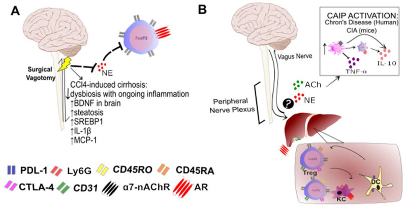
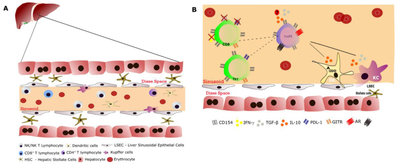
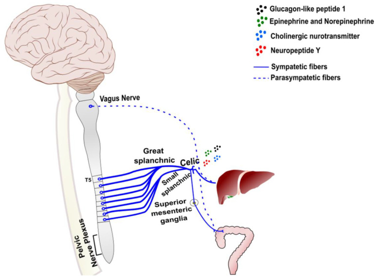
The transplantation world changed significantly following the introduction of immunosuppressants, with millions of people saved. Several physicians have noted that liver recipients that do not take their medication for different reasons became tolerant regarding kidney, heart, and lung transplantations at higher frequencies. Most studies have attempted to explain this phenomenon through unique immunological mechanisms and the fact that the hepatic environment is continuously exposed to high levels of pathogen-associated molecular patterns (PAMPs) or non-pathogenic microorganism-associated molecular patterns (MAMPs) from commensal flora. These components are highly inflammatory in the periphery but tolerated in the liver as part of the normal components that arrive via the hepatic portal vein. These immunological mechanisms are discussed herein based on current evidence, although we hypothesize the participation of neuroendocrine-immune pathways, which have played a relevant role in autoimmune diseases. Cells found in the liver present receptors for several cytokines, hormones, peptides, and neurotransmitters that would allow for system crosstalk. Furthermore, the liver is innervated by the autonomic system and may, thus, be influenced by the parasympathetic and sympathetic systems. This review therefore seeks to discuss classical immunological hepatic tolerance mechanisms and hypothesizes the possible participation of the neuroendocrine-immune system based on the current literature.

Keywords: adrenergic receptor; cholinergic receptor; immunological tolerance; liver transplantation; neuroendocrine-immune interaction; regulatory microenvironment.


Conflict of interest statement

The authors declare no conflict of interest.

Figures

Figure 1
Figure 1
Possible influence of the nervous system on the liver immune system after liver transplantation. (A) Among the interactions of neurotransmitters in liver injury by CCl4-induced cirrhosis, a decrease in norepinephrine (NE) is observed after sympathectomy, followed by a decrease in regulatory T cell (Treg) and increases in inflammatory cytokines, such as interleukin (IL)-1β and monocyte chemoattractant protein-1 (MCP-1), in addition to increases in hepatic steatosis and brain inflammation markers. (B) Collectively, evidence for neuroendocrine-immune interactions in the liver, mainly collected in clinical trials, shows that, after liver transplantation (LT), the Treg cell pool is heterogeneous and may present phenotypes indicative of different origins. Thus, these cells may express CD45RO or CD45RA, in addition to presenting CD31 on the cell surface. In addition, dendritic cells (DCs) contribute to the regulatory microenvironment, leading to a tolerogenic response after LT. In the peritoneal microenvironment, it is possible that these cells are modulated by β-adrenergic receptors in Kupffer cells (KC) via NE, as observed in other conditions associated with the peritoneal microenvironment or acetylcholine (ACh) release induced by cholinergic anti-inflammatory pathway (CAIP) activation. These changes are able to induce the anti-inflammatory macrophage profile via Treg cells and thus modulate the inflammatory response. BNDF: brain-derived neurotrophic factor; SREBP1: sterol regulatory element-binding protein-1; PDL-1: programmed death-ligand 1; CTLA-1: cytotoxic T-lymphocyte-associated antigen 4; α7nAChR: alpha7-nicotinic acetylcholine receptor; AR: adrenergic receptor.
Figure 2
Figure 2
Liver cells in immunity: hepatic cells in homeostasis and the regulatory microenvironment in the immunosuppressive response after LT. (A) The liver is an organ composed of parenchymal (hepatocyte) and nonparenchymal cells that exert different functions. During homeostasis, nonparenchymal cells such as Kupffer cells, dendritic cells, NK cells, NKT cells, and HSCs constitute the immunological liver microenvironment, responding to most gut-derived antigens. In addition to these resident cells, there are transient lymphocytes in the sinusoidal space. In addition, hepatocytes can play an immunological role in the context of innate protein release. (B) Regulatory T (Treg) cells play an important role in the mechanisms of allogeneic response suppression after LT. Kupffer cells (KCs) and dendritic cells (DCs) have important roles in this regulatory microenvironment mediated through IL-10 production and TGF-β release. These molecules contribute to the induction of Treg cells, which probably act directly to decrease the response via effects on CD4+ (Th1 cells) and CD8+ T cells, minimizing the rejection process. In CD8+ T cells, there is a decrease in CD154, an important protein expressed on the surface of activated cells in humans. Furthermore, in a mouse model, it was observed that cytotoxic T-lymphocyte-associated antigen 4 (CTLA-4) induction in Treg cells promoted an increase in glucocorticoid-induced TNFR-related protein (GITR) on CD8+ and CD4+ T cells, decreasing the rejection responses of these T cells through the induction of apoptosis in these cells, mainly via increased expression of programmed death-ligand1 (PDL-1). Thus, the production of inflammatory cytokines, such as IFN-γ, was decreased. LSEC: liver sinusoidal endothelial cells; IDO: indoleamine 2,3-dioxygenase.
Figure 3
Figure 3
Arrangement of autonomic fibers in the liver. The interaction of the liver with the nervous system takes place through communication between the peripheral nerves and the vagus nerve, which largely innervates the peritoneal cavity. Sympathetic fibers that innervate liver tissue originate from neurons in the celiac and superior mesenteric ganglia. Furthermore, parasympathetic fibers originate mainly from preganglionic neurons located in the dorsal motor nucleus of the vagus nerve. Both are capable of interactions mediated by the release of adrenergic and cholinergic neurotransmitters and neuropeptides, such as neuropeptide Y, (continue on next page) leading to neuroendocrine-immune interactions that contribute to the modulation of inflammatory responses.

Similar articles

Cited by

References

    1. Matzinger P. The Danger Model: A Renewed Sense of Self. Science. 2002;296:301–305. doi: 10.1126/science.1071059. - DOI - PubMed
    1. Venkatesh H.S., Morishita W., Geraghty A.C., Silverbush D., Gillespie S.M., Arzt M., Tam L.T., Espenel C., Ponnuswami A., Ni L., et al. Electrical and Synaptic Integration of Glioma into Neural Circuits. Nature. 2019;573:539–545. doi: 10.1038/s41586-019-1563-y. - DOI - PMC - PubMed
    1. Kaufmann S.H.E. Immunology’s Coming of Age. Front. Immunol. 2019;10:684. doi: 10.3389/fimmu.2019.00684. - DOI - PMC - PubMed
    1. Westerhof N. A Short History of Physiology. Acta Physiol. 2011;202:601–603. doi: 10.1111/j.1748-1716.2011.02286.x. - DOI - PubMed
    1. Hadden J.W., Hadden E.M., Middleton E. Lymphocyte Blast Transformation: I. Demonstration of Adrenergic Receptors in Human Peripheral Lymphocytes. Cell. Immunol. 1970;1:583–595. doi: 10.1016/0008-8749(70)90024-9. - DOI - PubMed

Publication types