. 2022 Jul 29;11(15):2334.
doi: 10.3390/cells11152334.

Smooth Muscle Myosin Localizes at the Leading Edge and Regulates the Redistribution of Actin-regulatory Proteins during Migration

Affiliations

Smooth Muscle Myosin Localizes at the Leading Edge and Regulates the Redistribution of Actin-regulatory Proteins during Migration

Ruping Wang et al. Cells. .

Abstract

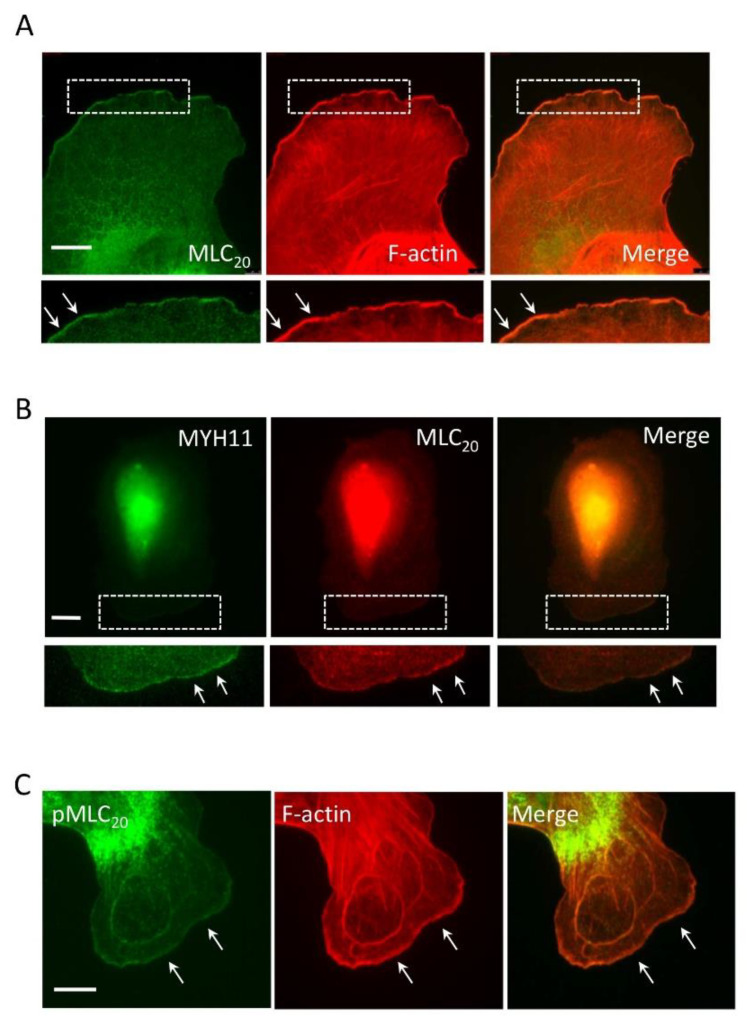
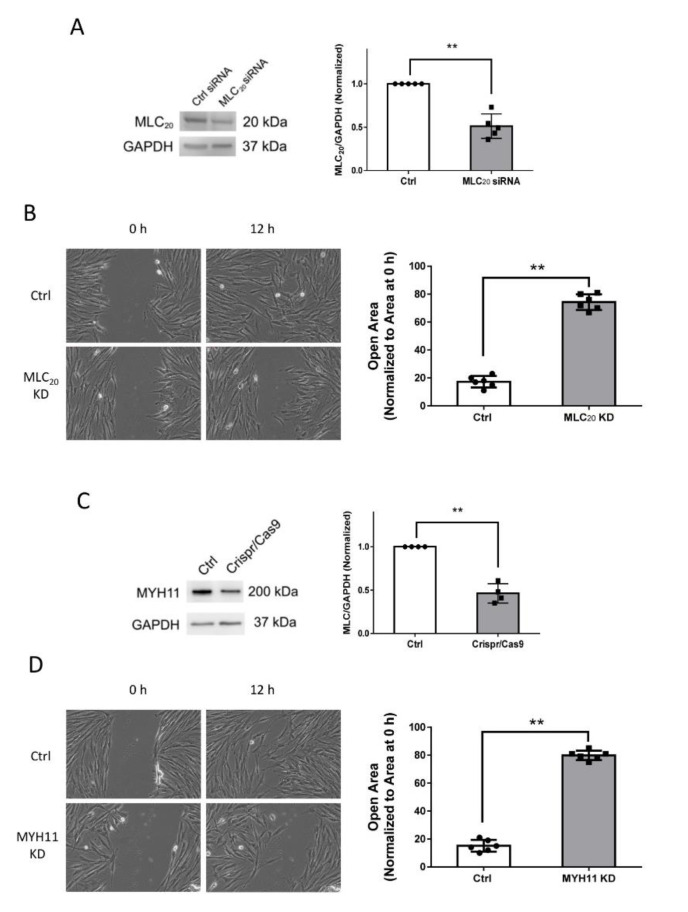
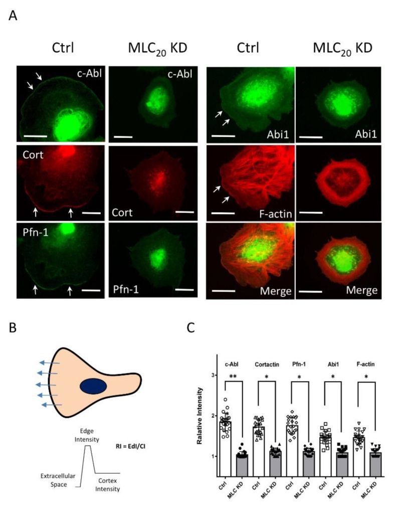
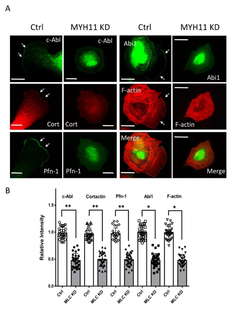
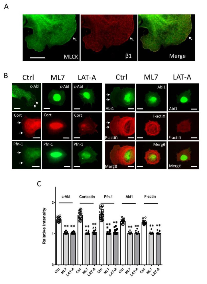
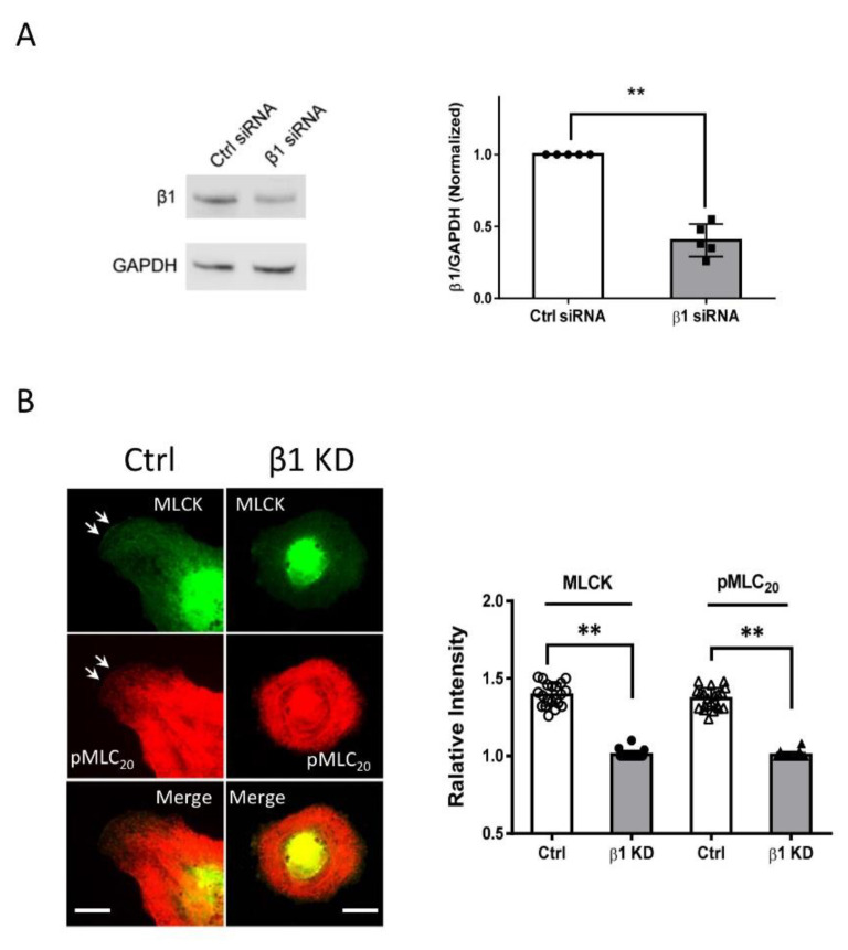
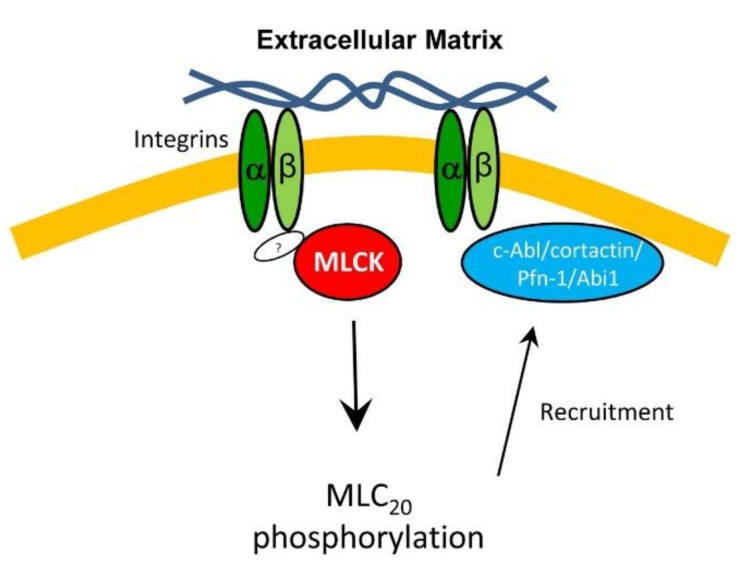
Airway smooth muscle cell migration plays an essential role in airway development, repair, and remodeling. Smooth muscle myosin II has been traditionally thought to localize in the cytoplasm solely and regulates cell migration by affecting stress fiber formation and focal adhesion assembly. In this study, we unexpectedly found that 20-kDa myosin light chain (MLC20) and myosin-11 (MYH11), important components of smooth muscle myosin, were present at the edge of lamellipodia. The knockdown of MLC20 or MYH11 attenuated the recruitment of c-Abl, cortactinProfilin-1 (Pfn-1), and Abi1 to the cell edge. Moreover, myosin light chain kinase (MLCK) colocalized with integrin β1 at the tip of protrusion. The inhibition of MLCK attenuated the recruitment of c-Abl, cortactin, Pfn-1, and Abi1 to the cell edge. Furthermore, MLCK localization at the leading edge was reduced by integrin β1 knockdown. Taken together, our results demonstrate that smooth muscle myosin localizes at the leading edge and orchestrates the recruitment of actin-regulatory proteins to the tip of lamellipodia. Mechanistically, integrin β1 recruits MLCK to the leading edge, which catalyzes MLC20 phosphorylation. Activated myosin regulates the recruitment of actin-regulatory proteins to the leading edge, and promotes lamellipodial formation and migration.

Keywords: actin-associated proteins; leading edge; migration; myosin; smooth muscle.


Conflict of interest statement

The authors declare that they have no competing interest.

Figures

Figure 1
Figure 1
Smooth muscle myosin localizes at the tip of lamellipodia. (A) Human airway smooth muscle (HASM) cells were plated onto collagen-coated coverslips for 30 min and stained for 20-kDa myosin light chain (MLC20) and F-actin. MLC20 is found at the leading cell edge. In addition, F-actin is also localized at the cell edge. (B) Myosin-11 (MYH11) colocalizes with MLC20 at the leading edge. (C) Phosphorylated MLC20 (pMLC20) is found to position at the leading edge. The arrows point to the leading edge. Scale bar: 10 µm.
Figure 2
Figure 2
Knockdown (KD) of MLC20 or MYH11 attenuates cell migration. (A) Human airway smooth muscle (HASM) cells treated with control (Ctrl) siRNA or MLC20 siRNA were evaluated by immunoblot analysis. MLC siRNA reduces the expression of MLC20 in HASM cells. Data are mean values of experiments from five cultures from three donors. Error bars indicate SD. (B) The migration of HASM cells was examined by using the wound healing assay. n = six experiments from three donors. Error bars indicate SD. (C) Cells were treated with Ctrl or Crispr/Cas9 constructs followed by immunoblot analysis. Data are the mean values of experiments from four cultures from three donors. Error bars indicate SD. (D) MYH11 KD reduces the migration of HASM cells. n = six experiments from three donors. Error bars indicate SD. ** p < 0.01. Student’s t-test was used for statistical analysis.
Figure 3
Figure 3
MLC20 regulates the recruitment of c-Ab, cortactin, Pfn-1, and Abi1 to the cell edge. (A) Ctrl and MLC20 KD cells were plated onto collagen-coated coverslips for 30 min followed by immunofluorescence and fluorescence analysis. MLC20 KD attenuates the localization of c-Abl, cortactin (cort), Pfn-1, Abi1, and F-actin at the cell edge. The arrows point to the leading edge. Scale bar: 10 µm. (B) Illustration of quantitative analysis. An average of at least 5 line scans across each cell is used for analysis. Relative Intensity (RI) = Edge Intensity (EdI)/Cortex Intensity (CI). (C) Data are mean values of experiments from at least 20 cells for each group. Error bars indicate SD. One-way ANOVA was used for statistical analysis. ** p < 0.01; * p < 0.05.
Figure 4
Figure 4
MYH11 orchestrates the positioning of c-Ab, cortactin, Pfn-1, and Abi1 to the cell edge. (A) Ctrl and MYH11 KD cells were plated onto collagen-coated coverslips for 30 min followed by immunofluorescence and fluorescence analysis. The arrows point to the leading edge. Scale bar: 10 µm. (B) Data are mean values of experiments from at least 20 cells for each group. Error bars indicate SD. One-way ANOVA was used for statistical analysis. ** p < 0.01; * p < 0.05.
Figure 5
Figure 5
MLC20 phosphorylation and actin polymerization regulate the recruitment of actin-regulatory proteins to the cell edge. (A) Myosin light chain kinase (MLCK) colocalizes with integrin β1 at the cell edge. Cells were immunostained for MLCK and integrin β1. The arrows point to the leading edge. Scale bar: 10 µm. (B) Cells were plated onto collagen-coated coverslips in the absence or presence of 1 µM ML7 or 10 nM latrunculin A (LAT-A) for 30 min. They were then stained for the indicated proteins or F-actin. Scale bar, 10 µm. Ctrl, control cells. The arrows point to the leading edges. (C) Data are mean values of experiments from at least 20 cells for each group. Error bars indicate SD. ** p < 0.01. One-way ANOVA was used for statistical analysis.
Figure 6
Figure 6
Integrin β1 controls the positioning of MLCK at the tip of the protrusion. (A) Human airway smooth muscle (HASM) cells treated with control (Ctrl) siRNA or β1 siRNA were evaluated by immunoblot analysis. Data are mean values of experiments from five cultures from three donors. Error bars indicate SD. (B) β1 KD attenuates the positioning of MLCK and pMLC20 at the cell edge. The arrows point to the leading edge. Scale bar: 10 µm. Data are mean values of experiments from at least 20 cells for each group. Error bars indicate SD. ** p < 0.01. The t-test was used for statistical analysis.
Figure 7
Figure 7
Proposed mechanism: In response to extracellular cues (e.g., the ECM), integrin β1 locates at the tip of lamellipodia, which recruits MLCK to the leading cell edge via an unknown mechanism. The kinase then catalyzes MLC20 phosphorylation, which promotes the recruitment of c-Abl, cortactin, Pfn-1, and Abi1 to the leading edge, lamellipodial formation, and migration.

Similar articles

Cited by

References

    1. Walsh M.P. Vascular smooth muscle myosin light chain diphosphorylation: Mechanism, function, and pathological implications. IUBMB Life. 2011;63:987–1000. doi: 10.1002/iub.527. - DOI - PubMed
    1. Gao N., Huang J., He W., Zhu M., Kamm K.E., Stull J.T. Signaling through myosin light chain kinase in smooth muscles. J. Biol. Chem. 2013;288:7596–7605. doi: 10.1074/jbc.M112.427112. - DOI - PMC - PubMed
    1. Petrov D., Dahan I., Cohen-Kfir E., Ravid S. Apkczeta affects directed cell migration through the regulation of myosin light chain phosphorylation. Cell Adhes. Migr. 2017;11:347–359. doi: 10.1080/19336918.2016.1225631. - DOI - PMC - PubMed
    1. Freed D.H., Chilton L., Li Y., Dangerfield A.L., Raizman J.E., Rattan S.G., Visen N., Hryshko L.V., Dixon I.M.C. Role of myosin light chain kinase in cardiotrophin-1-induced cardiac myofibroblast cell migration. Am. J. Physiol. Circ. Physiol. 2011;301:H514–H522. doi: 10.1152/ajpheart.01041.2010. - DOI - PubMed
    1. Peng Y., Chen Z., He Y., Li P., Chen Y., Chen X., Jiang Y., Qin X., Li S., Li T., et al. Non-muscle myosin II isoforms orchestrate substrate stiffness sensing to promote cancer cell contractility and migration. Cancer Lett. 2021;524:245–258. doi: 10.1016/j.canlet.2021.10.030. - DOI - PubMed

Publication types

MeSH terms

LinkOut - more resources