Review
. 2022 Jul 29;11(15):2336.
doi: 10.3390/cells11152336.

TGF-β Superfamily Signaling in the Eye: Implications for Ocular Pathologies

Affiliations
Review

TGF-β Superfamily Signaling in the Eye: Implications for Ocular Pathologies

Soumaya Hachana et al. Cells. .

Abstract

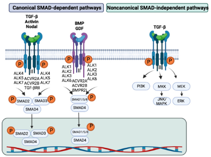
The TGF-β signaling pathway plays a crucial role in several key aspects of development and tissue homeostasis. TGF-β ligands and their mediators have been shown to be important regulators of ocular physiology and their dysregulation has been described in several eye pathologies. TGF-β signaling participates in regulating several key developmental processes in the eye, including angiogenesis and neurogenesis. Inadequate TGF-β signaling has been associated with defective angiogenesis, vascular barrier function, unfavorable inflammatory responses, and tissue fibrosis. In addition, experimental models of corneal neovascularization, diabetic retinopathy, proliferative vitreoretinopathy, glaucoma, or corneal injury suggest that aberrant TGF-β signaling may contribute to the pathological features of these conditions, showing the potential of modulating TGF-β signaling to treat eye diseases. This review highlights the key roles of TGF-β family members in ocular physiology and in eye diseases, and reviews approaches targeting the TGF-β signaling as potential treatment options.

Keywords: BMP; TGF-β; age-related macular degeneration; ocular diseases.


Conflict of interest statement

The authors declare no conflict of interest.

Figures

Figure 1
Figure 1
TGF-β superfamily signaling pathways. TGF-β superfamily proteins exert their cellular activity via canonical and noncanonical TGF-β signaling pathways. The activation of the heterotetrameric complex of TGF-β type I and type II receptors leads to phosphorylation and activation of SMAD proteins. In addition, TGF-β superfamily ligands can also activate noncanonical signaling pathways, such as the phosphatidylinositol 3-kinase (PI3K)/AKT, extracellular signal-regulated kinases (ERK), Jun N-terminal kinase family (JNK), and mitogen-activated protein kinase (MAPK) signaling pathways.
Figure 2
Figure 2
Graphic representation of the implications of TGF-β signaling in the pathological processes occurring during the progression of age-related macular degeneration. Schematic representation summarizing the TGF-β1, TGF-β2, and TGF-β3 expression in healthy condition in human eye structures and cells (left) and proangiogenic and antiangiogenic expression of TGF-β1, TGF-β2, and TGF-β3 in nAMD of human eye structures (right). The squares in blue or red indicate, respectively, down-regulation and up-regulation of different types of TGF-β with the related site in the eye. nAMD: neovascular age-related macular degeneration. EMT: epithelial to mesenchymal transition. RPE: retinal pigment epithelium. CNV: choroidal neovascularization.
Figure 3
Figure 3
Functional implications of TGF-β signaling in pathological processes underlying age-related macular degeneration. Description of the mechanisms of TGF-β superfamily in CNV and wet AMD, including the modulation of angiogenesis-related factors, inflammation, vascular fibrosis, and immune responses. EC: endothelial cell. ECM: extracellular matrix proteins. MET: mesenchymal transition. RPE: retinal pigment epithelium. CNV: choroidal neovascularization.

Similar articles

Cited by

References

    1. Choudhary M., Malek G. A Review of Pathogenic Drivers of Age-Related Macular Degeneration, Beyond Complement, with a Focus on Potential Endpoints for Testing Therapeutic Interventions in Preclinical Studies. Adv. Exp. Med. Biol. 2019;1185:9–13. doi: 10.1007/978-3-030-27378-1_2. - DOI - PMC - PubMed
    1. Wong W.L., Su X., Li X., Cheung C.M., Klein R., Cheng C.Y., Wong T.Y. Global prevalence of age-related macular degeneration and disease burden projection for 2020 and 2040: A systematic review and meta-analysis. Lancet Glob. Health. 2014;2:e106–e116. doi: 10.1016/S2214-109X(13)70145-1. - DOI - PubMed
    1. El-Amir A.N., Sagoo M.S., da Cruz L. Age-related macular degeneration. Br. J. Hosp. Med. 2005;66:677–681. doi: 10.12968/hmed.2005.66.12.20206. - DOI - PubMed
    1. Keane P.A., Sadda S.R. Development of Anti-VEGF Therapies for Intraocular Use: A Guide for Clinicians. J. Ophthalmol. 2012;2012:483034. doi: 10.1155/2012/483034. - DOI - PMC - PubMed
    1. Saint-Geniez M., Maharaj A.S., Walshe T.E., Tucker B.A., Sekiyama E., Kurihara T., Darland D.C., Young M.J., D’Amore P.A. Endogenous VEGF is required for visual function: Evidence for a survival role on muller cells and photoreceptors. PLoS ONE. 2008;3:e3554. doi: 10.1371/journal.pone.0003554. - DOI - PMC - PubMed

Publication types

Substances

Grants and funding