. 2022 Jul 29;11(15):2343.
doi: 10.3390/cells11152343.

β-Glucocerebrosidase Deficiency Activates an Aberrant Lysosome-Plasma Membrane Axis Responsible for the Onset of Neurodegeneration

Affiliations

β-Glucocerebrosidase Deficiency Activates an Aberrant Lysosome-Plasma Membrane Axis Responsible for the Onset of Neurodegeneration

Giulia Lunghi et al. Cells. .

Abstract

β-glucocerebrosidase is a lysosomal hydrolase involved in the catabolism of the sphingolipid glucosylceramide. Biallelic loss of function mutations in this enzyme are responsible for the onset of Gaucher disease, while monoallelic β-glucocerebrosidase mutations represent the first genetic risk factor for Parkinson's disease. Despite this evidence, the molecular mechanism linking the impairment in β-glucocerebrosidase activity with the onset of neurodegeneration in still unknown. In this frame, we developed two in vitro neuronal models of β-glucocerebrosidase deficiency, represented by mouse cerebellar granule neurons and human-induced pluripotent stem cells-derived dopaminergic neurons treated with the specific β-glucocerebrosidase inhibitor conduritol B epoxide. Neurons deficient for β-glucocerebrosidase activity showed a lysosomal accumulation of glucosylceramide and the onset of neuronal damage. Moreover, we found that neurons react to the lysosomal impairment by the induction of their biogenesis and exocytosis. This latter event was responsible for glucosylceramide accumulation also at the plasma membrane level, with an alteration in lipid and protein composition of specific signaling microdomains. Collectively, our data suggest that β-glucocerebrosidase loss of function impairs the lysosomal compartment, establishing a lysosome-plasma membrane axis responsible for modifications in the plasma membrane architecture and possible alterations of intracellular signaling pathways, leading to neuronal damage.

Keywords: GBA1; Gaucher disease; glucosylceramide; lipid rafts; lysosomes; plasma membrane.


Conflict of interest statement

The authors declare no conflict of interest.

Figures

Figure 1
Figure 1
Effect of conduritol B epoxide treatment on β-glucocerebrosidase activity. β-glucocerebrosidase activity was evaluated in (a) mouse cerebellar granule neurons treated or not with 0.5 mM CBE for 7 or 14 days and (b) in human iPSC-derived dopaminergic neurons treated or not with 0.5 mM CBE for 14 or 29 days. β-glucocerebrosidase activity is expressed as nmoles of formed product/hour/mg cell proteins. All data are shown as mean ± SEM of three different experiments. *** p < 0.001, **** p < 0.0001, two-tailed Student’s t-test vs. CBE-untreated cells.
Figure 2
Figure 2
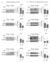
Effect of β-glucocerebrosidase inhibition on neuronal markers’ expression. Immunoblotting analysis of the expression of Neurofilament H (NF-H), MAP2, TAU and PSD95 in: (a) mouse cerebellar granule neurons (CGN) treated or not with conduritol B epoxide (CBE) 0.5 mM for 7 and 14 days and (b) human iPSC-derived dopaminergic neurons treated or not with CBE 0.5 mM for 14 and 29 days. Optical densities of the individual bands were quantified using NIH ImageJ and normalized to GAPDH. Data are expressed as fold change with respect to CBE-untreated cells (dashed lined) and are the mean ± SEM of three different experiments. * p < 0.05; ** p < 0.01; *** p < 0.001. Two-tailed Student’s t-test vs. CBE-untreated cells.
Figure 3
Figure 3
Effect of β-glucocerebrosidase inhibition on glucosylceramide and glucosylsphingosine levels. Representative digital autoradiography of sphingolipid pattern and quantification of the radioactivity associated with glucosylceramide (GlcCer), glucosylsphingosine (GlcSph), ceramide (Cer) and sphingomyelin (SM) in: (a) mouse cerebellar granule neurons fed with radioactive sphingosine and treated or not (−) with 0.5 mM conduritol B epoxide (CBE) for 7 and 14 days and (b) human iPSC-derived dopaminergic neurons fed with radioactive sphingosine and treated or not with CBE for 14 and 29 days. Data are expressed as fold change with respect to CBE-untreated cells (−) and are the mean ± SEM of three different experiments. * p < 0.05; ** p < 0.01; *** p < 0.001; **** p < 0.0001. Two-tailed Student’s t-test vs. CBE-untreated cells.
Figure 4
Figure 4
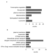
Effect of β-glucocerebrosidase inhibition on proteomic profile. Bar chart showing the cluster enriched annotation groups of proteins (a) upregulated and (b) downregulated in CBE-treated DA neurons with respect to untreated cells obtained by David bioinformatics analysis. The x-axis indicates the number of proteins involved in each term.
Figure 5
Figure 5
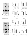
Effect of glucosylceramide accumulation on lysosomal-associated membrane protein-1 expression. Immunoblotting analyses of lysosomal-associated membrane protein (LAMP-1) in (a) mouse cerebellar granule neurons and (b) human iPSC-derived dopaminergic neurons treated or not with 0.5 mM conduritol B epoxide (CBE) for 14 and 29 days, respectively. Data are expressed as fold change with respect to CBE-untreated cells (CTRL) and are the mean ± SEM of three experiments; * p < 0.05, two-tailed Student’s t-test vs. CBE-untreated cells.
Figure 6
Figure 6
Activity of plasma membrane glycohydrolases and extracellular release of glucosylceramide upon its accumulation. Specific enzyme activity of plasma membrane-associated β-galactosidase and β-hexosaminidase and representative image and quantification of extracellular release of glucosylceramide (GlcCer) in (a) mouse cerebellar granule neurons and (b) human iPSC-derived dopaminergic neurons fed with radioactive sphingosine followed by treatment in presence or in absence of 0.5 mM conduritol B epoxide (CBE) for 14 and 29 days, respectively. Enzyme activities are expressed as nmoles/ h normalized to 106 cells and are the mean ± SEM of three experiments (* p < 0.05, *** p < 0.001, two-tailed Student’s t-test vs. CBE-untreated cells (CTRL)). Extracellular release of radioactive GlcCer was determined after the lipid extraction of cell culture medium and HPTLC separation (Cer = ceramide, PE = phosphatidylethanolamine, SM = sphingomyelin, ganglio = gangliosides). Data are expressed as fold change with respect to CBE-untreated cells (CTRL) ad are the mean ± SEM of three experiments; * p < 0.05, ** p < 0.01, *** p < 0.001, two-tailed Student’s t-test vs. CBE-untreated cells.
Figure 7
Figure 7
Accumulation of glucosylceramide at the cell surface. Representative HPTLC image of radioactive lipids of cell fractions obtained after selective cell surface protein biotinylation followed by magnetic separation using Dynabeads™ streptavidin magnetic beads; lipids were extracted from the supernatants containing non biotinylated proteins (SN) and the pellets containing the biotinylated cell surface proteins (P); (a) mouse cerebellar granule neurons and (b) human iPSC-derived dopaminergic neurons treated or not (−) with 0.5 mM conduritol B epoxide (CBE), for 14 and 29 days, respectively. (Cer = ceramide; GlcCer = glucosylceramide; PE = phosphatidylethanolammine; SM = sphingomyelin). Data are expressed as fold change of GlcCer with respect to CBE-untreated cell fraction (−) and are the mean ± SEM of three experiments (*** p < 0.001; **** p < 0.0001; one-way two-tailed Student’s t-test vs. CBE-untreated cells). GlcCer content was normalized nCi/ mg of proteins of the sample.
Figure 8
Figure 8
p416 c-Src expression and lipid distribution in detergent-resistant plasma membrane fractions of iPSCs-derived dopaminergic neurons treated or not with conduritol B epoxide. (a) Representative image of HPTLC of radioactive lipids of detergent-resistant membrane domain fraction (DRM) of human iPSCs-derived dopaminergic neurons treated or not (−) with 0.5 mM conduritol B epoxide (CBE) for 29 days. (b) Representative image of HPTLC of radioactive lipids of detergent-resistant membrane domain fraction (DRM) after cell surface biotinylation of human iPSCs-derived dopaminergic neurons treated or not (−) with 0.5 mM conduritol B epoxide (CBE) for 29 days. Detergent-resistant plasma membrane domain fraction (PM-DRM) was obtained from DRM by streptavidine magnetic beads separation. Data are expressed as nCi and are the mean ± SEM from at least three experiments (* p < 0.05; ** p < 0.01; *** p < 0.001; **** p < 0.0001; two-tailed Student’s t-test). (Cer = ceramide; GlcCer = glucosylceramide; PE = phosphatidylethanolamine; Sulf = sulphatides; Gb3 = globo-triaosylceramide; SM = sphingomyelin). (c) Immunoblotting analysis of Src and pTyr416 c-Src expression in DRM fraction and non-DRM high density fractions (HD) of dopaminergic neurons treated or not with CBE for 29 days; analysis of pTyr416 c-Src activation: pTyr416 c-Src band intensity was normalized to c-Src signal of the same sample. Data are expressed as fold change with respect to CBE-untreated cell fraction of and are the mean ± SEM of at least three experiments; ** p < 0.01; two-tailed Student’s t-test.

References

    1. Stirnemann J., Belmatoug N., Camou F., Serratrice C., Froissart R., Caillaud C., Levade T., Astudillo L., Serratrice J., Brassier A., et al. A Review of Gaucher Disease Pathophysiology, Clinical Presentation and Treatments. Int. J. Mol. Sci. 2017;18:441. doi: 10.3390/ijms18020441. - DOI - PMC - PubMed
    1. Chiricozzi E., Aureli M., Mauri L., Di Biase E., Lunghi G., Fazzari M., Valsecchi M., Carsana E.V., Loberto N., Prinetti A., et al. Glycosphingolipids. Adv. Exp. Med. Biol. 2021;1325:61–102. doi: 10.1007/978-3-030-70115-4_3. - DOI - PubMed
    1. Lwin A., Orvisky E., Goker-Alpan O., LaMarca M.E., Sidransky E. Glucocerebrosidase mutations in subjects with parkinsonism. Mol. Genet. Metab. 2004;81:70–73. doi: 10.1016/j.ymgme.2003.11.004. - DOI - PubMed
    1. Goker-Alpan O., Schiffmann R., LaMarca M.E., Nussbaum R.L., McInerney-Leo A., Sidransky E. Parkinsonism among Gaucher disease carriers. J. Med. Genet. 2004;41:937–940. doi: 10.1136/jmg.2004.024455. - DOI - PMC - PubMed
    1. Eblan M.J., Walker J.M., Sidransky E. The glucocerebrosidase gene and Parkinson’s disease in Ashkenazi Jews. N. Engl. J. Med. 2005;352:728–731. doi: 10.1056/NEJM200502173520719. author reply 728–731. - DOI - PubMed

Publication types