

Nuclear Receptor Atlases of Choroidal Tissues Reveal Candidate Receptors Associated with Age-Related Macular Degeneration
- PMID: 35954227
- PMCID: PMC9367936
- DOI: 10.3390/cells11152386
Nuclear Receptor Atlases of Choroidal Tissues Reveal Candidate Receptors Associated with Age-Related Macular Degeneration
Erratum in
-
Correction: Peavey et al. Nuclear Receptor Atlases of Choroidal Tissues Reveal Candidate Receptors Associated with Age-Related Macular Degeneration. Cells 2022, 11, 2386.Cells. 2022 Dec 7;11(24):3948. doi: 10.3390/cells11243948. Cells. 2022. PMID: 36552901 Free PMC article.
Abstract
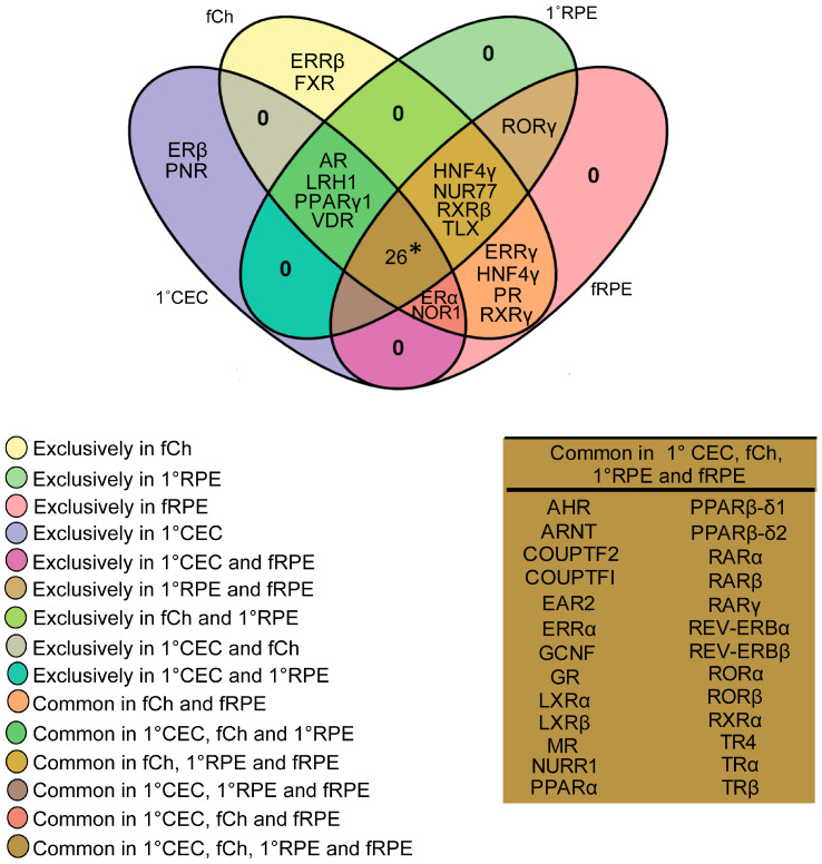
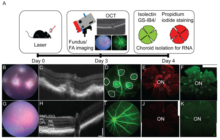
The choroid is a vulnerable tissue site in the eye, impacted in several blinding diseases including age related macular degeneration (AMD), which is the leading cause of central vision loss in the aging population. Choroidal thinning and choriocapillary dropout are features of the early form of AMD, and endothelial dysfunction and vascular changes are primary characteristics of the neovascular clinical sub-type of AMD. Given the importance, the choroidal endothelium and outer vasculature play in supporting visual function, a better understanding of baseline choroidal signaling pathways engaged in tissue and cellular homeostasis is needed. Nuclear receptors are a large family of transcription factors responsible for maintaining various cellular processes during development, aging and disease. Herein we developed a comprehensive nuclear receptor atlas of human choroidal endothelial cells and freshly isolated choroidal tissue by examining the expression levels of all members of this transcription family using quantitative real time PCR. Given the close relationship between the choroid and retinal pigment epithelium (RPE), this data was cross-referenced with the expression profile of nuclear receptors in human RPE cells, to discover potential overlap versus cell-specific nuclear receptor expression. Finally, to identify candidate receptors that may participate in the pathobiology of AMD, we cataloged nuclear receptor expression in a murine model of wet AMD, from which we discovered a subset of nuclear receptors differentially regulated following neovascularization. Overall, these databases serve as useful resources establishing the influence of nuclear receptor signaling pathways on the outer vascular tissue of the eye, while providing a list of receptors, for more focused investigations in the future, to determine their suitability as potential therapeutic targets for diseases, in which the choroid is affected.
Keywords: age-related macular degeneration; choroidal endothelial cells; choroidal injury; nuclear receptor atlas.
Conflict of interest statement
The authors have declared that no conflict of interest exist.
Figures


Similar articles
-
Potential therapeutic targets for age-related macular degeneration: The nuclear option.Prog Retin Eye Res. 2023 May;94:101130. doi: 10.1016/j.preteyeres.2022.101130. Epub 2022 Oct 8. Prog Retin Eye Res. 2023. PMID: 36220751 Free PMC article. Review.
-
Research resource: nuclear receptor atlas of human retinal pigment epithelial cells: potential relevance to age-related macular degeneration.Mol Endocrinol. 2011 Feb;25(2):360-72. doi: 10.1210/me.2010-0392. Epub 2011 Jan 14. Mol Endocrinol. 2011. PMID: 21239617 Free PMC article.
-
Characterization of a tissue-engineered choroid.Acta Biomater. 2019 Jan 15;84:305-316. doi: 10.1016/j.actbio.2018.11.033. Epub 2018 Nov 23. Acta Biomater. 2019. PMID: 30476582
-
Single-cell RNA sequencing reveals transcriptional changes of human choroidal and retinal pigment epithelium cells during fetal development, in healthy adult and intermediate age-related macular degeneration.Hum Mol Genet. 2023 May 5;32(10):1698-1710. doi: 10.1093/hmg/ddad007. Hum Mol Genet. 2023. PMID: 36645183 Free PMC article.
-
Anaphylatoxin Signaling in Retinal Pigment and Choroidal Endothelial Cells: Characteristics and Relevance to Age-Related Macular Degeneration.Adv Exp Med Biol. 2018;1074:45-51. doi: 10.1007/978-3-319-75402-4_6. Adv Exp Med Biol. 2018. PMID: 29721926 Review.
Cited by
-
Potential therapeutic targets for age-related macular degeneration: The nuclear option.Prog Retin Eye Res. 2023 May;94:101130. doi: 10.1016/j.preteyeres.2022.101130. Epub 2022 Oct 8. Prog Retin Eye Res. 2023. PMID: 36220751 Free PMC article. Review.
-
Potential Role of NUR77 in the Aging Retinal Pigment Epithelium and Age-Related Macular Degeneration.Adv Exp Med Biol. 2025;1468:165-169. doi: 10.1007/978-3-031-76550-6_27. Adv Exp Med Biol. 2025. PMID: 39930190 Review.
-
Does senescence play a role in age-related macular degeneration?Exp Eye Res. 2022 Dec;225:109254. doi: 10.1016/j.exer.2022.109254. Epub 2022 Sep 21. Exp Eye Res. 2022. PMID: 36150544 Free PMC article. Review.
-
Correction: Peavey et al. Nuclear Receptor Atlases of Choroidal Tissues Reveal Candidate Receptors Associated with Age-Related Macular Degeneration. Cells 2022, 11, 2386.Cells. 2022 Dec 7;11(24):3948. doi: 10.3390/cells11243948. Cells. 2022. PMID: 36552901 Free PMC article.
-
Estrogen related receptor alpha: Potential modulator of age-related macular degeneration.Curr Opin Pharmacol. 2024 Apr;75:102439. doi: 10.1016/j.coph.2024.102439. Epub 2024 Mar 5. Curr Opin Pharmacol. 2024. PMID: 38447458 Free PMC article. Review.
References
-
- Hogan M.J., Alvarado J.A., Weddell J.E. Histology of the Human Eye; An Atlas and Textbook. Saunders; Philadelphia, PA, USA: 1971.
-
- Campbell M., Humphries P. The blood-retina barrier: Tight junctions and barrier modulation. Adv. Exp. Med. Biol. 2012;763:70–84. - PubMed
Publication types
MeSH terms
Substances
Grants and funding
LinkOut - more resources
Full Text Sources
Medical
