. 2022 Aug 3;11(15):2388.
doi: 10.3390/cells11152388.

Adipocyte-Derived Extracellular Vesicles Promote Prostate Cancer Cell Aggressiveness by Enabling Multiple Phenotypic and Metabolic Changes

Affiliations

Adipocyte-Derived Extracellular Vesicles Promote Prostate Cancer Cell Aggressiveness by Enabling Multiple Phenotypic and Metabolic Changes

Fabrizio Fontana et al. Cells. .

Abstract

Background: In recent decades, obesity has widely emerged as an important risk factor for prostate cancer (PCa). Adipose tissue and PCa cells have been shown to orchestrate a complex interaction network to support tumor growth and evolution; nonetheless, the study of this communication has only been focused on soluble factors, although increasing evidence highlights the key role of extracellular vesicles (EVs) in the modulation of tumor progression.

Methods and results: In the present study, we found that EVs derived from 3T3-L1 adipocytes could affect PC3 and DU145 PCa cell traits, inducing increased proliferation, migration and invasion. Furthermore, conditioning of both PCa cell lines with adipocyte-released EVs resulted in lower sensitivity to docetaxel, with reduced phosphatidylserine externalization and decreased caspase 3 and PARP cleavage. In particular, these alterations were paralleled by an Akt/HIF-1α axis-related Warburg effect, characterized by enhanced glucose consumption, lactate release and ATP production.

Conclusions: Collectively, these findings demonstrate that EV-mediated crosstalk exists between adipocytes and PCa, driving tumor aggressiveness.

Keywords: Warburg effect; adipocytes; chemoresistance; extracellular vesicles; metastasis; obesity; prostate cancer.


Conflict of interest statement

The authors declare no conflict of interest.

Figures

Figure 1
Figure 1
Characterization of adipocyte-derived extracellular vesicles. (A) EV size measured by nanoparticle tracking analysis. (B) EV morphology visualized by transmission electron microscopy. Scale bar is 100 nm. (C) Western blot analysis was performed to investigate the expression levels of TSG101, Alix, Hsc70, calnexin and cytochrome c in 3T3-L1 adipocytes and in their EVs. One representative of three experiments performed is shown.
Figure 2
Figure 2
Adipocyte-released EVs promote prostate cancer cell proliferation. (A) PC3 and DU145 cells were incubated with adipocyte-released EVs (30 μg/mL) for 96 h. Cell proliferation was then evaluated by Trypan Blue exclusion assay. Each experiment was repeated three times. Data represent mean values ± SEM and were analyzed using a t-test. * p < 0.05 vs. PC3 or DU145 (control). (B) Cells were incubated with adipocyte-released EVs (30 μg/mL) for 24 h. Cell cycle distribution was then evaluated by cytofluorimetric analysis after staining with Invitrogen™ FxCycle™ PI/RNase Staining Solution (according to the manufacturer’s protocol). Each experiment was repeated three times. Data represent mean values ± SEM and were analyzed using a t-test. * p < 0.05 vs. PC3 or DU145 (control), ** p < 0.01 vs. PC3 or DU145 (control), *** p < 0.001 vs. PC3 or DU145 (control).
Figure 3
Figure 3
Adipocyte-associated EVs stimulate prostate cancer cell migration and invasion. (A) PC3 and DU145 cells were incubated with adipocyte-associated EVs (30 μg/mL) for 24 h. Cell migration was then evaluated by wound healing assay. Each experiment was repeated three times. Scale bar is 200 μm. (B) Cells were incubated with adipocyte-associated EVs (30 μg/mL) for 24 h. Cell invasion was then evaluated by Boyden chamber assay. Each experiment was repeated three times. Scale bar is 100 μm. Data represent mean values ± SEM and were analyzed using a t-test. *** p < 0.001 vs. PC3 or DU145 (control).
Figure 4
Figure 4
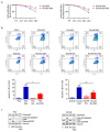
Adipocyte-secreted EVs enhance prostate cancer cell chemoresistance. (A) PC3 and DU145 cells were pre-treated with adipocyte-secreted EVs (30 μg/mL) for 24 h and then treated with docetaxel (100 nM) for 48 h. Cell proliferation was then evaluated by Trypan Blue exclusion assay. Each experiment was repeated three times. Data represent mean values ± SEM and were analyzed using a t-test. * p < 0.05 vs. PC3 DOC or DU145 DOC (control). (B) Cells were pre-treated with adipocyte-secreted EVs (30 μg/mL) for 24 h and then treated with docetaxel (100 nM) for 48 h. Apoptotic rate was then evaluated by cytofluorimetric analysis after staining with eBioscience™ Annexin V-FITC Apoptosis Detection Kit (according to the manufacturer’s protocol). One representative of three experiments performed is shown. Data represent mean values ± SEM and were analyzed using a Bonferroni’s test after one-way analysis of variance. ** p < 0.01 vs. PC3 or DU145, *** p < 0.001 vs. PC3 or DU145, # p < 0.05 vs. PC3 DOC or DU145 DOC, ## p < 0.01 vs. PC3 DOC or DU145 DOC. (C) After adipocyte-secreted EV pre-treatment (30 μg/mL, 24 h) and docetaxel treatment (100 nM, 48 h), Western blot analysis was performed to investigate the expression levels of cleaved caspase 3 and PARP. Tubulin expression was evaluated as a loading control. One representative of three experiments performed is shown.
Figure 5
Figure 5
Extracellular vesicles from adipocytes reprogram prostate cancer cell metabolism. (A) PC3 and DU145 cells were incubated with adipocyte-derived EVs (30 μg/mL) for 24 h. Glucose consumption was then evaluated by cytofluorimetric analysis after staining with 2-(N-(7-Nitrobenz-2-oxa-1,3-diazol-4-yl)amino)-2-Deoxyglucose (100 μM) for 30 min. Each experiment was repeated three times. Data represent mean values ± SEM and were analyzed using a t-test. *** p < 0.001 vs. PC3 or DU145 (control). (B) Cells were incubated with adipocyte-derived EVs (30 μg/mL) for 24 h. Lactate production was then evaluated by using a lactate assay kit (according to the manufacturer’s protocol). Each experiment was repeated three times. Data represent mean values ± SEM and were analyzed using a t-test. *** p < 0.001 vs. PC3 or DU145 (control). (C) Cells were incubated with adipocyte-derived EVs (30 μg/mL) for 24 h. ATP synthesis was then evaluated by using an ATP assay kit (according to the manufacturer’s protocol). Each experiment was repeated three times. Data represent mean values ± SEM and were analyzed using a t-test. *** p < 0.001 vs. PC3 or DU145 (control). (D) After adipocyte-derived EV treatment (30 μg/mL, 24 h), Western blot analysis was performed to investigate the expression levels of p-Akt and HIF-1α in PC3 and DU145 cells. Tubulin expression was evaluated as a loading control. One representative of three experiments performed is shown.

Similar articles

Cited by

References

    1. Siegel R.L., Miller K.D., Fuchs H.E., Jemal A. Cancer statistics, 2022. CA. Cancer J. Clin. 2022;72:7–33. doi: 10.3322/caac.21708. - DOI - PubMed
    1. Litwin M.S., Tan H.-J. The Diagnosis and Treatment of Prostate Cancer. JAMA. 2017;317:2532. doi: 10.1001/jama.2017.7248. - DOI - PubMed
    1. Fontana F., Marzagalli M., Montagnani Marelli M., Raimondi M., Moretti R., Limonta P. Gonadotropin-Releasing Hormone Receptors in Prostate Cancer: Molecular Aspects and Biological Functions. Int. J. Mol. Sci. 2020;21:9511. doi: 10.3390/ijms21249511. - DOI - PMC - PubMed
    1. Fontana F., Limonta P. Dissecting the Hormonal Signaling Landscape in Castration-Resistant Prostate Cancer. Cells. 2021;10:1133. doi: 10.3390/cells10051133. - DOI - PMC - PubMed
    1. Freedland S.J., Aronson W.J. Examining the relationship between obesity and prostate cancer. Rev. Urol. 2004;6:73–81. - PMC - PubMed

Publication types

Supplementary concepts