. 2022 Aug 5;11(15):2421.
doi: 10.3390/cells11152421.

High Levels of Tumor miR-187-3p-A Potential Tumor-Suppressor microRNA-Are Correlated with Poor Prognosis in Colorectal Cancer

Affiliations

High Levels of Tumor miR-187-3p-A Potential Tumor-Suppressor microRNA-Are Correlated with Poor Prognosis in Colorectal Cancer

Lui Ng et al. Cells. .

Abstract

Background: The microRNA miR-187-3p plays antitumor roles in a variety of cancers. We and others have previously identified miR-187-3p as a potential tumor suppressor in colorectal cancer (CRC), but there are also reports revealing that high miR-187-3p levels are associated with poor prognosis among CRC patients. This study further investigated the clinicopathological significance of miR-187-3p in CRC.

Methods: MiR-187-3p levels in paired polyp/CRC/normal specimens or primary CRC/liver metastasis specimens were determined by qPCR, and correlated with the patient's clinicopathological and postoperative survival data. The clinical findings were validated using our validation cohort and data obtained from the TCGA or GEO databases. The functional effects of miR-187-3p were investigated through its overexpression in CRC cell lines.

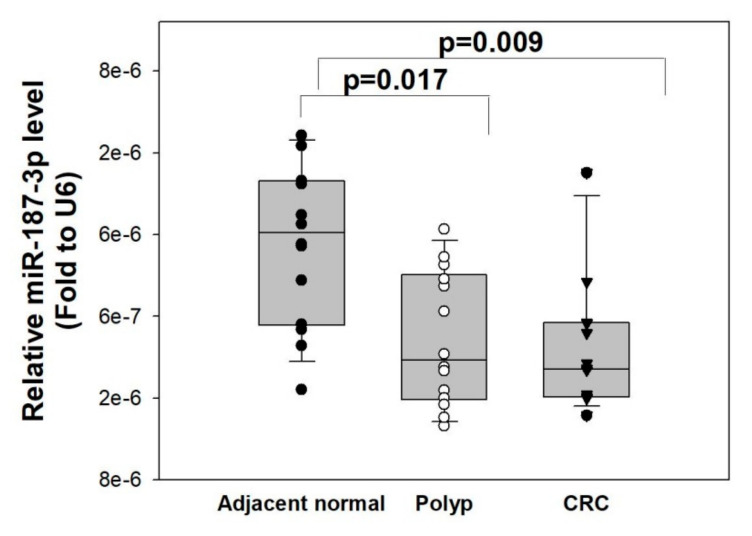
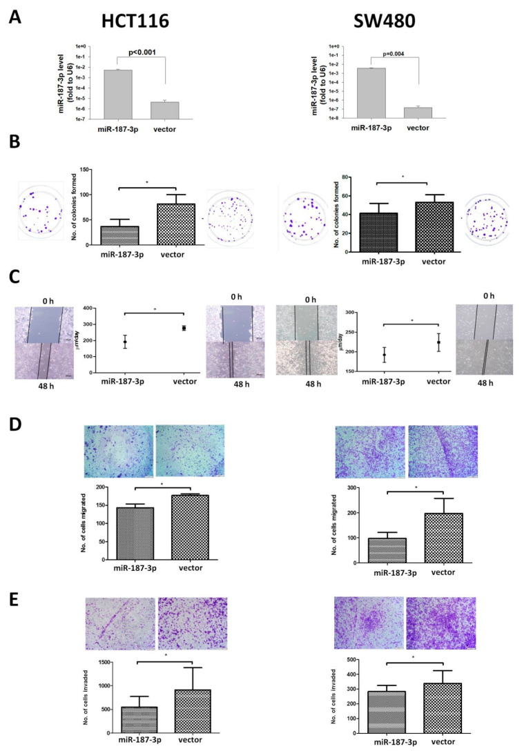
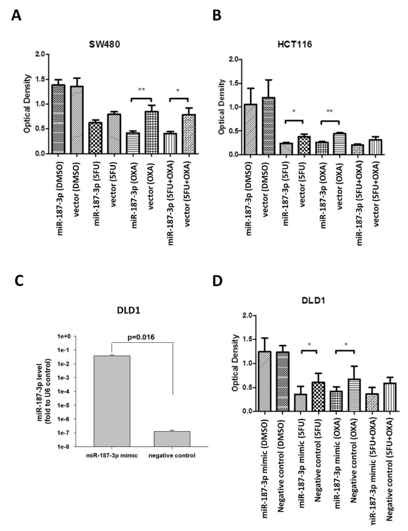
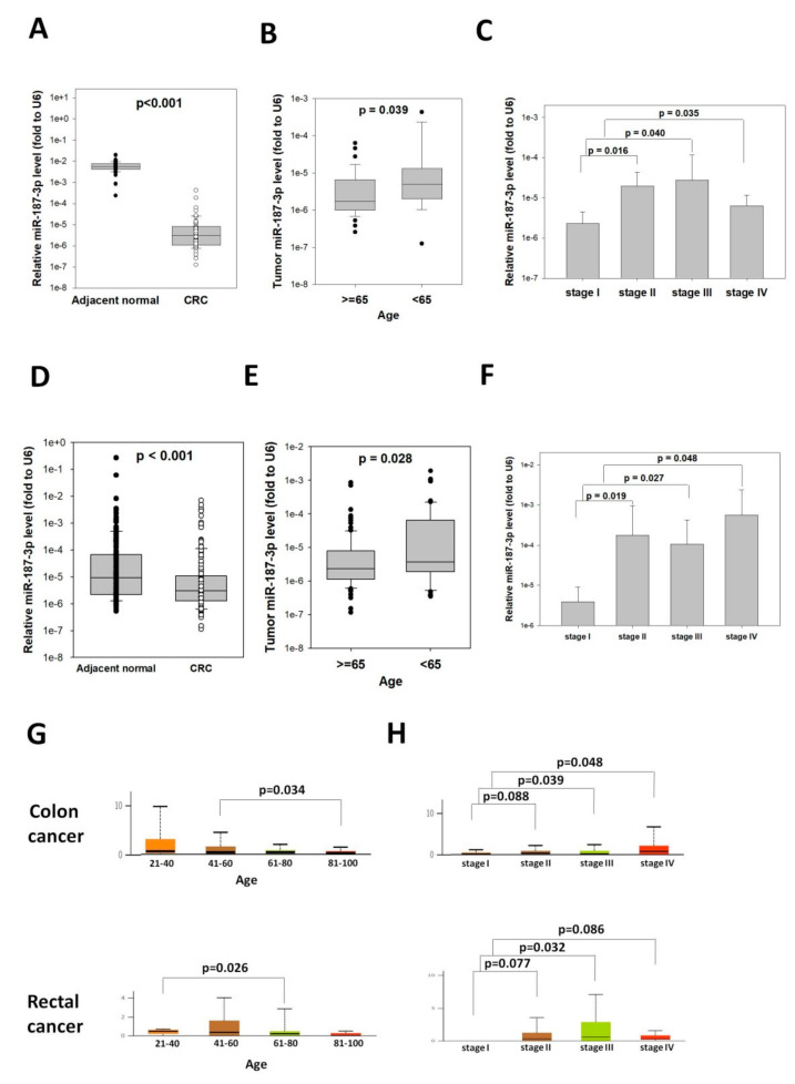
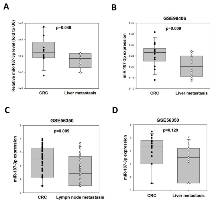
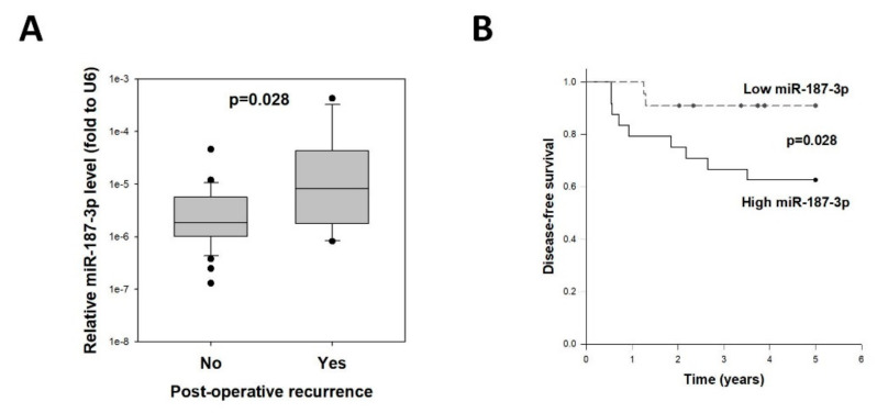
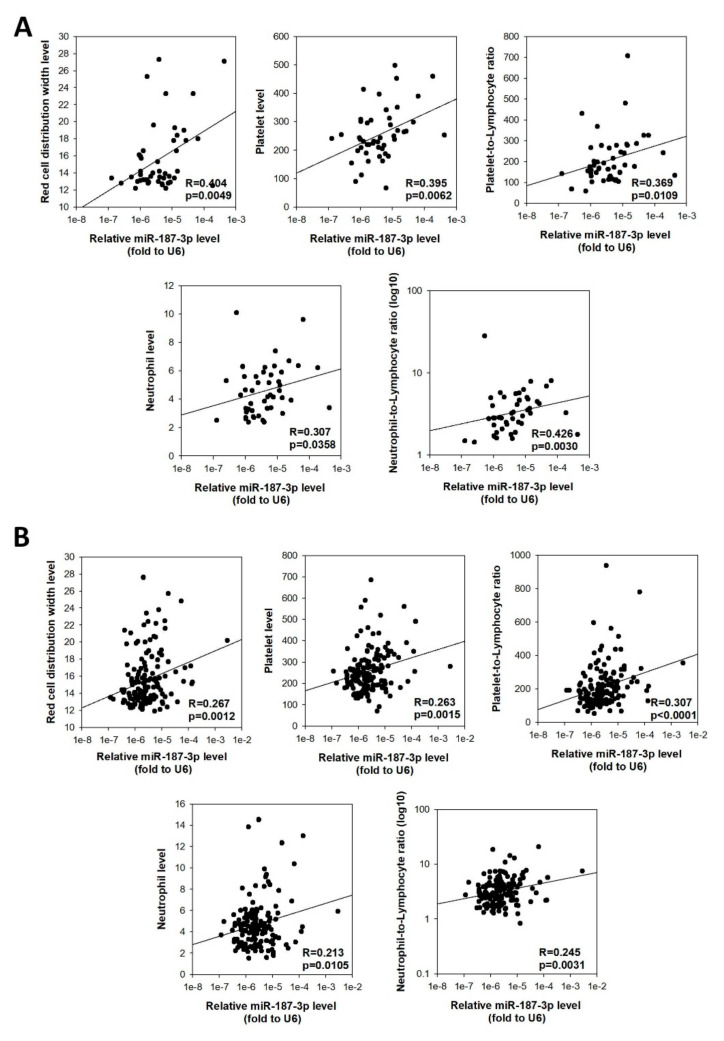
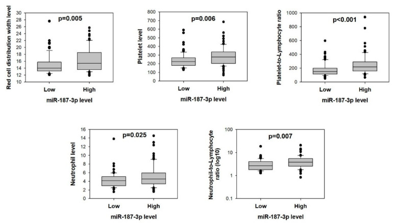
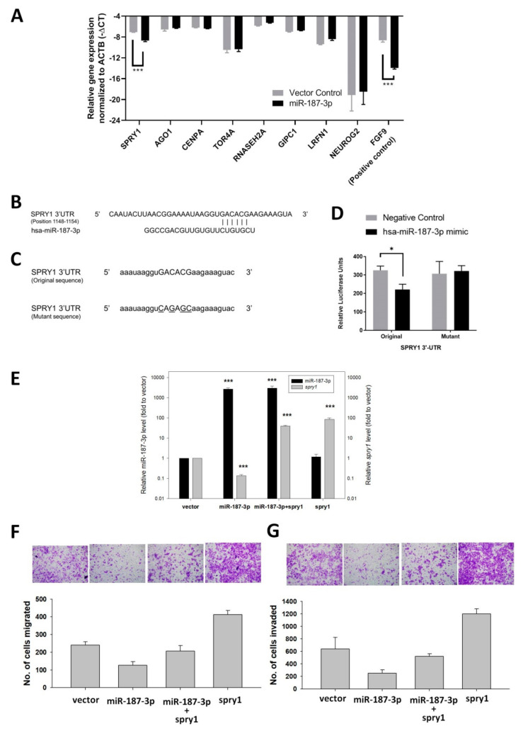
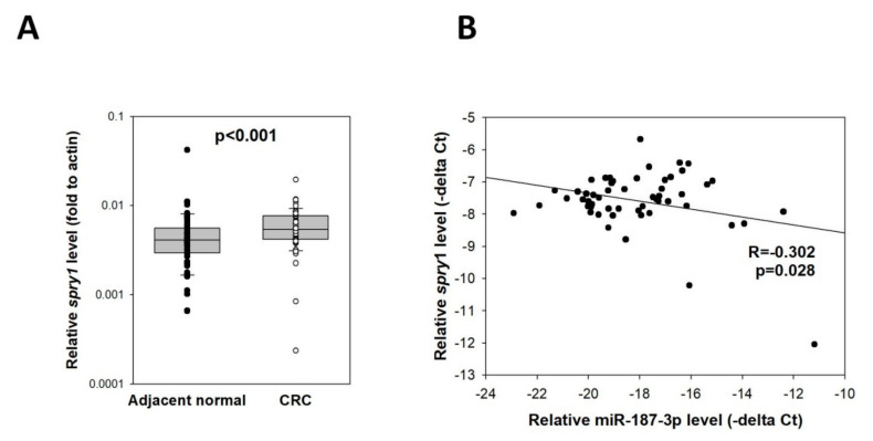
Results: MiR-187-3p was significantly repressed in colorectal polyps and CRC when compared to adjacent normal tissue. Overexpression of miR-187-3p in CRC cell lines impaired colony formation, cell migration, and invasion, and induced chemosensitivity. Clinical analysis revealed that despite miR-187-3p being repressed in CRC, high tumor miR-187-3p levels were positively correlated with tumor stage and disease recurrence. Further analysis showed that miR-187-3p levels were lower in metastatic specimens when compared to paired primary CRC, suggesting that high tumor miR-187-3p levels resulted from the dissemination of metastatic tumor cells. Tumor miR-187-3p levels were positively correlated with peripheral inflammation-related blood markers. Finally, SPRY1 was identified as a novel target gene of miR-187-3p, and was involved in miR-187-3p-impaired CRC metastasis.

Conclusions: This study demonstrated that in spite of its repression and role as a tumor suppressor in CRC, high levels of miR-187-3p in tumors were correlated with poor prognosis and higher levels of peripheral inflammation-related blood markers.

Keywords: colorectal cancer; miR-187-3p; neutrophil; platelet; tumor suppressor.


Conflict of interest statement

The authors declare no conflict of interest.

Figures

Figure 1
Figure 1
MiR-187-3p was repressed in colorectal polyp and CRC tissues: MiR-187-3p levels were determined in synchronous polyp, CRC, and normal mucosa samples from 14 patients by quantitative RT-PCR. MiR-187-3p was significantly repressed in polyp (p = 0.017) and CRC (p = 0.009) tissues when compared to adjacent normal tissue. No significant difference was observed between polyp and CRC tissues. The level of miR-187-3p was expressed as 2(−delta Ct[miR-187-3p–U6]). Experiments were performed in duplicate, and the data are expressed as the mean ± SEM of three independent experiments.
Figure 2
Figure 2
MiR-187-3p overexpression inhibited CRC cell migration and colony formation: (A) MiR-187-3p plasmids or control vectors were stably overexpressed in HCT116 or SW480 cells. Quantitative PCR was performed to determine the expression of miR-187-3p in miR-187-3p or control vector clones. (B) Colony-formation assay was performed to compare the clonogenic ability of stable miR-187-3p and control vector HCT116 or SW480 cells. (C) Wound-healing assay was performed to compare the cell migration rates of stable HCT116 or SW480 cells with miR-187-3p or the control vector 48 h after wound creation. (D) Cell migration assay was performed to compare the migration ability of stable HCT116 or SW480 cells with miR-187-3p or the control vector. (E) Cell invasion assay was performed to compare the invasion ability of stable HCT116 or SW480 cells with miR-187-3p or the control vector. All experiments were performed in duplicate, and the data are expressed as the mean ± SEM of three independent experiments; * indicates that the difference is statistically significant (p < 0.05).
Figure 3
Figure 3
MiR-187-3p overexpression induced response of CRC cells to chemotherapeutic drugs: MTT assay was performed to determine the effect of 72 h treatment with vehicle control (DMSO), 5-fluorouracil (5-FU), oxaliplatin (OXA), or combined 5-FU + OXA on (A) stable HCT116-miR-139-3p or control vector cells, (B) stable HCT116-miR-139-3p or control vector cells, and (C) DLD1 transiently overexpressed miR-187-3p mimic or negative control cells. The left panel shows the miR-187-3p levels in the transfected cells as determined by quantitative PCR. (D) The optical density in terms of absorbance at 570 nm was determined after 72 h drug or vehicle treatment. All experiments were performed in triplicate, and the data are expressed as the mean ± SEM of three independent experiments; * and ** indicate that the difference is statistically significant (p < 0.05 and p < 0.01, respectively).
Figure 4
Figure 4
MiR-187-3p levels were associated with the age and tumor stage of CRC patients: (A) MiR-187-3p levels in paired CRC and adjacent normal tissues of 56 CRC patients in the study cohort. (B) The miR-187-3p levels were significantly lower in patients aged 65 years or above compared to those aged below 65. (C) The miR-187-3p levels were significantly higher in stage II–IV CRCs compared to stage I CRCs. (D) MiR-187-3p levels in paired CRC and adjacent normal tissues of 160 CRC patients in the validation cohort. (E) The miR-187-3p levels were significantly lower in patients aged 65 years or above compared to those aged below 65. (F) The miR-187-3p levels were significantly higher in stage II–IV CRCs compared to stage I CRCs. (G) TCGA analysis showed that miR-187-3p levels were significantly different between different age groups in patients with colon cancer (upper panel) or rectal cancer (lower panel). (H) TCGA analysis showed that miR-187-3p levels were significantly higher or tended to be higher in stage II–IV CRCs compared to stage I CRCs for patients with colon cancer (upper panel) or rectal cancer (lower panel). ∙ and ○ indicated the individual data points of adjacent normal tissues and CRC, respectively.
Figure 5
Figure 5
MiR-187-3p levels were lower in metastatic cancer cells compared to primary CRC cells: (A) MiR-187-3p levels in paired liver metastasis and primary CRC specimens of 10 patients in this study. (B) Data obtained from the GEO dataset GSE98406 showing that miR-187-3p levels were significantly lower in liver metastases comparing to primary CRC cells (n = 14). (C) Data obtained from the GEO dataset GSE56350 showing that miR-187-3p levels were significantly lower in lymph node metastases compared to primary CRC cells (n = 41). (D) Data obtained from the GEO dataset GSE56350 showing that there was a trend of lower miR-187-3p levels in paired liver metastases comparing to primary CRC cells (n = 14). ∙ and ○ indicated the individual data points of adjacent normal tissues and CRC, respectively.
Figure 6
Figure 6
High tumor miR-187-3p levels were correlated with postoperative recurrence: (A) Tumor miR-187-3p levels in CRC patients with no recurrence (n = 35) or with recurrent disease (n = 11) within 5 years following R0 resection for stage I–III CRC. (B) Disease-free survival curves for CRC patients with high or low tumor miR-187-3p levels.
Figure 7
Figure 7
Tumor miR-187-3p levels were correlated with preoperative peripheral inflammation-related blood parameters: Positive correlation was found between tumor miR-187-3p levels and levels of red cell distribution width, platelet count, platelet-to-lymphocyte ratio, neutrophil count, and neutrophil-to-lymphocyte ratio for patients in the (A) study cohort (n = 54) and (B) validation cohort (n = 160).
Figure 8
Figure 8
Higher preoperative peripheral inflammation-related blood parameters in CRC patients with higher tumor miR-187-3p levels: Comparison of red cell distribution width, platelet count, platelet-to-lymphocyte ratio, neutrophil count, and neutrophil-to-lymphocyte ratio between CRC patients with high or low tumor miR-187-3p levels in the validation cohort (n = 160).
Figure 9
Figure 9
Identification of SPRY1 as a target gene of miR-187-3p in vitro: (A) Quantitative RT-PCR-based expression analysis of putative target genes within HCT116 stably transfected with miR-187-3p or the control vector. Expression of target genes was normalized to ACTB and reported as –delta Ct (negative delta Ct). FGF9 was used as a positive control. (B) Predicted binding site of miR-187-3p within the 3′-UTR region of the SPRY1 gene. (C) Design of SPRY1 (original and mutant) insert sequence within the reporter construct. Uppercase: potential binding site; underline: mutated bases within the potential binding site; lowercase: flanking 3′-UTR bases. (D) HCT116 cells were co-transfected with SPRY1 3′-UTR original or mutant sequence reporter constructs with has-miR-187-3p mimic or negative control (empty vector) and Renilla luciferase control vector. The firefly luciferase signal for each transfectant was normalized by its corresponding Renilla luciferase signal and expressed in relative luciferase units. (E) Gene expression levels of miR-187-3p and SPRY1 within the HCT116 control vector cells (vector), stable miR-187-3p cells (miR-187-3p)), stable miR-187-3p cells transiently transfected with the SPRY1 expression plasmid (miR-187-3p+SPRY1), or control vector cells transfected with the SPRY1 expression plasmid (SPRY1). (F,G) Rescue experiment demonstrating the effects of SPRY1 overexpression in HCT116 control vector cells or stable miR-187-3p cells on (F) cell migration and (G) invasion. Data are expressed as the mean ± SEM of three independent experiments; * and *** indicate that the difference is statistically significant (p < 0.05 and p < 0.001, respectively).
Figure 10
Figure 10
Expression of SPRY1 and its correlation with miR-187-3p in CRC tissues: (A) Quantitative RT-PCR was performed to determine the SPRY1 levels in 54 paired CRC specimens and adjacent normal mucosa. (B) Pearson’s correlation analysis in the CRC cohort showed that there was a significant inverse correlation between miR-187-3p and SPRY1 (R = −0.302, p = 0.028). The levels of miR-187-3p were expressed as –delta Ct (miR-187-3p–U6), whereas levels of SPRY1 were expressed as –delta Ct (spry1-actin). ∙ and ○ indicated the individual data points of adjacent normal tissues and CRC, respectively.

References

    1. Bray F., Ferlay J., Soerjomataram I., Siegel R.L., Torre L.A., Jemal A. Global cancer statistics 2018: GLOBOCAN estimates of incidence and mortality worldwide for 36 cancers in 185 countries. CA Cancer J Clin. 2018;68:394–424. doi: 10.3322/caac.21492. - DOI - PubMed
    1. Weitz J., Koch M., Debus J., Hohler T., Galle P.R., Buchler M.W. Colorectal cancer. Lancet. 2005;365:153–165. doi: 10.1016/S0140-6736(05)17706-X. - DOI - PubMed
    1. de la Chapelle A. Genetic predisposition to colorectal cancer. Nat Rev Cancer. 2004;4:769–780. doi: 10.1038/nrc1453. - DOI - PubMed
    1. Oliveto S., Mancino M., Manfrini N., Biffo S. Role of microRNAs in translation regulation and cancer. World J Biol Chem. 2017;8:45–56. doi: 10.4331/wjbc.v8.i1.45. - DOI - PMC - PubMed
    1. Wan T.M., Iyer D.N., Ng L. Roles of microRNAs as non-invasive biomarker and therapeutic target in colorectal cancer. Histol Histopathol. 2020;35:225–237. doi: 10.14670/HH-18-171. - DOI - PubMed

Publication types