. 2022 Jul 27;14(15):3647.
doi: 10.3390/cancers14153647.

Preliminary Discovery of Small Molecule Inhibitors of Epidermal Growth Factor Receptor (EGFR) That Bind to the Extracellular Domain

Affiliations

Preliminary Discovery of Small Molecule Inhibitors of Epidermal Growth Factor Receptor (EGFR) That Bind to the Extracellular Domain

Rosa Di Liddo et al. Cancers (Basel). .

Abstract

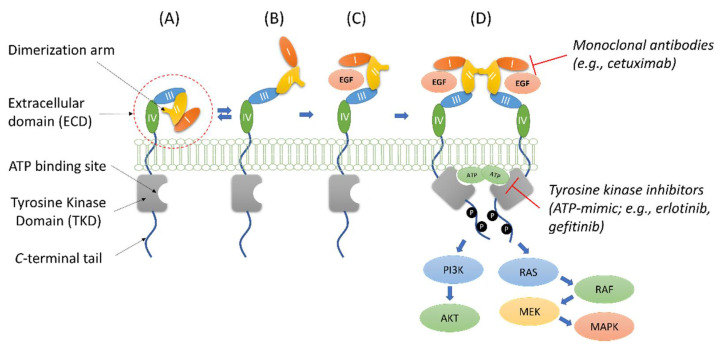
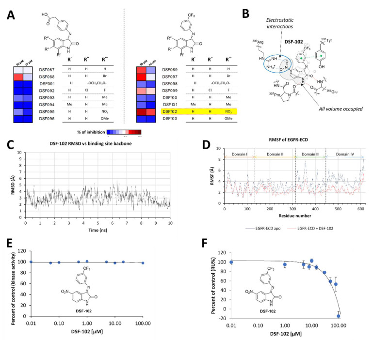
The Epidermal Growth Factor Receptor (EGFR) is a transmembrane glycoprotein belonging to the protein kinase superfamily. It is composed of an extracellular domain, a transmembrane anchoring region and a cytoplasmic region endowed with tyrosine kinase activity. Genetic mutations of EGFR kinase cause higher activity thereby stimulating downstream signaling pathways that, in turn, impact transcription and cell cycle progression. Due to the involvement of mutant EGFR in tumors and inflammatory diseases, in the past decade, several EGFR inhibitory strategies have been extensively studied, either targeting the extracellular domain (through monoclonal antibodies) or the intracellular kinase domain (through ATP-mimic small molecules). Monoclonal antibodies impair the binding to growth factor, the receptor dimerization, and its activation, whereas small molecules block the intracellular catalytic activity. Herein, we describe the development of a novel small molecule, called DSF-102, that interacts with the extracellular domain of EGFR. When tested in vitro in KRAS mutant A549 cells, it impairs EGFR activity by exerting (i) dose-dependent toxicity effects; (ii) a negative regulation of ERK, MAPK p38 and AKT; and (iii) a modulation of the intracellular trafficking and lysosomal degradation of EGFR. Interestingly, DSF-102 exerts its EGFR inhibitory activity without showing interaction with the intracellular kinase domain. Taken together, these findings suggest that DSF-102 is a promising hit compound for the development of a novel class of anti-EGFR compounds, i.e., small molecules able to interact with the extracellular domain of EGFR and useful for overcoming the KRAS-driven resistance to TKI treatment.

Keywords: EGFR-ECD; inhibition; isatin; trafficking.


Conflict of interest statement

The authors declare no conflict of interest.

Figures

Figure 1
Figure 1
Structure and simplified dynamics representation of EGFR. The auto-tethered fully inactive conformation of EGFR (A) is in equilibrium with a partially opened state (B) that can be stabilized by the binding with EGF (C). The EGF-activated conformation can dimerize (D) leading to receptor autophosphorylation and activation of the AKT and MAPK cascades that mediate cell survival and proliferation. The target regions for monoclonal antibodies and ATP-mimic compounds are also reported in (D).
Figure 2
Figure 2
(A) Heat-map representing the percentage of EGFR dimerization inhibition at 50 and 10 µM. Structures and percentage of inhibition at 50 µM for the most interesting isatin Schiff bases are reported. (B) Top: binding site predicted for DSF-069 (highlighted with magenta circles) on EGFR-ECD (surface representation). Glycans are depicted as gray spheres. Bottom: schematic representation of the binding site for isatin Schiff bases on EGFR-ECD and proposed mechanism of action, i.e., block of the receptor in the inactive/closed conformation. (C) Details of the interactions (docking simulation) between DSF-069 and EGFR-ECD. Hydrogen bonds are depicted as red dashed lines; π-π interaction is depicted as green dashed line. The volume unoccupied by DSF-069 is highlighted in gray. The possibility to functionalize the molecule with an electron-dense group is also shown.
Scheme 1
Scheme 1
Synthesis of isatin Schiff-base derivatives. Reaction conditions: (a) chloral hydrate, NH2OH·HCl, Na2SO4, 1M HCl, 80 °C, 2 h; (b) conc. H2SO4, 60 °C, 15 min; (c) (4-aminophenyl)acetic acid or 3-aminobenzotrifluoride, AcOH, ethanol, reflux or MW irradiation. See Supplementary Materials for details. The structures of the initial hit (DSF-069) and of the most active compound in the series (DSF-102; vide post) are explicitly reported on the right.
Figure 3
Figure 3
(A) Heat-map representing the percentage of EGFR dimerization inhibition at 50 and 10 µM. (B) Binding mode proposed for DSF-102 and EGFR-ECD. Hydrogen bonds are depicted as red dashed lines; π-π interaction are depicted as green dashed lines; electrostatic interactions are depicted as blue dashed lines. The binding mode of DSF-069 (light gray) is also reported to show how DSF-102 resulted slightly shifted with respect to the parent compound, thus leading to a higher complementarity with the binding pocket. (C) Root mean squared deviation (RMSD, Å) for DSF-102 during 10 ns of MD simulation. (D) Comparison of per-residue root mean squared fluctuation (RMSF, Å) obtained during 10 ns of MD simulation of apo EGFR-ECD (blue line) and DSF-102 bound EGFR-ECD (red line). (E) Residual catalytic activity (%) of the isolated kinase domain of EGFR upon treatment with DSF-102. Please note that no inhibition was observed up to 100 µM. (F) DSF-102 dependent inhibition of the binding between EGF and isolated EGFR-ECD as measured by SPR analysis.
Figure 4
Figure 4
Study of the biological activity of DSF-102 (2 µM or 50 µM) on A549 cell line. Assessment of cell death by (A) optical microscopy analysis (scale bar: 25 µm) and (B) apoptosis analysis by flow cytometry after 24 h of stimulation. A549 cells kept under resting conditions or treated with cetuximab (0.07 µM) (B1, B2), or primary dermal fibroblasts (NDFa) (B3) were used as controls. Percentage of apoptotic cells were discriminated using TO/PI assay according to the manufacturer’s instructions. (C) Evaluation of phospho AKT 1(Ser473) expression by flow cytometry at different time points (15, 30, 60 min) from stimulation with DSF-102 (2 µM or 50 µM). Resting cells and samples treated with cetuximab (10 µg/mL; 0.07 µM) were used as reference. Flow cytometry data were expressed as MFI ± SEM. * (p ≤ 0.05); ** (p ≤ 0.01).
Figure 5
Figure 5
(A) Confocal microscopy images showing the trafficking kinetics of membrane EGFR pH (green) under resting conditions and after treatment (from 15 to 60 min) with DSF-102 (50 µM) or cetuximab. Cellular nuclei were counterstained with DAPI (blue). Scale bar: 10 µm. (B) Immunofluorescence analysis of EEA1, Rab7 and LAMP1 (red) in fixed and permeabilized cultures compared to resting cells, at 60 min after treatment with DSF-102 (50 µM). Scale bar: 10 µm.
Figure 6
Figure 6
Flow cytometry analysis of (A) membrane EGFR (green line) and (B) intracellular EGF (red line) at 60 min of stimulation with 50 µM DSF-102 alone or combined with brefeldin A (BFA/DSF-102). Resting cells and BFA-treated samples were used as controls. To detect only intracellular EGF, flow cytometry was coupled with acid washing. Data were expressed as MFI ± SEM. ** (p ≤ 0.01).
Figure 7
Figure 7
Flow cytometry analysis of DSF-102 activity on EGFR signaling. (A) p44/42 ERK and (B) p38 MAPK activation at 30 and 60 min following the co-administration of DSF-102 (50 µM) and EGF (100 ng/mL). Samples treated with only EGF or DSF-102, and resting cells were used as controls. Data were expressed as MFI ± SEM. * (p ≤ 0.05); ** (p ≤ 0.01).
Figure 8
Figure 8
Confocal microscopy evaluation. Images show the localization of EGFR (green) and lysosomal marker LAMP1 (red) in A549 cells resting (time 0′) (A) or treated for 60 min with DSF-102 (50 µM) and/or EGF (100 ng/mL) (B). For each picture, a region of interest of was included at higher magnification. Nuclei were counterstained with DAPI (blue). Scale bar: 10 µm.

Similar articles

Cited by

References

    1. Lemmon M.A., Schlessinger J. Cell Signaling by Receptor Tyrosine Kinases. Cell. 2010;141:1117–1134. doi: 10.1016/j.cell.2010.06.011. - DOI - PMC - PubMed
    1. Lemmon M.A., Schlessinger J., Ferguson K.M. The EGFR family: Not so prototypical receptor tyrosine kinases. Cold Spring Harb. Perspect. Biol. 2014;6:a020768. doi: 10.1101/cshperspect.a020768. - DOI - PMC - PubMed
    1. Low-Nam S.T., Lidke K.A., Cutler P.J., Roovers R.C., van Bergen en Henegouwen P.M., Wilson B.S., Lidke D.S. ErbB1 dimerization is promoted by domain co-confinement and stabilized by ligand binding. Nat. Struct. Mol. Biol. 2011;18:1244–1249. doi: 10.1038/nsmb.2135. - DOI - PMC - PubMed
    1. Kaszuba K., Grzybek M., Orłowski A., Danne R., Róg T., Simons K., Coskun Ü., Vattulainen I. N-Glycosylation as determinant of epidermal growth factor receptor conformation in membranes. Proc. Natl. Acad. Sci. USA. 2015;112:4334–4339. doi: 10.1073/pnas.1503262112. - DOI - PMC - PubMed
    1. Kaplan M., Narasimhan S., de Heus C., Mance D., van Doorn S., Houben K., Popov-Čeleketić D., Damman R., Katrukha E.A., Jain P., et al. EGFR Dynamics Change during Activation in Native Membranes as Revealed by NMR. Cell. 2016;167:1241–1251.e1211. doi: 10.1016/j.cell.2016.10.038. - DOI - PubMed