Review
. 2022 Jul 27;14(15):3652.
doi: 10.3390/cancers14153652.

Leydig Cell Tumors of the Testis: An Update of the Imaging Characteristics of a Not So Rare Lesion

Affiliations
Review

Leydig Cell Tumors of the Testis: An Update of the Imaging Characteristics of a Not So Rare Lesion

Florian Maxwell et al. Cancers (Basel). .

Abstract

Pre-operative testicular tumor characterization is a challenge for radiologists and urologists. New data concerning imaging approaches or immunochemistry markers improve the management of patients presenting with a testicular tumor, sometimes avoiding radical orchiectomy. In the past 20 years, imaging modalities, especially ultrasound (US) and magnetic resonance imaging (MRI), improved, allowing for great progress in lesion characterization. Leydig cell tumors (LCT) are rare testicular tumors developing from the stromal tissue, with relatively scarce literature, as most of the studies focus on the much more frequent germ cell tumors. However, with the increase in testicular sonography numbers, the incidence of LCT appears much higher than expected, with some studies reporting up to 22% of small testicular nodules. Multimodal ultrasound using Doppler, Elastography, or injection of contrast media can provide crucial arguments to differentiate LCT from germ cell tumors. Multiparametric MRI is a second intention exam, but it allows for extraction of quantifiable data to assess the diagnosis of LCT. The aims of this article are to review the latest data regarding LCT imaging features, using multimodal ultrasound and multiparametric MRI, and to focus on the peculiar aspect of the testis of patients with Klinefelter's syndrome. The possibility of an LCT should be raised in front of a small hypoechoic tumor with a marked corbelling hypervascularization in an otherwise normal testicular pulp. Ultrasonographic modules, such as ultrasensitive Doppler, contrast-enhanced ultrasonography, or elastography, can be used to reinforce the suspicion of LCT. MRI provides objective data regarding vascularization and enhancement kinetics.

Keywords: Doppler; Klinefelter’s syndrome (KS); Leydig cell hyperplasia; Leydig cell tumors (LCT); contrast-enhanced ultrasound (CEUS); elastography; magnetic resonance imaging (MRI); testicular stromal tumors; ultrasound (US).


Conflict of interest statement

The Authors declare no conflict of interest.

Figures

Figure 1
Figure 1
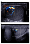
28-year-old patient presenting with left testicular pain (significant left varicocele during the exam). Typical LCT 12 × 9 mm on B mode us (a) and color Doppler (b) discovered on the right testis. Well-defined lobulated solid lesion moderately hypoechoic and homogeneous echo structure with normal adjacent pulp and absence of microlithiasis. The lesion is hyper vascularized with a mixed peripheral and internal pattern. (c) From left to right: Macroscopic view of the patient’s LCT after enucleation. The typical “golden brown” color of the lesion often allows the surgeon and the pathologist to confirm the diagnosis during surgery. HE × 30 Hematein–Eosin coloration showing a high cellular density with no necrosis. HE × 40 with an endothelial cell marker anti CD-31, showing a rich vascularization of the tumor. Courtesy of Pr S. Ferlicot, Department of Anatomo-pathology, Bicêtre Hospital.
Figure 1
Figure 1
28-year-old patient presenting with left testicular pain (significant left varicocele during the exam). Typical LCT 12 × 9 mm on B mode us (a) and color Doppler (b) discovered on the right testis. Well-defined lobulated solid lesion moderately hypoechoic and homogeneous echo structure with normal adjacent pulp and absence of microlithiasis. The lesion is hyper vascularized with a mixed peripheral and internal pattern. (c) From left to right: Macroscopic view of the patient’s LCT after enucleation. The typical “golden brown” color of the lesion often allows the surgeon and the pathologist to confirm the diagnosis during surgery. HE × 30 Hematein–Eosin coloration showing a high cellular density with no necrosis. HE × 40 with an endothelial cell marker anti CD-31, showing a rich vascularization of the tumor. Courtesy of Pr S. Ferlicot, Department of Anatomo-pathology, Bicêtre Hospital.
Figure 2
Figure 2
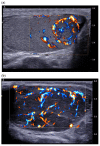
(a) Same lesion as Figure 1 with ultrasensitive Doppler allowing better characterization of corbelling vascular architecture. (b) Small LCT of 7 mm, marked hyper vascularization compared with adjacent pulp, with main feeding vessel pattern.
Figure 3
Figure 3
(a) Typical LCT 16 × 14 mm with mixed corbelling and central vascularization pattern in a 42-year-old patient presenting for infertility work up. The large size of the lesion helps to highlight this typical pattern. (b) 31-year-old patient presenting for right testicular mass: 38 mm typical seminoma with a radial trans-lesional « anarchic » vascularization pattern.
Figure 4
Figure 4
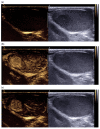
Contrast-enhanced ultrasound using Sonovue® of a typical LCT (a) t = 0 s (b) t = 30 s (c) t = 60 s found in a 45-year-old patient addressed for infertility work up.
Figure 5
Figure 5
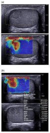
Same patient as Figure 1: Shear wave elastography of typical LCT, qualitative (a) and quantitative (b) evaluation of stiffness.
Figure 6
Figure 6
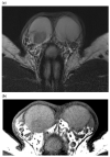
44-year-old patient, infertility work up, right testicular tumor on first line sonography. MRI (3T) aspect of a typical LCT. (a) T2 weighted sequence, Axial: LCT of the right testis appears as a solid well-circumscribed lesion with a sharp demarcation with the rest of the testis and a homogeneous marked hypo T2 signal. Note the absence of alteration of the signal of the testicular pulp apart from the LCT. (b) T1 weighted sequence, Axial: LCT has a homogeneous hypo T1 signal, with no cystic or hemorrhagic component.
Figure 7
Figure 7
Same patient as Figure 6. Diffusion-weighted sequence b800 and ADC map.
Figure 8
Figure 8
(a) Same patient as Figure 6. T1 weighted sequence with fat suppression and after dynamic gadolinium contrast media injection. Fast and intense uptake of contrast media before the rest of the testis and wash-out on the late phase (6 min post contrast media injection). (b) Post treatment using Syngovia® workstation: Dynamic enhancement curve shows a shorter and a higher time to peak, followed by a prolonged wash-out and a higher area under curve compared to normal pulp.
Figure 9
Figure 9
26-year-old patient addressed for infertility work up/Klinefelter’s syndrome follow up. Leydig cell hyperplasia in a patient with Klinefelter’s syndrome. (a) Two patients with major testicular hypotrophy (2 mL), with coarse echostructure and micronodules mostly hyperechoic. (b) Color Doppler ultrasound showing diffuse hypervascularization of the testicle, which contrasts with the relatively hypovascularized aspect of undescended testicles.

References

    1. Kim I., Young R.H., Scully R.E. Leydig cell tumors of the testis. A clinicopathological analysis of 40 cases and review of the literature. Am. J. Surg. Pathol. 1985;9:177–192. doi: 10.1097/00000478-198503000-00002. - DOI - PubMed
    1. Isidori A.M., Pozza C., Gianfrilli D., Giannetta E., Lemma A., Pofi R., Barbagallo F., Manganaro L., Martino G., Lombardo F., et al. Differential Diagnosis of Nonpalpable Testicular Lesions: Qualitative and Quantitative Contrast-enhanced US of Benign and Malignant Testicular Tumors. Radiology. 2014;273:606–618. doi: 10.1148/radiol.14132718. - DOI - PubMed
    1. Colecchia M., Nistal M., Gonzalez-Peramato P., Carmignani L., Salvioni R., Nicolai N., Regadera J. Leydig cell tumor and hyperplasia: A review. Anal. Quant. Cytol. Histol. 2007;29:139–147. - PubMed
    1. Hendry J., Fraser S., White J., Rajan P., Hendry D.S. Retroperitoneal lymph node dissection (RPLND) for malignant phenotype Leydig cell tumours of the testis: A 10-year experience. SpringerPlus. 2015;4:20. doi: 10.1186/s40064-014-0781-x. - DOI - PMC - PubMed
    1. Ruf C.G., Sanatgar N., Isbarn H., Ruf B., Simon J., Fankhauser C.D., Dieckmann K.-P. Leydig-cell tumour of the testis: Retrospective analysis of clinical and therapeutic features in 204 cases. World J. Urol. 2020;38:2857–2862. doi: 10.1007/s00345-020-03079-1. - DOI - PubMed

LinkOut - more resources