. 2022 Jul 31;14(15):3737.
doi: 10.3390/cancers14153737.

Cancer Associated Fibroblast (CAF) Regulation of PDAC Parenchymal (CPC) and CSC Phenotypes Is Modulated by ECM Composition

Affiliations

Cancer Associated Fibroblast (CAF) Regulation of PDAC Parenchymal (CPC) and CSC Phenotypes Is Modulated by ECM Composition

Stefania Cannone et al. Cancers (Basel). .

Abstract

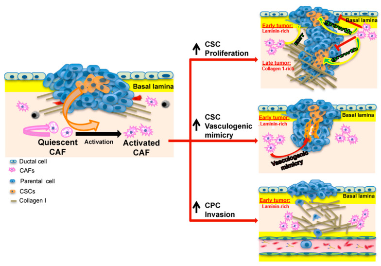
Background: Pancreatic ductal adenocarcinoma (PDAC) is one of the deadliest of all cancers, having one of the lowest five-year survival rates. One of its hallmarks is a dense desmoplastic stroma consisting in the abnormal accumulation of extracellular matrix (ECM) components, especially Collagen I. This highly fibrotic stroma embeds the bulk cancer (parenchymal) cells (CPCs), cancer stem cells (CSCs) and the main producers of the stromal reaction, the Cancer Associated Fibroblasts (CAFs). Little is known about the role of the acellular ECM in the interplay of the CAFs with the different tumor cell types in determining their phenotypic plasticity and eventual cell fate.

Methods: Here, we analyzed the role of ECM collagen I in modulating the effect of CAF-derived signals by incubating PDAC CPCs and CSCs grown on ECM mimicking early (low collagen I levels) and late (high collagen I levels) stage PDAC stroma with conditioned medium from primary cultured CAFs derived from patients with PDAC in a previously described three-dimensional (3D) organotypic model of PDAC.

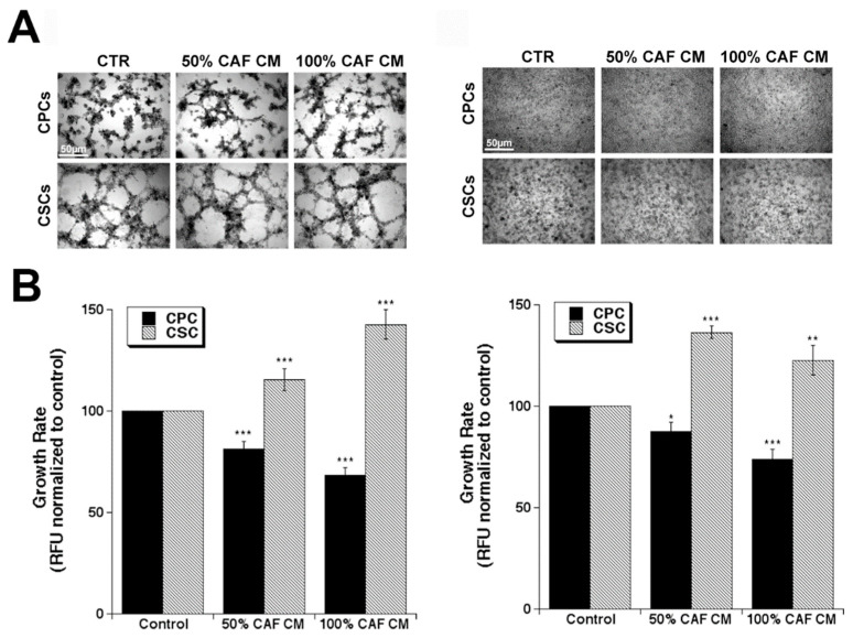
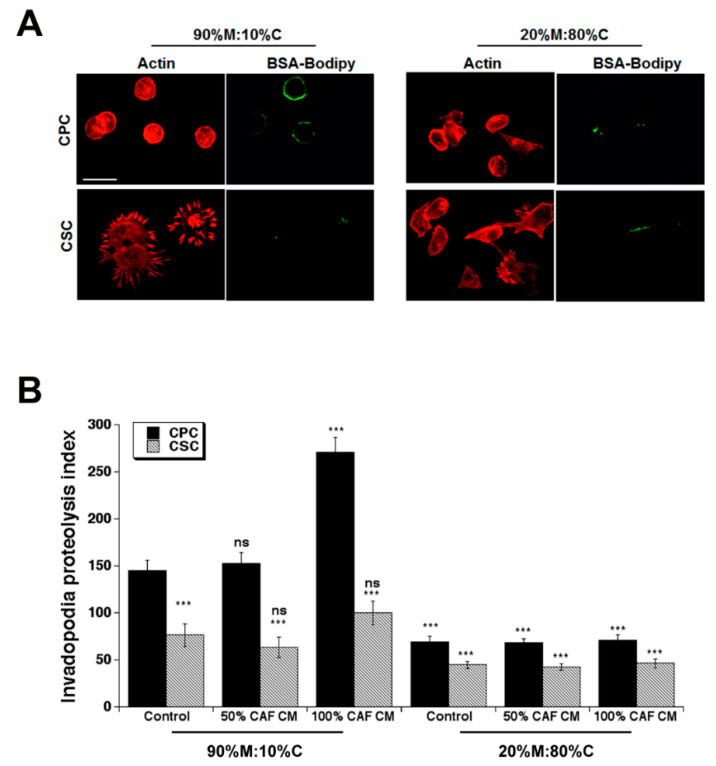
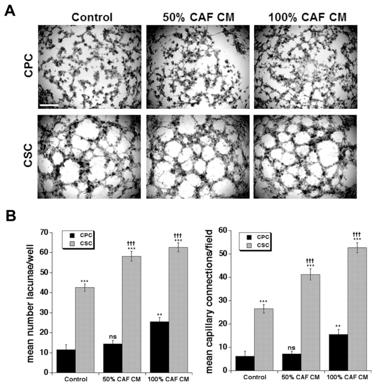
Results: We found that CAFs (1) reduced CPC growth while favoring CSC growth independently of the ECM; (2) increased the invasive capacity of only CPCs on the ECM mimicking the early tumor; and (3) favored vasculogenic mimicry (VM) especially of the CSCs on the ECM mimicking an early tumor.

Conclusions: We conclude that the CAFs and acellular stromal components interact to modulate the tumor behaviors of the PDAC CPC and CSC cell types and drive metastatic progression by stimulating the phenotypic characteristics of each tumor cell type that contribute to metastasis.

Keywords: 3D organotypic cultures; desmoplastic reaction; invadopodia; pancreatic ductal adenocarcinoma; vasculogenic mimicry.


Conflict of interest statement

The authors declare no conflict of interest. The funders had no role in the design of the study; in the collection, analyses, or interpretation of data; in the writing of the manuscript; or in the decision to publish the results.

Figures

Figure 1
Figure 1
CAF conditioned medium inhibits CPC viability and stimulates CSC viability on both ECM compositions. (A) Representative microscopic images of growth morphology of Panc1 CPCs and their derived CSCs after 5 days of culture on organotypic cultures composed of 90% Matrigel:10% Collagen I (left panel) and 20% Matrigel: 80% Collagen I (right panel). Scale bar represents 50 µm for all images. (B) CPC and CSC viability in organotypic cultures of 90% Matrigel:10% Collagen I (left panel) and 20% Matrigel:80% Collagen I (right panel) were calculated from Resazurin reduction assays and are normalized to the control as 100 as described in Materials and Methods. Data are mean ± SEM, n = 5, * p < 0.05, ** p < 0.01, *** p < 0.001 to the control of each cell type on each matrix.
Figure 2
Figure 2
ECM composition modifies the effect of CAF conditioned medium on parenchymal (CPC) and CSC invadopodia ECM degradation. (A) Representative microscopic images of actin (red) and invadopodia-dependent proteolysis of BSA-Bodipy dissolved in the ECM (green) as described in the Methods. Scale bar represents 50 µm for all images. (B) CPC and CSC invadopodial proteolysis rates in organotypic cultures of 90% Matrigel:10% Collagen I and 20% Matrigel: 80% Collagen I calculated as described in Methods. Data are mean ± SEM, n = 5, ns non significant, *** p < 0.001 compared to the control for each cell line on 90% Matrigel:10% collagen I.
Figure 3
Figure 3
CAF conditioned medium stimulates the vascular-like morphology (VM) of both CSCs and CPCs grown on 90% Matrigel:10% Collagen I. ECM composition modifies the effect of CAF CM on vasculogenic mimicry in CPCs and CSCs. Cells were grown on 90% Matrigel: 10% Collagen I and after 24 h to permit their adherence the cultures were incubated with CAF conditioned medium and the cells cultured an additional 5 days and VM measured as described in Materials and Methods. (A) Representative microscopic images of growth morphology of CPCs and their derived CSCs cultured on organotypic cultures composed of 90% Matrigel:10% Collagen I for 5 days with their growth medium or with either 50% or 100% of the CAF conditioned medium. Scale bar represents 50 µm for all images. (B) After 6 days in these growth conditions, vascular channel networks were analyzed as described in Material and Methods for mean number of lacunae per well (left panel) and mean number of capillary connections per field (right panel). Mean ± SEM from three independent experiments, ns, non significant, ** p < 0.01, *** p < 0.001 compared to the CPC control; ††† p < 0.001 CSCs compared to their control.
Figure 4
Figure 4
Model of Influence of the ECM composition on the CAF-dependent regulation of CPC and CSC plasticity. Matrigel induces the formation of autocrine loops in CSCs. Indeed, on Matrigel CSCs secrete a high amount of potent proangiogenic and growth factors (i.e., PDGF, MMP9, IL8, EGF, HGF, bFGF, ET-1) [5], which, via paracrine mechanisms, stimulate CAF growth [20], which in turn secrete factors that increase CSC growth/self-renewal and vasculogenic mimicry while decreasing CPC growth [24,28] but increasing CPC invasion [24,25,26,27]. This creates a vicious positive-feedback growth cycle between the CAFs and CSCs to increase the stemness of the tumor while exacerbating the aggressive angiogenesis phenotype of the CSCs and invasive phenotype of the CPCs.

References

    1. Hong S.M., Park J.Y., Hruban R.H., Goggins M. Molecular signatures of pancreatic cancer. Arch. Pathol. Lab. Med. 2011;135:716–727. doi: 10.5858/2010-0566-RA.1. - DOI - PMC - PubMed
    1. Di Marco M., Grassi E., Durante S., Vecchiarelli S., Palloni A., Macchini M., Casadei R., Ricci C., Panzacchi R., Santini D., et al. State of the art biological therapies in pancreatic cancer. World J. Gastrointest. Oncol. 2016;8:55–66. doi: 10.4251/wjgo.v8.i1.55. - DOI - PMC - PubMed
    1. Siegel R.L., Miller K.D., Jemal A. Cancer Statistics. CA Cancer J. Clin. 2017;67:7–30. doi: 10.3322/caac.21387. - DOI - PubMed
    1. Schober M., Jesenofsky R., Faissner R., Weidenauer C., Hagmann W., Michl P., Heuchel R.L., Haas S.L., Löhr J.-M. Desmoplasia and chemoresistance in pancreatic cancer. Cancers. 2014;6:2137–2154. doi: 10.3390/cancers6042137. - DOI - PMC - PubMed
    1. Biondani G., Zeeberg K., Greco M.R., Cannone S., Dando I., Pozza E.D., Mastrodonato M., Forciniti S., Casavola V., Palmieri M., et al. Extracellular Matrix composition modulates PDAC parenchymal and stem cell plasticity and behavior through the secretome. FEBS J. 2018;285:2104–2124. doi: 10.1111/febs.14471. - DOI - PubMed

LinkOut - more resources