. 2022 Aug 2;14(15):3769.
doi: 10.3390/cancers14153769.

Refining the Role of Pyruvate Dehydrogenase Kinases in Glioblastoma Development

Affiliations

Refining the Role of Pyruvate Dehydrogenase Kinases in Glioblastoma Development

Claire M Larrieu et al. Cancers (Basel). .

Abstract

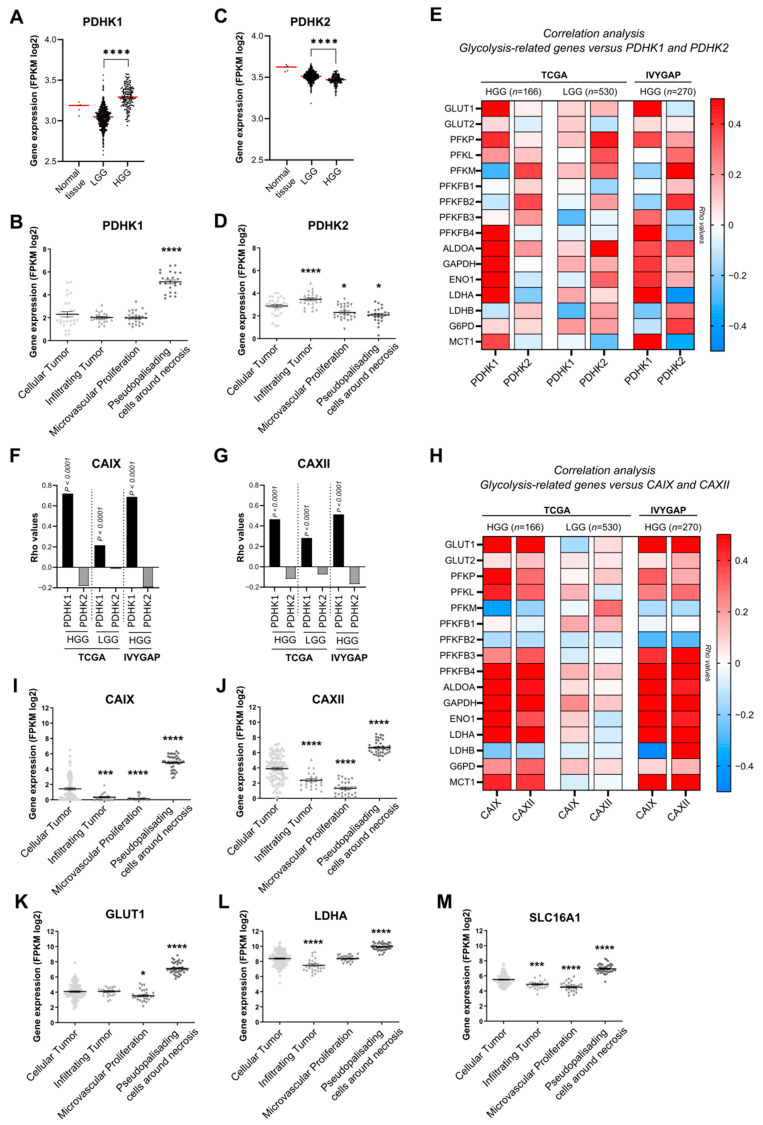
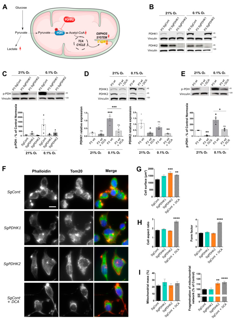
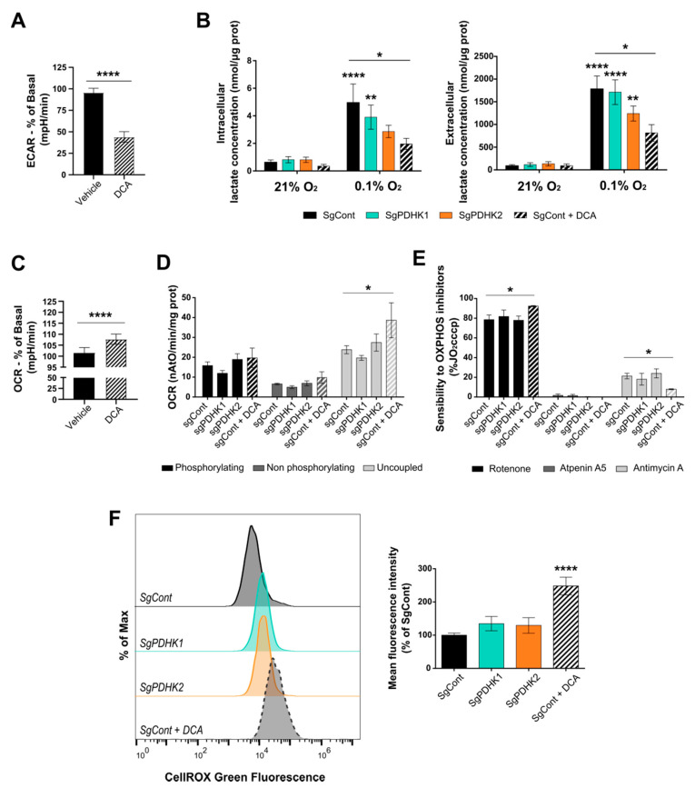
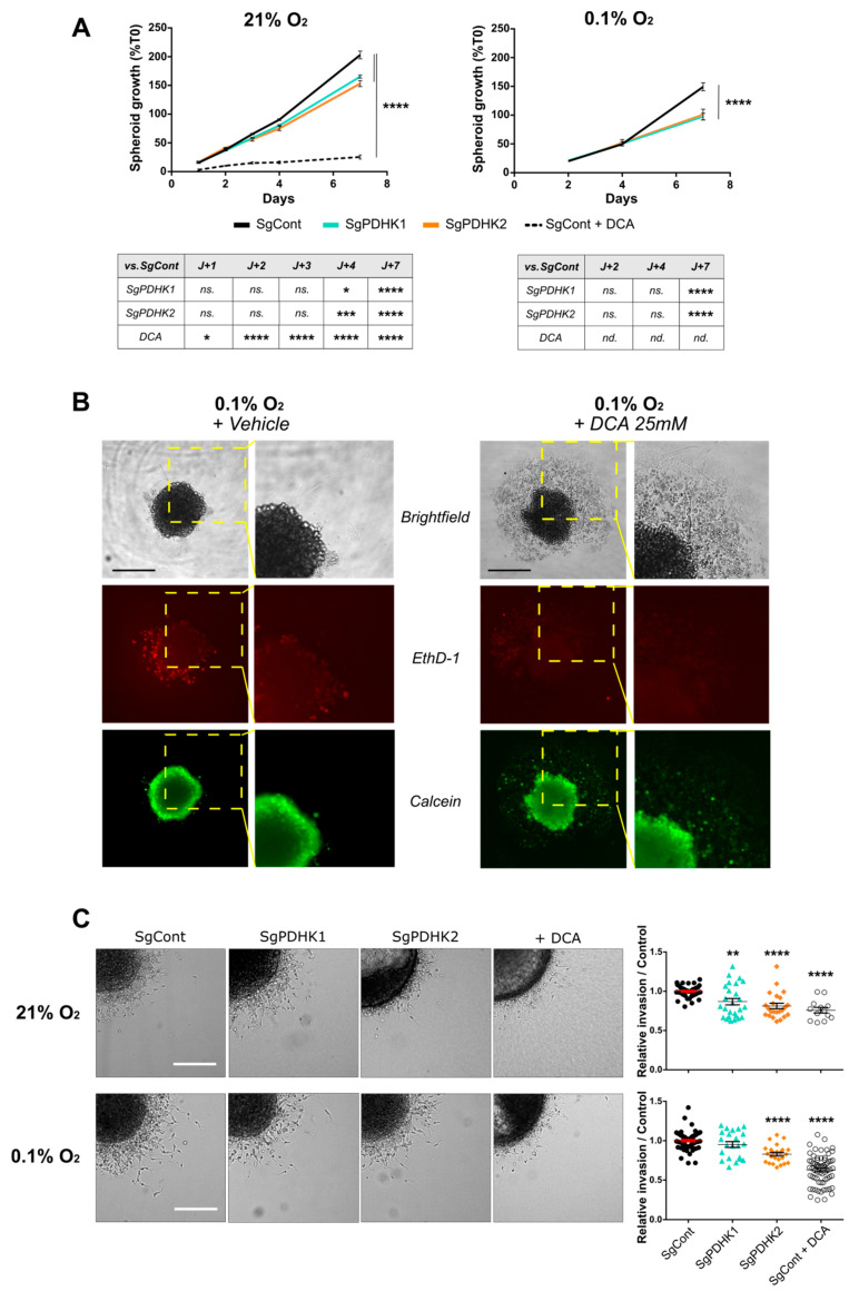
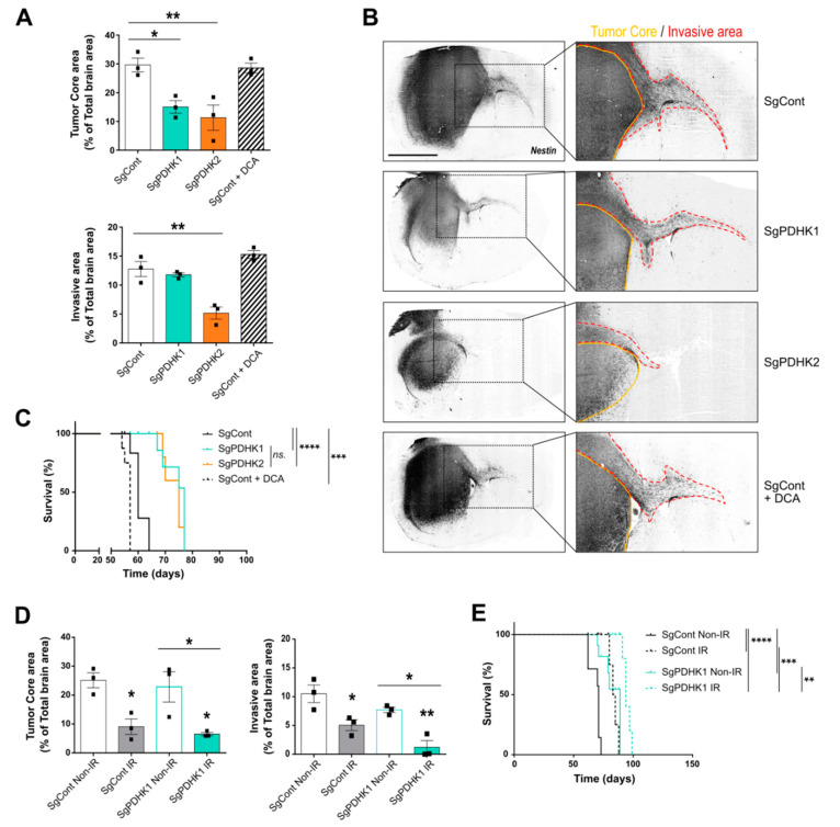
Glioblastoma (GB) are the most frequent brain cancers. Aggressive growth and limited treatment options induce a median survival of 12-15 months. In addition to highly proliferative and invasive properties, GB cells show cancer-associated metabolic characteristics such as increased aerobic glycolysis. Pyruvate dehydrogenase (PDH) is a key enzyme complex at the crossroads between lactic fermentation and oxidative pathways, finely regulated by PDH kinases (PDHKs). PDHKs are often overexpressed in cancer cells to facilitate high glycolytic flux. We hypothesized that targeting PDHKs, by disturbing cancer metabolic homeostasis, would alter GB progression and render cells vulnerable to additional cancer treatment. Using patient databases, distinct expression patterns of PDHK1 and PDHK2 in GB tissues were obvious. To disturb protumoral glycolysis, we modulated PDH activity through the genetic or pharmacological inhibition of PDHK in patient-derived stem-like spheroids. Striking effects of PDHKs inhibition using dichloroacetate were observed in vitro on cell morphology and metabolism, resulting in increased intracellular ROS levels and decreased proliferation and invasion. In vivo findings confirmed a reduction in tumor size and better survival of mice implanted with PDHK1 and PDHK2 knockout cells. Adding a radiotherapeutic protocol further resulted in a reduction in tumor size and improved mouse survival in our model.

Keywords: DCA; glioblastoma; invasion; lactate; pyruvate dehydrogenase kinases.


Conflict of interest statement

The authors declare no conflict of interest.

Figures

Figure 1
Figure 1
Expression and correlation analysis of PDHK1, PDHK2 and genes involved in glucose metabolism in HGG and LGG. (A) Expression levels of PDHK1 in normal tissue, low-grade glioma (LGG) and high-grade glioma (HGG) (TCGA). (B) PDHK1 expression levels and localization in HGG tissue (IVYGAP). (C) Expression levels of PDHK2 in normal tissue, LGG and HGG (TCGA). (D) PDHK2 expression levels and localization in HGG tissue (IVYGAP). (E) Correlation analysis of genes involved in glycolysis with PDHK1 and PDHK2 in HGG and LGG from TCGA and HGG from TCGA + IVYGAP. (F) Correlation analysis of CAIX with PDHK1 and PDHK2 in HGG and LGG from TCGA and HGG from IVYGAP. (G) Correlation analysis of CAXII with PDHK1 and PDHK2 in HGG and LGG from TCGA and HGG (TCGA + IVYGAP). (H) Correlation analysis of genes involved in glycolysis with CAIX and CAXII (TCGA + IVYGAP). (I) CAIX and (J) CAXII expression levels and localization in HGG tissue (IVYGAP). (K) GLUT1, (L) LDHA and (M) SLC16A1 expression levels and localization in HGG tissue defined by the IVYGAP database. Data are expressed as the mean ± SEM and p-values were obtained using unpaired t-tests (A,B), Spearman’s correlation coefficient tests (EH) and one-way ANOVA (B,D,IM); * p < 0.05; *** p < 0.001; **** p < 0.0001.
Figure 2
Figure 2
Dichloroacetate (DCA) reduces levels of phosphorylated PDH in a primary glioblastoma in vitro cell model, impacting cell and mitochondrial organization. (A) Illustration of the two alternative pyruvate utilization pathways and the impact of PDHK activity on the balance between lactic fermentation or mitochondrial oxidation of pyruvate. (BE) Western blotting and corresponding quantification of PDHK1, PDHK2 expression or phospho-PDH status in P3 cells control, knockout for PDHK1, PDHK2 or treated with DCA 25 mM for 3 days; all cells were exposed to either 21% O2 or 0.1% O2. Graphs represent densitometry quantification normalized with vinculin (n = 3 or 4). (F) Epifluorescence images of immunofluorescence stainings on P3 SgCont, SgPDHK1, SgPDHK2 or treated with DCA 25 mM for 3 days (Phalloidin, red; Tom20, green; Hoechst, blue). Scale bar: 20 µm. (G,H) Immunofluorescence image analysis (120 to 240 cells per group): (G) cell surface quantification; (H, left) cell aspect ratio corresponding to the ratio of the major axis to minor axis of the cells; (H, right) form factor imaging branching of the cells. (I) Mitochondrial network analysis based on immunofluorescence pictures; (left) mitochondrial mass expressed as the percentage of total cell area (39 to 99 cells per group); (right) quantification of mitochondrial network fragmentation corresponding to the ratio of the number of mitochondria per cell on the area of the full mitochondrial network, normalized by the mean of control group and expressed as the percentage of control. Data are expressed as the mean ± SEM and p-values were obtained using one-way ANOVA (D,G,H,I left) or unpaired t-tests (C,E,I right); * p < 0.05; ** p < 0.01; *** p < 0.001; **** p < 0.0001; ns.: not significant.
Figure 3
Figure 3
DCA disturbs GB cell metabolism and induces ROS production. (A) The extracellular acidification rate (ECAR) was measured in real time upon the addition of 20 mM DCA/vehicle to P3 wt cells. Values are presented as the percentage change compared with basal ECAR prior to DCA addition. (B) Intra- (left) and extracellular (right) lactate measurement in P3 SgCont, SgPDHK1, SgPDHK2 or with DCA treatment (25 mM for 3 days), upon 21% or 0.1% O2 exposure; n = 7 to 8 for each condition; when not noticed, significance was assessed by comparing with control cells 21% O2. (C) The oxygen consumption rate (OCR) was measured in real time upon the addition of 20 mM DCA/vehicle to P3 wt cells. Values are presented as the percentage change compared with basal OCR prior to DCA addition. (D) Oxygraphy analysis of P3 cells in a phosphorylating state (basal cell respiration), non-phosphorylating state (oligomycin addition) or uncoupled state (CCCP addition); n = 4. (E) Sensitivity in P3 cells to specific OxPhos complex inhibitors rotenone (complex I), atpeninA5 (complex II) and antimycin A (complex III); n = 4. The addition of inhibitors was performed upon uncoupled respiration conditions and results are expressed as the percentage of uncoupled respiration prior inhibitor addition (%JO2cccp). (F) ROS production assessed in triplicates with CellROX Green probe in PDHK1 or PDHK2 KO cells, or in control cells exposed to 3 days of DCA treatment (25 mM). Left panel: Representative histograms in modal scaling (percentage of Max). Right panel: Quantification of mean fluorescence intensity, expressed as the percentage of control cells. Data are expressed as the mean ± SEM, and p-values were obtained using unpaired t-tests (A,C), one-way ANOVA (F) or two-way ANOVA (B,D,E); * p < 0.05; ** p < 0.01; **** p < 0.0001.
Figure 4
Figure 4
Genetic or pharmacologic PDHK disruption impairs GB cell proliferation and invasion. (A) Left panel: P3 spheroid growth recorded over 7 days in normoxic conditions (21% O2, 8–12 spheroids per condition, n = 3). Statistics versus control condition are presented in the table. Right panel: the same experiment was processed in hypoxic condition (0.1% O2). (B) Evaluation of live/dead cell status of spheroids exposed to both hypoxic conditions and DCA treatment. Upper panel: brightfield image of P3 SgCont spheroid before and after being exposed to vehicle or DCA 25 mM treatment for 7 days (left: vehicle; right: DCA). Middle and lower panels: viability assessment of spheroids, incubated with calcein (green) and ethidium homodimer (red), after a 7-day DCA or vehicle treatment. Scale bar: 150 µm. (C) Left panel: representative images of invasive spheroids SgCont, SgPDHK1, SgPDHK2 or treated with DCA, included in a collagen I matrix and incubated either at 21% or 0.1% O2 for 24 h. Scale bar: 150 µm. Right panel: relative invasion rate compared with the control condition (8–10 spheroids per condition, n = 3 to 5) upon 21% or 0.1% O2 exposure. Data are expressed as the mean ± SEM, and p-values were obtained using two-way ANOVA (A,C); * p < 0.05; ** p < 0.01; *** p < 0.001; **** p < 0.0001; ns.: not significant; nd.: not determined.
Figure 5
Figure 5
PDHK1 and PDHK2 KO reduce in vivo tumor growth and improve mice survival. (A) Tumor core and invasive areas were calculated in control, PDHK1 KO, PDHK2 KO and DCA-treated tumors based on mouse brain slices. (B) Right panel: histological analysis and staining with a human anti-nestin antibody (dark grey staining); left panel: magnified images showing contralateral invasion (red dashed lines). (C) Kaplan–Meier survival curves of xenotransplanted mice with P3 cells KO for PDHK1 (green), PDHK2 (orange), DCA-treated (dashed line) or control (black) (n = 5 to 8 mice per group). (D) Tumor core and invasive areas were calculated in control and PDHK1 KO tumors, irradiated (IR, 3 × 2 Gy) or not (Non-IR). (E) Kaplan–Meier survival curves of xenotransplanted mice with P3 cells KO for non-irradiated PDHK1 (green), or irradiated PDHK1 (green dashed line), or non-irradiated control (black) or irradiated control (black dashed line) (n = 4 to 7 mice per group). Data are expressed as the mean ± SEM, and p-values were obtained using one-way ANOVA (A,D) or log-rank (Mantel–Cox) test (C,E); * p < 0.05; ** p < 0.01; *** p < 0.001; **** p < 0.0001; ns.: not significant.

Similar articles

Cited by

References

    1. Hanahan D. Hallmarks of Cancer: New Dimensions. Cancer Discov. 2022;12:31–46. doi: 10.1158/2159-8290.CD-21-1059. - DOI - PubMed
    1. Warburg O. On the Origin of Cancer Cells. Science. 1956;123:309–314. doi: 10.1126/science.123.3191.309. - DOI - PubMed
    1. Benej M., Pastorekova S., Pastorek J. Carbonic Anhydrase IX: Regulation and Role in Cancer. Subcell. Biochem. 2014;75:199–219. doi: 10.1007/978-94-007-7359-2_11. - DOI - PubMed
    1. Corbet C., Feron O. Tumour Acidosis: From the Passenger to the Driver’s Seat. Nat. Rev. Cancer. 2017;17:577–593. doi: 10.1038/nrc.2017.77. - DOI - PubMed
    1. Lunt S.Y., Vander Heiden M.G. Aerobic Glycolysis: Meeting the Metabolic Requirements of Cell Proliferation. Annu. Rev. Cell Dev. Biol. 2011;27:441–464. doi: 10.1146/annurev-cellbio-092910-154237. - DOI - PubMed