. 2022 Aug 3;14(15):3776.
doi: 10.3390/cancers14153776.

Exosomes from EGFR-Mutated Adenocarcinoma Induce a Hybrid EMT and MMP9-Dependant Tumor Invasion

Affiliations

Exosomes from EGFR-Mutated Adenocarcinoma Induce a Hybrid EMT and MMP9-Dependant Tumor Invasion

Amina Jouida et al. Cancers (Basel). .

Abstract

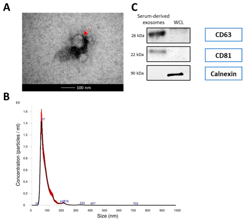
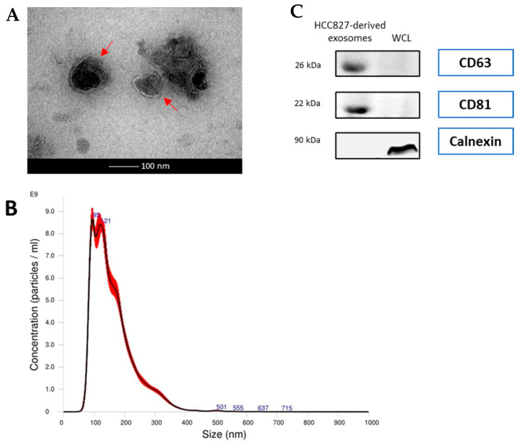
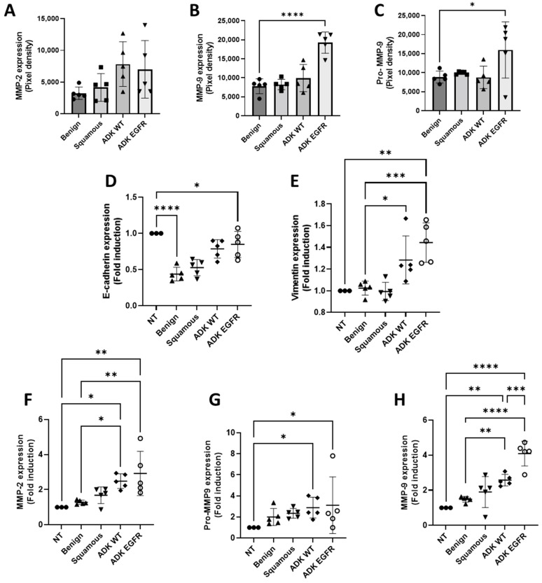
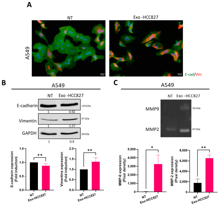
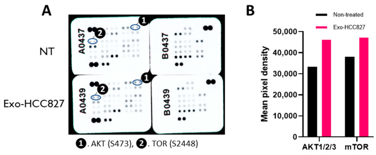
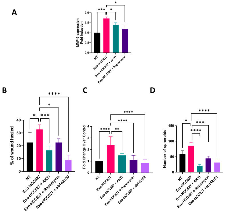
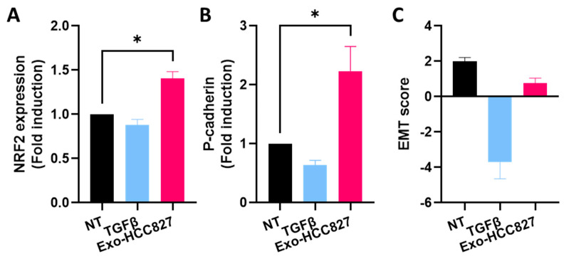
Exosomes, a class of extra cellular nano-sized vesicles (EVs), and their contents have gained attention as potential sources of information on tumor detection and regulatory drivers of tumor progression and metastasis. The effect of exosomes isolated from patients with an Epidermal Growth Factor Receptor (EGFR)-mutated adenocarcinoma on the promotion of epithelial-mesenchymal transition (EMT) and invasion were examined. Exosomes derived from serum of patients with EGFR-mutated non-small cell lung cancer (NSCLC) mediate the activation of the Phosphoinositide 3-kinase (PI3K)/AKT/ mammalian target of rapamycin (mTOR) pathway and induce an invasion through the up-regulation of matrix metalloproteinase-9 (MMP-9) in A549 cells. We observed a significant increase in the expression of vimentin, a mesenchymal marker, while retaining the epithelial characteristics, as evidenced by the unaltered levels of E-cadherin and Epithelial cell adhesion molecule (EPCAM). We also observed an increase of nuclear factor erythroid 2-related factor 2 (NFR2) and P-cadherin expression, markers of hybrid EMT. Exosomes derived from EGFR-mutated adenocarcinoma serum could be a potential mediator of hybrid EMT and tumor invasion. Understanding how cancerous cells communicate and interact with their environment via exosomes will improve our understanding of lung cancer progression and metastasis formation.

Keywords: EGFR; exosome; lung cancer; partial EMT.


Conflict of interest statement

The authors declare no conflict of interest.

Figures

Figure 1
Figure 1
Characterization of serum exosomes (A) Transmission electron microscope (TEM) images of exosomes isolated from serum. The bar represents 100 nm. Red arrows indicate exosomes. (B) The size distribution of serum exosomes determined by nanoparticle tracking analysis (NTA). (C) Western blot analysis of exosome markers (CD63 and CD81) and negative markers (calnexin) in equivalent amounts of protein from serum exosomes and A549 whole cell lysates (WCL) (as a control). (Original whole Western-blot Images and densitometry reading in Figure 1 and Table S2).
Figure 2
Figure 2
Characterization of exosomes from the HCC827 cell line (A) Transmission electron microscope (TEM) images of exosomes isolated from HCC827 cells. The bar represents 100 nm. Red arrows indicate exosomes. (B) The size distribution of serum exosomes determined by nanoparticle tracking analysis (NTA). (C) Western blot analysis of exosome markers (CD63 and CD81) and negative markers (calnexin) in equivalent amounts of protein from HCC827-derived exosomes and A549 whole cell lysates (WCL) (as a control). (Original whole Western-blot Images and densitometry reading in Figure 2 and Table S3).
Figure 3
Figure 3
Exosomes from serum of EGFR-mutated patients promote extracellular matrix degradation and EMT-like process (AC) Quantification analysis of expression of MMPs obtained by zymography in exosomes isolated from serum (mean ± SEM, n = 5). (D,E) Quantification analysis of vimentin and E-cadherin expression obtain by Western blotting of protein extracts of A549 cells treated with exosomes isolated from serum. GAPDH is used as a loading control (mean ± SEM, n = 5). (FH) Quantification analysis of expression of MMPs obtained by zymography of supernatant of A549 cells treated with exosomes isolated from serum (mean ± SEM, n = 5). Significance was set at * p < 0.05, ** p < 0.01, *** p < 0.001 and **** p < 0.0001.
Figure 4
Figure 4
Exosomes derived from the HCC827 cell line promoted extracellular matrix degradation and EMT (A) Immunofluorescence detection of vimentin (red) and E-cadherin (green) in A549 cells treated with exosomes isolated from HCC827 supernatant. Nuclei were stained with DAPI (blue). Scale bar = 20 μm. (B) Western blot analysis and quantification of vimentin and E-cadherin expression in A549 cells treated with exosomes isolated from HCC827 supernatant. GAPDH was used as a loading control (mean ± SEM, n = 4; statistical analysis was performed using unpaired t-test). (Original whole Western-blot Images and densitometry reading in Figure 3 and Table S4). (C) Zymography analysis and quantification of A549 cells (right) treated with exosomes isolated from HCC827 supernatant (mean ± SEM, n = 4; statistical analysis was performed using unpaired t-test). Significance was set at * p < 0.05 and ** p < 0.01.
Figure 5
Figure 5
Exosomes from HCC827 cell line activates the PI3K/AKT/mTOR signaling pathway. (A) Phosphokinase antibody array blots using lysates of A549 treated with 50 µg of exosomes isolated from HCC827 supernatant. Circled spots indicated positive signal for ❶ AKT (S473) and ❷. TOR (S2448) (B) Quantification of the relative levels of the phosphorylation of indicated kinases.
Figure 6
Figure 6
Exosomes from HCC827 cell line induced MMP-9-dependent invasion and migration. (A) Quantification analysis of MMP-9 expression obtained by RT-qPCR of protein extracts of A549 cells treated with exosomes isolated from HCC827 supernatant with or without AKTi or rapamycin (mean ± SEM, n = 3; statistical analysis was performed using two-way ANOVA). (B) Analysis of invasive capacities of A549 cells treated with exosomes isolated from HCC827 supernatant with or without AKTi, rapamycin, or ab142180 by wound healing assay (mean ± SEM, n = 3; statistical analysis was performed using two-way ANOVA). (C) Analysis of invasive capacities of A549 cells treated with exosomes isolated from HCC827 supernatant with or without AKTi, rapamycin, or ab142180 in a modified Boyden chamber invasion assay (mean ± SEM, n = 3; statistical analysis was performed using two-way ANOVA). (D) Analysis of 3D outgrowth of A549 cells treated with exosomes isolated from HCC827 supernatant with or without AKTi, rapamycin, or ab142180 by 3D organotypic growth assay (mean ± SEM, n = 4; statistical analysis was performed using two-way ANOVA). Significance was set at * p < 0.05, ** p < 0.01, *** p < 0.001 and **** p < 0.0001.
Figure 7
Figure 7
Exosomes from the HCC827 cell line induced hybrid EMT. Quantification analysis of NFR2 (A) and P-cadherin (B) expression obtained by RT-qPCR of protein extracts of A549 cells treated with TGFβ and exosomes isolated from HCC827 supernatant (mean ± SEM, n = 3; statistical analysis was performed using unpaired t-test). Significance was set at * p < 0.05. (C) EMT score calculated by using RT-qPCR levels of two epithelial and three mesenchymal markers. (Chae et al., modified formula).

References

    1. van Niel G., D’Angelo G., Raposo G. Shedding Light on the Cell Biology of Extracellular Vesicles. Nat. Rev. Mol. Cell Biol. 2018;19:213–228. doi: 10.1038/nrm.2017.125. - DOI - PubMed
    1. Kalluri R. The Biology and Function of Exosomes in Cancer. J. Clin. Investig. 2016;126:1208–1215. doi: 10.1172/JCI81135. - DOI - PMC - PubMed
    1. Skog J., Würdinger T., van Rijn S., Meijer D.H., Gainche L., Sena-Esteves M., Curry W.T., Carter B.S., Krichevsky A.M., Breakefield X.O. Glioblastoma Microvesicles Transport RNA and Proteins That Promote Tumour Growth and Provide Diagnostic Biomarkers. Nat. Cell Biol. 2008;10:1470–1476. doi: 10.1038/ncb1800. - DOI - PMC - PubMed
    1. Valadi H., Ekström K., Bossios A., Sjöstrand M., Lee J.J., Lötvall J.O. Exosome-Mediated Transfer of MRNAs and MicroRNAs Is a Novel Mechanism of Genetic Exchange between Cells. Nat. Cell Biol. 2007;9:654–659. doi: 10.1038/ncb1596. - DOI - PubMed
    1. Melo S.A., Sugimoto H., O’Connell J.T., Kato N., Villanueva A., Vidal A., Qiu L., Vitkin E., Perelman L.T., Melo C.A., et al. Cancer Exosomes Perform Cell-Independent MicroRNA Biogenesis and Promote Tumorigenesis. Cancer Cell. 2014;26:707–721. doi: 10.1016/j.ccell.2014.09.005. - DOI - PMC - PubMed