. 2022 Aug 5;14(15):3810.
doi: 10.3390/cancers14153810.

Cancer-Associated Fibroblasts in a 3D Engineered Tissue Model Induce Tumor-like Matrix Stiffening and EMT Transition

Affiliations

Cancer-Associated Fibroblasts in a 3D Engineered Tissue Model Induce Tumor-like Matrix Stiffening and EMT Transition

Martial Millet et al. Cancers (Basel). .

Abstract

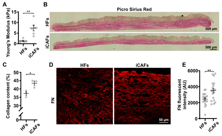
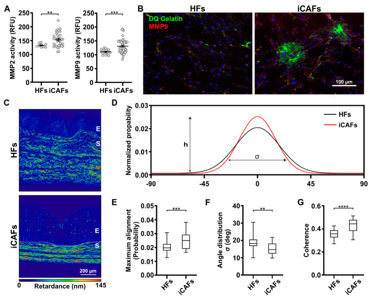
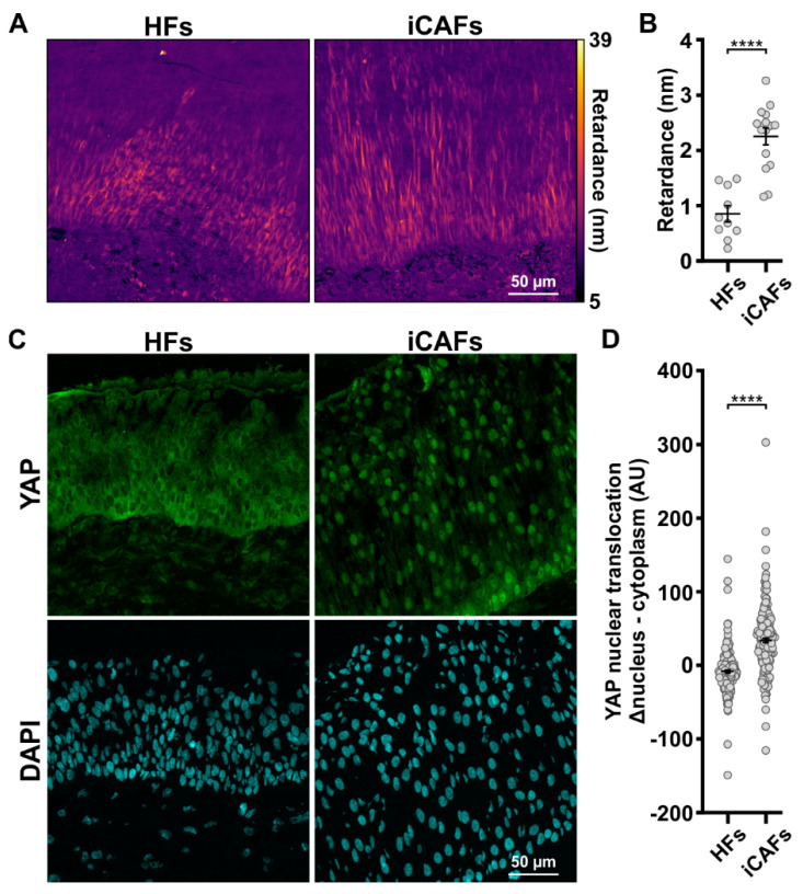
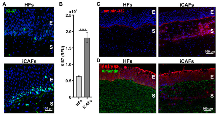
A tumor microenvironment is characterized by its altered mechanical properties. However, most models remain unable to faithfully recreate the mechanical properties of a tumor. Engineered models based on the self-assembly method have the potential to better recapitulate the stroma architecture and composition. Here, we used the self-assembly method based on a bladder tissue model to engineer a tumor-like environment. The tissue-engineered tumor models were reconstituted from stroma-derived healthy primary fibroblasts (HFs) induced into cancer-associated fibroblast cells (iCAFs) along with an urothelium overlay. The iCAFs-derived extracellular matrix (ECM) composition was found to be stiffer, with increased ECM deposition and remodeling. The urothelial cells overlaid on the iCAFs-derived ECM were more contractile, as measured by quantitative polarization microscopy, and displayed increased YAP nuclear translocation. We further showed that the proliferation and expression of epithelial-to-mesenchymal transition (EMT) marker in the urothelial cells correlate with the increased stiffness of the iCAFs-derived ECM. Our data showed an increased expression of EMT markers within the urothelium on the iCAFs-derived ECM. Together, our results demonstrate that our tissue-engineered tumor model can achieve stiffness levels comparable to that of a bladder tumor, while triggering a tumor-like response from the urothelium.

Keywords: 3D tumor models; ECM remodeling; EMT; bladder cancer; cancer-associated fibroblasts; cell contractility; engineered tumor microenvironment; matrix stiffness; mechanotransduction.


Conflict of interest statement

The authors declare no conflict of interest.

Figures

Figure 1
Figure 1
Altered ECM protein content in iCAFs-derived stroma. HFs and iCAFs were cultured in order to produce ECM sheets. Three cell sheets were stacked together, and urothelial cells were seeded on top to create 3D vesical models. (A) Young’s elastic modulus computed from tensile deformation test of HFs and iCAFs-derived stroma. N = 6 independent biological replicates. An unpaired t-test was performed. (B) Tile images of histological sections from HFs and iCAFs-derived models stained with Picrosirius red showing the collagen fibers. (C) Corresponding quantification of collagen content (N = 3 independent biological replicates). (D) Representative confocal images of HFs-derived and iCAFs-derived constructs stained for fibronectin (FN). (E) Corresponding quantification of FN signal based on the background normalized intensity (N = 3 independent biological replicates, 15 images per condition). Data are presented as mean ± SEM. * p < 0.05, ** p < 0.01.
Figure 2
Figure 2
iCAFs-derived 3D vesical model displays increased remodeling of collagen fibers. (A) Quantification in relative fluorescence units (RFU) of MMP2 and MMP9 activity secreted by HFs and iCAFs. The data are represented as mean ± SEM. N = 4 independent biological replicates. (B) Fluorescence images of the matrix degradation by the HFs and iCAFs visualized with the DQTM gelatin. The green fluorescence corresponds to gelatin degradation and the red fluorescence to MMP9. Nuclei were counterstained with DAPI. (C) Retardance images from HFs-derived and iCAFs-derived constructs stained with Picrosirius red. (D) Measured distribution of the collagen fibers’ spatial orientation over a range of 180°, (E) along with the corresponding quantification of the peak distribution (h) representing the proportion of collagen fibrils at 0°, (F) and the distribution width of the fiber angle (σ). (G) Quantification of the coherence of the Picrosirius red signal as a readout of local ECM alignment in the stroma (N = 3 per condition, 30 images per condition). ** p ≤ 0.01, *** p ≤ 0.001, and **** p ≤ 0.0001.
Figure 3
Figure 3
Mechanical forces induced through iCAFs-derived urothelium correlate with YAP nuclear translocation. (A) Retardance image of the urothelium on the HFs-derived and iCAFs-derived stroma. Imaging of the optical retardance was performed on label-free cryosections with the QPOL system. (B) Corresponding quantification of cell contractility within the urothelium (N = 3 per condition, 30 images per condition). (C) Representative confocal images of the urothelial cells seeded on HFs-derived and iCAFs-derived stroma stained for YAP. Nuclei were counterstained with DAPI. (D) Corresponding quantification of YAP nuclear translocation by measuring the difference between nuclear expression and cytoplasm expression in urothelial cells. (N = 3 per condition, 15 images per condition). The data are represented as mean ± SEM. **** p ≤ 0.0001.
Figure 4
Figure 4
The presence of iCAFs in the stroma of 3D vesical models induces the proliferation and infiltration of urothelial cells. (A) Representative fluorescent images of the HFs-derived and iCAFs-derived models stained for Ki67 (green). Nuclei were counterstained with Hoechst (blue). (B) Corresponding quantification in relative fluorescence units (RFU) of the Ki67 signal. N = 4 independent biological replicates. (C) Representative fluorescent images of the HFs-derived and iCAFs-derived models of laminin-332. (D) Representative fluorescent images of vimentin- and epithelium-specific cytokeratin marker AE1/AE3 in the HFs-derived and iCAFs-derived constructs. The images show infiltration of epithelial cells within the stroma in the iCAFs-derived stroma. E = epithelium; S = stroma. The data are represented as mean ± SEM. **** p < 0.0001.
Figure 5
Figure 5
The iCAFs-derived ECM promotes an EMT state in urothelial cells. (A) Representative confocal images of the urothelium seeded on HFs-derived and iCAFs-derived stroma and stained for vimentin. (B) Corresponding quantification of the vimentin signal across the urothelium. The basal lamina was set as the origin and positive values go toward the top of the urothelium. (C) Representative confocal images of the urothelium seeded on HFs-derived and iCAFs-derived stroma and stained for ZEB1. (D) Corresponding quantification of ZEB1 signal by measuring the difference between nuclear expression and cytoplasm expression in urothelial cells. Data are presented as scatter plots (N = 3 per condition, 15 images per condition). Nuclei were counterstained with DAPI. The data are represented as mean ± SEM. **** p ≤ 0.0001.

References

    1. Levental K.R., Yu H., Kass L., Lakins J.N., Egeblad M., Erler J.T., Fong S.F.T.T., Csiszar K., Giaccia A., Weninger W., et al. Matrix crosslinking forces tumor progression by enhancing integrin signaling. Cell. 2009;139:891–906. doi: 10.1016/j.cell.2009.10.027. - DOI - PMC - PubMed
    1. DuFort C.C., Paszek M.J., Weaver V.M. Balancing forces: Architectural control of mechanotransduction. Nat. Rev. Mol. Cell Biol. 2011;12:308–319. doi: 10.1038/nrm3112. - DOI - PMC - PubMed
    1. Pathak A., Kumar S. Transforming potential and matrix stiffness co-regulate confinement sensitivity of tumor cell migration. Integr. Biol. 2013;5:1067–1075. doi: 10.1039/c3ib40017d. - DOI - PMC - PubMed
    1. Paszek M.J., Zahir N., Johnson K.R., Lakins J.N., Rozenberg G.I., Gefen A., Reinhart-King C.A., Margulies S.S., Dembo M., Boettiger D., et al. Tensional homeostasis and the malignant phenotype. Cancer Cell. 2005;8:241–254. doi: 10.1016/j.ccr.2005.08.010. - DOI - PubMed
    1. Shin J.-W., Mooney D.J. Extracellular matrix stiffness causes systematic variations in proliferation and chemosensitivity in myeloid leukemias. Proc. Natl. Acad. Sci. USA. 2016;113:12126–12131. doi: 10.1073/pnas.1611338113. - DOI - PMC - PubMed

LinkOut - more resources