Review
. 2022 Aug 8;14(15):3840.
doi: 10.3390/cancers14153840.

Theranostics Using Indocyanine Green Lactosomes

Affiliations
Review

Theranostics Using Indocyanine Green Lactosomes

Masaki Kaibori et al. Cancers (Basel). .

Abstract

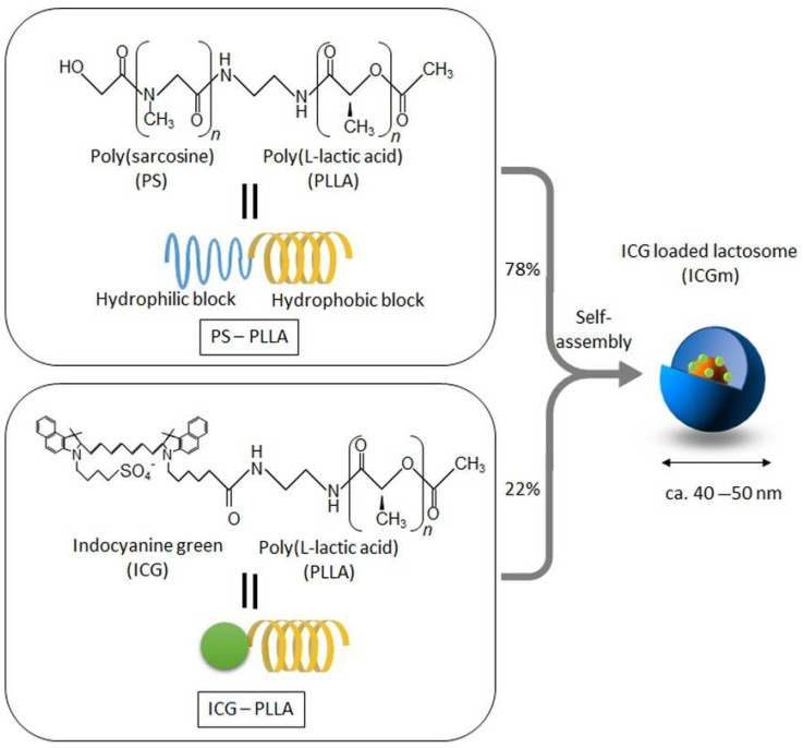
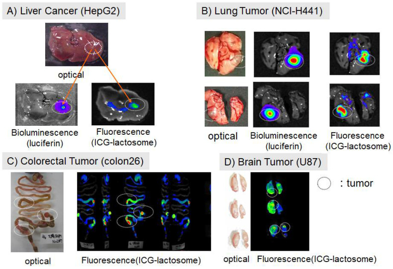
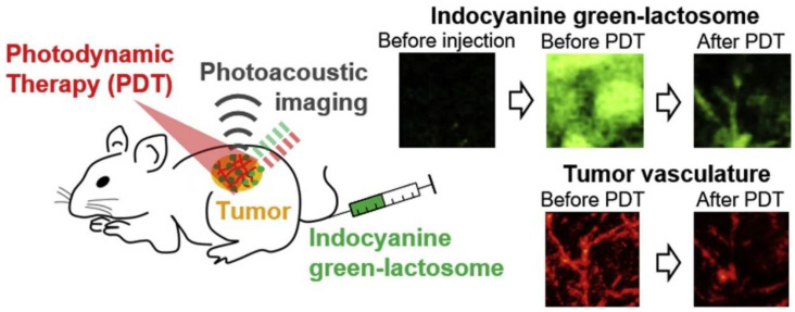
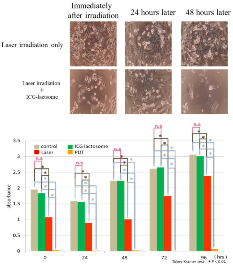
Lactosomes™ are biocompatible nanoparticles that can be used for cancer tissue imaging and drug delivery. Lactosomes are polymeric micelles formed by the self-assembly of biodegradable amphiphilic block copolymers composed of hydrophilic polysarcosine and hydrophobic poly-L-lactic acid chains. The particle size can be controlled in the range of 20 to 100 nm. Lactosomes can also be loaded with hydrophobic imaging probes and photosensitizers, such as indocyanine green. Indocyanine green-loaded lactosomes are stable for long-term circulation in the blood, allowing for accumulation in cancer tissues. Such lactosomes function as a photosensitizer, which simultaneously enables fluorescence diagnosis and photodynamic therapy. This review provides an overview of lactosomes with respect to molecular design, accumulation in cancer tissue, and theranostics applications. The use of lactosomes can facilitate the treatment of cancers in unresectable tissues, such as glioblastoma and head and neck cancers, which can lead to improved quality of life for patients with recurrent and unresectable cancers. We conclude by describing some outstanding questions and future directions for cancer theranostics with respect to clinical applications.

Keywords: indocyanine green lactosome; photodynamic diagnosis; photodynamic therapy; tumor accumulation.


Conflict of interest statement

The authors declare no conflict of interest.

Figures

Figure 1
Figure 1
The structure of indocyanine green (ICG)-loaded lactosomes (ICGm’s). ICGm’s are formed by molecular assembly with hydrophobic helical poly-L-lactic acid (PLLA) and hydrophilic polysarcosine (PS) amphiphilic block polydepsipeptide, which include ICG-labeled PLLA in the hydrophobic inner core. (Reprinted from Tsujimoto et al. [9]).
Figure 2
Figure 2
Accumulation of ICG-lactosomes in mouse cancer models. Images were obtained at 24 h after intravenous administration of ICG-lactosomes via the tail vein. The cancer cells carried the luciferase reporter gene. The ICG fluorescence sites overlapped at the luciferin bioluminescence sites, which are a marker of cancer cells. Cancer tissue was confirmed by luciferin bioluminescence and macroscopic findings. (Reproduced from Ozeki and Hara [17]).
Figure 3
Figure 3
Schematic of photodynamic therapy (PDT) and photoacoustic imaging with ICG-lactosomes. Photoacoustic images (3.9 × 3.9 mm2) show the accumulation of ICG-lactosomes in the tumor (before PDT). The photoacoustic probe was automatically scanned over a 3.9 mm × 3.9 mm region of interest with a step size of 50 and 150 μm for imaging of blood vessels and ICG distribution, respectively. The wavelength of the short laser pulse was 532 nm for photoacoustic imaging of oxy- and deoxy-hemoglobin in blood vessels. The laser pulse at 796 nm was used for ICG imaging. Results of the fluorescence imaging of ICG showed that the photoacoustic imaging decreased by only 0.78% in average fluorescence intensity originating from ICG in the tumor (data not shown), indicating a negligible photobleaching by the laser light for photoacoustic imaging. After PDT, the photoacoustic signal was drastically decreased, suggesting photobleaching in the tumor. (Reprinted from Tsunoi et al. [13] with permission from Elsevier).
Figure 4
Figure 4
Tumor cell proliferation of the human breast cancer cell line MDA-MB-231 is suppressed by ICG-lactosomes. ICG-lactosomes combined with laser irradiation abolished cells (lower panels). Mean absorbance values indicate cell viability in the WST-1 assay. The laser-irradiated group (red) showed significantly lower cell viability than the control and ICG-lactosome alone (green) groups. The fluence rate and irradiation period were set to 298 mW/cm2 and 60 s, respectively, corresponding to a fluence of 17.9 J/cm2. Furthermore, the photodynamic therapy (PDT) group (yellow) showed drastically lower cell viability compared to the laser group [20]. n.s, not significant; * p < 0.01. (Reproduced from Ozeki and Hara [17]).
Figure 5
Figure 5
Photodynamic therapy (PDT) in bone metastases. The photodynamic therapy group underwent laser irradiation 24 h after ICG-lactosome administration at 1, 3, and 5 weeks after tumor cell transplantation. The area of osteolysis was compared using computed tomography imaging at 7 weeks after tumor cell transplantation. The area surrounded by the dotted line indicates tumor cells. The PDT group showed smaller tumor areas than the laser group. (Reproduced from Ozeki and Hara [17]).
Figure 6
Figure 6
Photodynamic therapy of peritoneal dissemination in gastric cancer. (Left) Comparison between ICG and ICG-lactosomes in terms of luminescence and ICG-fluorescence from tumors. (Right) Survival rate after photodynamic therapy (PDT) in mice treated with ICG or ICG-lactosomes. PDT with ICG-lactosomes resulted in significantly improved survival rates compared to that with ICG alone. (Reproduced from Tsujimoto et al. [9]).
Figure 7
Figure 7
Photodynamic diagnosis of lymph node metastasis. (A) In vivo images of the popliteal lymph nodes (arrowheads). (B) Ex vivo images of the bilateral popliteal lymph nodes. (Reprinted from Tsujimoto et al. [22] with permission from Springer Nature).
Figure 8
Figure 8
Photodynamic therapy in gallbladder cancer. (A) Accumulation of fluorescence imaging in mouse subcutaneous tumors after administration of ICG or ICG-lactosomes was observed daily using in vivo imaging systems. On the second day following administration, PDT was performed in groups with ICG (n = 5, open circles) and single (ICG-Lact1, n = 5, closed squares) or double PDT with ICG-lactosomes (ICG-Lact2, n = 5, closed triangles). The second irradiation was performed on day 5. (B) Effect of PDT on tumor growth in mice with subcutaneous tumors treated with ICG and ICG-lactosomes. On the second day following administration, PDT was performed in the ICG (n = 8, open circles) and ICG-Lact1 (n = 8, closed squares) groups. The second PDT was performed 3 days after the first PDT in the ICG-Lact2 group (n = 8, closed triangles). * p < 0.001 between the ICG and ICG-Lact2 groups.  p < 0.05 between the ICG and ICG-Lact1 groups. p < 0.05 between the ICG-Lact1 and ICG-Lact2 groups. (Reproduced from Hishikawa et al. [24]).
Figure 9
Figure 9
(A1) An increase in temperature at the near-infrared (NIR) irradiation spot on the tumor and non-tumor sites in mice administered ICG-lactosomes. The irradiation points are indicated by black circles. (A2) The increase in temperature at the tumor site was greater than that at the non-tumor site. * p < 0.01. (B1) Black circles indicate the NIR irradiation points on the non-tumor site in mice with or without ICG-lactosome administration. (B2) There was no difference in temperature increase at the non-tumor site with or without ICG-lactosome administration. (Reprinted from Nomura et al. [26]).

References

    1. Matsumura Y., Maeda H. A new concept for macromolecular therapeutics in cancer chemotherapy: Mechanism of tumoritropic accumulation of proteins and the antitumor agent smancs. Cancer Res. 1986;46:6387–6392. - PubMed
    1. Lukyanov A.N., Hartner W.C., Torchilin V.P. Increased accumulation of PEG-PE micelles in the area of experimental myocardial infarction in rabbits. J. Control. Release. 2004;94:187–193. doi: 10.1016/j.jconrel.2003.10.008. - DOI - PubMed
    1. Torchilin V.P. Micellar nanocarriers: Pharmaceutical perspectives. Pharm. Res. 2007;24:1–16. doi: 10.1007/s11095-006-9132-0. - DOI - PubMed
    1. Makino A., Kizaka-Kondoh S., Yamahara R., Hara I., Kanzaki T., Ozeki E., Hiraoka M., Kimura S. Near-infrared fluorescence tumor imaging using nanocarrier composed of poly(L-lactic acid)-block-poly(sarcosine) amphiphilic polydepsipeptide. Biomaterials. 2009;30:5156–5160. doi: 10.1016/j.biomaterials.2009.05.046. - DOI - PubMed
    1. Makino A., Hara E., Hara I., Yamahara R., Kurihara K., Yamanoto F., Kimura S. Control of in vivo blood clearance time of polymeric micelle by stereochemistry of amphiphilic polydepsipeptides. J. Control. Release. 2012;161:821–825. doi: 10.1016/j.jconrel.2012.05.006. - DOI - PubMed

LinkOut - more resources